[Preprint]. 2024 May 25:2023.06.22.546088.
doi: 10.1101/2023.06.22.546088.

Single-molecule imaging reveals the kinetics of non-homologous end-joining in living cells

Affiliations

Single-molecule imaging reveals the kinetics of non-homologous end-joining in living cells

Mariia Mikhova et al. bioRxiv. .

Update in

Abstract

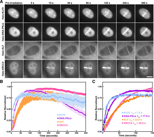
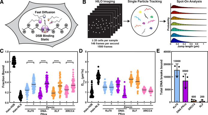
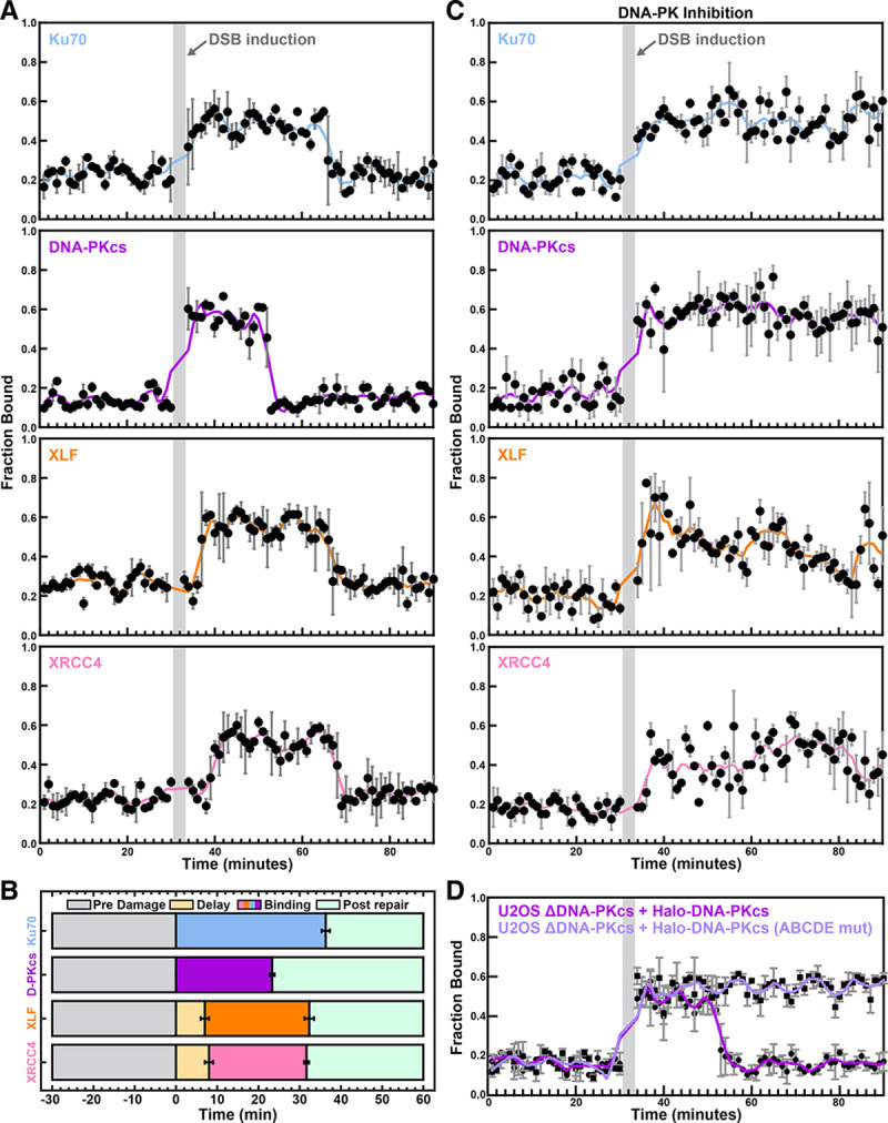
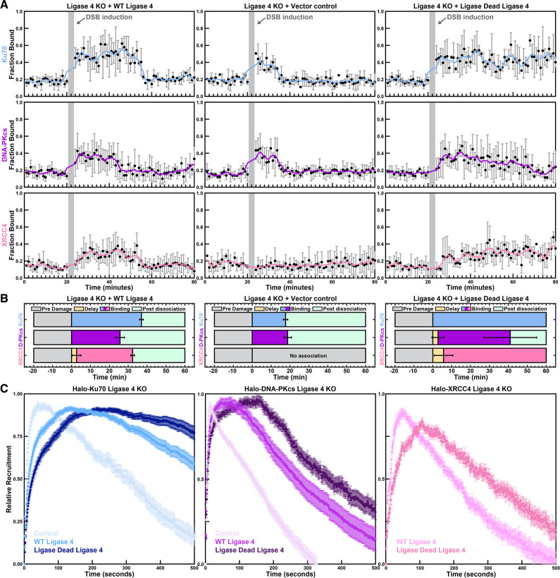
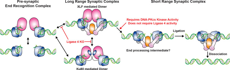
Non-homologous end joining (NHEJ) is the predominant pathway that repairs DNA double-stranded breaks (DSBs) in vertebrates. However, due to challenges in detecting DSBs in living cells, the repair capacity of the NHEJ pathway is unknown. The DNA termini of many DSBs must be processed to allow ligation while minimizing genetic changes that result from break repair. Emerging models propose that DNA termini are first synapsed ~115Å apart in one of several long-range synaptic complexes before transitioning into a short-range synaptic complex that juxtaposes DNA ends to facilitate ligation. The transition from long-range to short-range synaptic complexes involves both conformational and compositional changes of the NHEJ factors bound to the DNA break. Importantly, it is unclear how NHEJ proceeds in vivo because of the challenges involved in analyzing recruitment of NHEJ factors to DSBs over time in living cells. Here, we develop a new approach to study the temporal and compositional dynamics of NHEJ complexes using live cell single-molecule imaging. Our results provide direct evidence for stepwise maturation of the NHEJ complex, pinpoint key regulatory steps in NHEJ progression, and define the overall repair capacity NHEJ in living cells.


Conflict of interest statement

COMPETING INTERESTS None declared.

Figures

Figure 1.
Figure 1.. Generation and validation of HaloTagged core NHEJ proteins in U20S cells.
(A) Graphical depiction of the long-range XLF-mediated NHEJ synaptic complex and HaloTag (based on PDB: 7NFC and 6U32) ,. (B) Western blots of U2OS cells expressing HaloTagged NHEJ factors and parental U2OS cells probed with antibodies against Ku70 (top), XRCC4 (middle), and XLF (bottom). (C) Representative images of living cells expressing HaloTagged NHEJ factors and Halo-Rif1 labeled with JFX650 HaloTag ligand in the presence or absence of zeocin (Scale bar = 10 um). (D) Clonogenic survival assay of U2OS cells expressing HaloTagged NHEJ factors, parental U2OS cells, and U2OS cells with DNA-PKcs knocked out after challenge with Zeocin (N = 4, 3 technical replicates per biological replicate, Mean ± S.D.). (E) Fluorescence imaging of 2 clones of each HaloTagged NHEJ factor labeled with JF650 HaloTag ligand and separated on an SDS-PAGE gel. (F) Quantification of the total protein abundance based on recombinant 3X FLAG-HaloTag labeled with JF646 and cell lysates from a specific number of U2OS using in-gel fluorescence intensity values after applying a correction factor from western blots (see Fig. S1C–D, N = 3, Mean ± S.D.).
Figure 2.
Figure 2.. Recruitment kinetics of the core NHEJ factors to complex DNA lesions
(A) Representative images of HaloTagged NHEJ factor (JFX650) recruitment to laser-induced DNA lesions over time (Scale bar = 10 μm). (B) Normalized recruitment kinetics of HaloTagged DDR proteins to laser-induced DSBs. Data are presented as the average increase in fluorescence post-laser microirradiation normalized to the brightest frame for cell analyzed (N = 10 – 15 individual cells analyzed for each HaloTag cell line, Mean ± S.D.). (C) Half-times (t1/2) of each protein to laser-induced DSBs determined by nonlinear regression assuming one-phase association (without dissociation).
Figure 3.
Figure 3.. Zeocin-induced DSB formation promotes chromatin association of Halo-tagged NHEJ factors.
(A) Rational for analyzing DSB association of NHEJ factors using single-molecule imaging. DSB association proteins are static while unbound molecules diffuse rapidly through the nucleus. (B) Graphical representation of the workflow used for live-cell single-molecule imaging of core NHEJ proteins. (C) Plot of the Fraction Bound for each HaloTag NHEJ protein under untreated conditions and post-zeocin exposure that were analyzed using a two-state model of diffusion. Each data point represents the fraction bound of each protein in an individual cell (N = 3, n ≥ 20 cells for each protein per replicate and condition, Black bar = median). Data were analyzed by two-way ANOVA with Tukey’s posthoc test (**** = p < 0.0001). (D) Diffusion coefficients for freely diffusing HaloTagged NHEJ proteins. Each data point represents the DFree calculated from the tracks in an individual cell (N = 3, n ≥ 20 cells for each protein per replicate and condition, Black bar = median). (E) Total number of DNA breaks bound be each tagged DNA repair factor after treatment with zeocin (N = 3, Mean ± S.D.).
Figure 4.
Figure 4.. Time course analysis of the recruitment of Halo-tagged NHEJ factors to DNA breaks.
(A) Experimental approach to analyze the chromatin binding of the tagged DNA repair factors over time. (B) Plot of the static fraction of Halo-Ku70, Halo-DNA-PKcs, and Halo-XRCC4 before and after DNA damage induction (shaded area) with various concentrations of calicheamicin for 3 minutes. Graphs represent the rolling average over three consecutive timepoints (for individual graphs with experimental error see Figure S4A). (C) Quantification of the timing of the recruitment of Halo-Ku70, Halo-DNA-PKcs, and Halo-XRCC4 after DNA damage induction (N = 3, Mean ± S.D.). (D) Quantification of the number of break bound Halo-Ku70 (pooled data for 5 time points after DNA damage induction), Halo-DNA-PKcs (pooled data for 10 time points after DNA damage induction), and Halo-XRCC4 (pooled data for 10 time points after chromatin recruitment) molecules after DNA damage induction with various concentrations of calicheamicin (N = 3, Mean ± S.D.). (E) Quantification of the DNA repair rate by using static Halo-Ku70 as a marker for the initial number of DNA breaks (Fig. 4D) and the dissociation of XRCC4 (Fig. 4B) to mark the completion of DNA repair by NHEJ (N = 3, Mean ± S.D.)
Figure 5.
Figure 5.. Compositional changes of the NHEJ complex are controlled by DNA-PK activity.
(A) Plot of the static fraction of each HaloTag NHEJ protein over time. A single cell was imaged at each timepoint. After 30 minutes DSBs were induced with 40 nM calicheamicin for 3 minutes (shaded area) and cells were imaged for 60 minutes thereafter (N = 3, Mean ± S.D.). (B) Quantification of the timing of the recruitment of Halo-Ku70, Halo-DNA-PKcs, Halo-XLF, and Halo-XRCC4 after DNA damage induction (N = 3, Mean ± S.D.). (C) Plot of the static fraction of each HaloTag NHEJ protein over time in the presence of a DNA-PK inhibitor (Nu7441, 2 nM). A single cell was imaged at each timepoint. After 30 minutes DSBs were induced with 40 nM Calicheamicin for 3 minutes (shaded area) and cells were imaged for 60 minutes thereafter (N = 3, Mean ± S.D.). (D) Plot of the static fraction of wildtype Halo-DNA-PKcs or Halo-DNA-PKcs with mutated autophosphorylation sites (ABCDE mut.) expressing in U2OS cells in which DNA-PKcs was knocked out. A single cell was imaged at each timepoint. After 30 minutes DSBs were induced with 40 nM calicheamicin for 3 minutes (shaded area) and cells were imaged for 60 minutes thereafter (N = 3, Mean ± S.D.).
Figure 6.
Figure 6.. Ligase 4 is required for the retention of Ku70 and DNA-PKcs at DNA breaks.
(A) Plot of the static fraction of Halo-Ku70 (top row), Halo-DNA-PKcs (middle row), or Halo-XRCC4 (bottom row) over time in ligase 4 knockout cells, transiently expressing wildtype ligase 4 (left column), a vector control (middle column) or ligase dead ligase 4 (right column). After 20 minutes DSBs were induced with 40 nM Calicheamicin for 3 minutes (shaded area) and cells were imaged for 60 minutes thereafter (N = 3, Mean ± S.D.). (B) Quantification of the timing of the recruitment of Halo-Ku70, Halo-DNA-PKcs, Halo-XLF, and Halo-XRCC4 after DNA damage induction in Ligase 4 knockout cells expressing a control plasmid, or expression vectors for wildtype and catalytically inactive Ligase 4 (N = 3, Mean ± S.D.). (C) Laser micro irradiation of U2OS cells with ligase 4 knockout, expressing Halo-Ku70 (left), Halo-DNA-PKcs (middle), or Halo-XRCC4 (right), transiently expressing wildtype ligase 4, a vector control, or catalytically inactive ligase 4 (N = 30–45 cells per condition, Mean ± S.E.M.).
Figure 7.
Figure 7.. Model for NHEJ.
Stepwise maturation of the NHEJ complex throughout DNA break repair, highlighting key transition points controlled by DNA-PKcs catalytic activity and structural contributions of the XRCC4-Ligase 4 complex.

Similar articles

References

    1. Chapman J.R., Taylor M.R.G., and Boulton S.J. (2012). Playing the End Game: DNA Double-Strand Break Repair Pathway Choice. Mol Cell 47. 10.1016/j.molcel.2012.07.029. - DOI - PubMed
    1. Stinson B.M., and Loparo J.J. (2021). Repair of DNA Double-Strand Breaks by the Nonhomologous End Joining Pathway. Annu Rev Biochem 90. 10.1146/annurev-biochem-080320-110356. - DOI - PMC - PubMed
    1. Watanabe G., and Lieber M.R. (2023). The flexible and iterative steps within the NHEJ pathway. Prog Biophys Mol Biol 180–181, 105–119. 10.1016/j.pbiomolbio.2023.05.001. - DOI - PMC - PubMed
    1. Karanam K., Kafri R., Loewer A., and Lahav G. (2012). Quantitative Live Cell Imaging Reveals a Gradual Shift between DNA Repair Mechanisms and a Maximal Use of HR in Mid S Phase. Mol Cell 47. 10.1016/j.molcel.2012.05.052. - DOI - PMC - PubMed
    1. Jasin M., and Rothstein R. (2013). Repair of strand breaks by homologous recombination. Cold Spring Harb Perspect Biol 5. 10.1101/cshperspect.a012740. - DOI - PMC - PubMed

Publication types