[Preprint]. 2024 May 26:2024.05.22.594428.
doi: 10.1101/2024.05.22.594428.

Absence of c-Maf and IL-10 enables Type I IFN enhancement of innate responses to low-dose LPS in alveolar macrophages

Affiliations

Absence of c-Maf and IL-10 enables Type I IFN enhancement of innate responses to low-dose LPS in alveolar macrophages

Pamelia N Lim et al. bioRxiv. .

Update in

Abstract

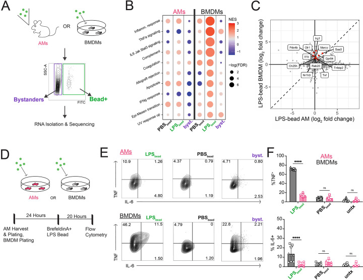
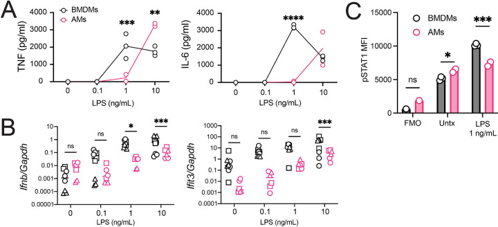
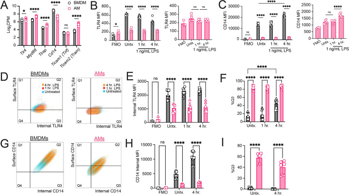
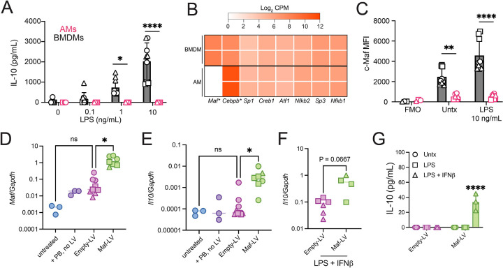
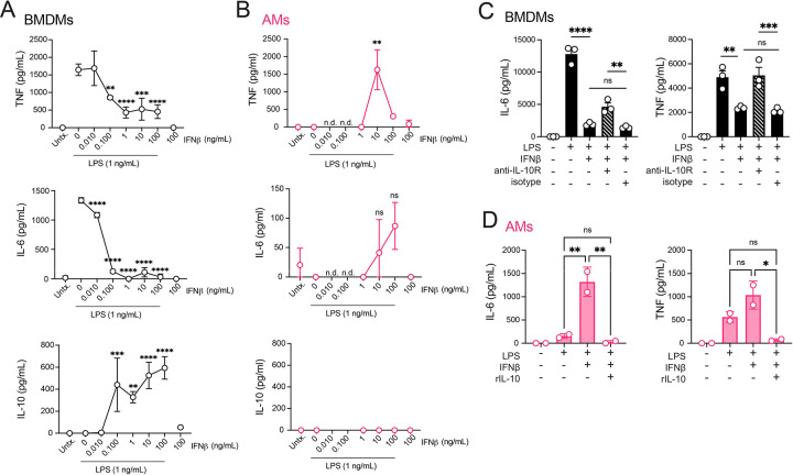
Alveolar macrophages (AMs) are lower-airway resident myeloid cells and are among the first to respond to inhaled pathogens. Here, we interrogate AM innate sensing to Pathogen Associated Molecular Patterns (PAMPs) and determine AMs have decreased responses to low-dose LPS compared to other macrophages, as measured by TNF, IL-6, Ifnb, and Ifit3. We find the reduced response to low-dose LPS correlates with minimal TLR4 and CD14 surface expression, despite sufficient internal expression of TLR4. Additionally, we find that AMs do not produce IL-10 in response to a variety of PAMPs due to low expression of transcription factor c-Maf and that lack of IL-10 production contributes to an enhancement of pro-inflammatory responses by Type I IFN. Our findings demonstrate that AMs have cell-intrinsic dampened responses to LPS, which is enhanced by type I IFN exposure. These data implicate conditions where AMs may have reduced or enhanced sentinel responses to bacterial infections.

Keywords: Alveolar macrophage; CD14; IL-10; TLR4; Type I IFN; c-Maf; innate response; lipopolysaccharide; myeloid cells.


Conflict of interest statement

Competing interests: Authors declare that they have no competing interests.

Figures

Figure 1:
Figure 1:. Alveolar macrophages do not mount a pro-inflammatory response to LPS-conjugated beads in vivo or ex vivo.
(A) Schematic representation of LPS-coated beads delivered to mice by nebulization or to BMDMs. Uncoated beads resuspended in PBS (PBS bead) delivered to controls. Cells were isolated and sorted into Bead+ or Bystander groups. (B) Gene Set Enrichment Analysis of the top ten differentially expressed pathways between LPSBead BMDMs and LPSBead AMs. (C) Scatter plot of log2 fold change values for AM LPSBead and BMDM LPSBead populations. Labeled genes are significantly up-regulated (FDR < 0.05, FC > 2) in only the BMDM LPSBead population. (D) Schematic representation of AMs or BMDMs stimulated with LPS-coated beads ex vivo and subsequently analyzed by flow cytometry for intracellular TNF and IL-6. (E) Representative dot plots of the conditions for BMDMs and AMs. (F) Intracellular Cytokine Staining of TNF and IL-6 for LPSbead, PBSbead, or untreated BMDMs and AMs. Data are representative of 3 independent experiments (E) or compiled from 2 independent experiments (B, C) or 3 independent experiments (F). Technical replicates within each experiment represented by unique shapes. ****P < 0.0001, ns not significant by two-way ANOVA with Sidak’s multiple comparison test.
Figure 2:
Figure 2:. Alveolar macrophages have a high LPS-specific activation threshold.
(A) TNF (left) and IL-6 (right) for AMs (pink, technical duplicate) and BMDMs (black, technical triplicate) either untreated or stimulated with 0.1, 1, or 10 ng/mL of LPS for 20 hours (B) Ifnb and Ifit3 gene expression for 0 – 10 ng/mL LPS for BMDMs (black) and AMs (pink) after 4 hours. (C) phospho-STAT1 intracellular staining MFI of BMDMs (black, technical triplicate) or AMs (pink, technical duplicate) after 20 hours of LPS (1 ng/mL) or fresh media (untreated). Data are representative of 3 independent experiments (A, C) or compiled from 3 independent experiments (B). Technical replicates within each experiment represented by unique shapes. (A-C) *P<0.05, ***P<0.001, ****P<0.0001, ns is not significant, Two-way ANOVA with Sidak’s multiple comparison test.
Figure 3:
Figure 3:. Alveolar macrophages have low surface expression of TLR4 and CD14 compared to other cell types.
(A) Gene expression (log2 CPM) of AMs (pink) and BMDMs (black) collected in Fig. 1A. (B) TLR4 MFI of AMs (pink, graphed independently to the right) and BMDMs (black) after 0, 1, and 4 hours of LPS (1 ng/mL) stimulation ex vivo. (C) CD14 MFI under the same conditions as (B). (D) Surface and internal TLR4 expression of BMDMs and AMs after no treatment (blue), 1 hour (red) or 4 hours (orange) post-LPS stimulation (1 ng/mL). Gates set based on cell-specific FMOs. (E) Internal TLR4 MFI for both BMDMs (black) and AMs (pink). (F) Frequency of Q3+ (internal TLR4+) BMDMs and AMs. (G) Surface and internal CD14 expression of AMs and BMDMs before and after stimulation with 4 hr. LPS (1 ng/mL). Gates set based on cell-specific FMOs. (H) Internal CD14 MFI for both BMDMs (black) and AMs (pink). (I) Frequency of Q3+ (internal CD14+) BMDMs and AMs. Data representative of 3 independent experiments (D, G) or compiled from 3 independent experiments (B, C, E, F, H, I). (A - I) *P<0.05, ****P < 0.001, ns, not significant. Two-way ANOVA with Sidak’s multiple comparison test.
Figure 4:
Figure 4:. Alveolar macrophages do not produce IL-10 in response to multiple stimuli due to low c-Maf expression.
(A) IL-10 for AMs (pink) and BMDMs (black) untreated or stimulated with 0.1, 1, or 10 ng/mL of LPS for 20 hours. (B) Expression of IL-10 promoter-associated genes (log2 CPM) from bulk RNA-sequencing of untreated AMs (left) and BMDMs (right) after 4 hours using data from Fig. 1A. *adj p-value < 0.01. (C) c-Maf MFI for BMDMs (black) or AMs (pink) after LPS (10 ng/mL) stimulation or untreated. (D) Maf gene expression relative to Gapdh for mexAMs treated with media (no polybrene (PB) no virus), PB only, Empty-lentivirus (LV), or Maf-LV. (E) Il10 gene expression relative to Gapdh for mexAMs under the same conditions for (D). (F) Il10 gene expression relative to Gapdh for mexAMs transduced with Empty or Maf LV then stimulated with LPS (10 ng/mL) and rIFNβ (100 ng/mL) for 4 hours. (G) IL-10 for mexAMs transduced with Empty or Maf-LV then stimulated with LPS (10 ng/mL) and IFNβ (100 ng/mL) for 24 hours. Data compiled from 3 independent experiments (A, C-E) or 2 independent experiments (F) or representative of 2 independent experiments (G). (A, C, I) *P<0.05, **P<0.01, ****P<0.0001 by two-way ANOVA with Sidak’s multiple comparison test. (F, G) *P<0.05 by one-way ANOVA with Tukey’s multiple comparisons test. (H) P-value reported by Mann-Whitney test.
Figure 5:
Figure 5:. IFNβ enhances alveolar macrophage TNF and IL-6 response to low-dose LPS in the absence of IL-10.
(A) TNF, IL-6, and IL-10 for BMDMs stimulated with LPS (1 ng/mL) with 0.01 – 100 ng/mL of rIFNβ. Data shown are mean with SD. with values (B) TNF, IL-6, and IL-10 for AMs stimulated with LPS (1 ng/mL) with 1 – 100 ng/mL of rIFNβ. (C) BMDMs stimulated with LPS (1 ng/mL), rIFNβ (10 ng/mL), anti-IL-10R (10 mg/mL), and/or isotype control (Rat IgG1k, 10 μg/mL). (D) AMs stimulated with LPS (1 ng/mL), rIFNβ (10 ng/mL), and/or rIL-10 (50 mg/mL) for 20 hours followed by ELISA for IL-6 (left) and TNF (right). Data are representative of 3 independent experiments (A-D). **P<0.01, ***P<0.001, ****P<0.0001, n.d. no data, ns not significant. (A, B) One-way ANOVA with Sidak’s multiple comparison test, compared to LPS only condition. (C, D) One-way ANOVA with Tukey’s multiple comparison test.

Similar articles

References

    1. Yu Y.-R.A., Hotten D.F., Malakhau Y., Volker E., Ghio A.J., Noble P.W., Kraft M., Hollingsworth J.W., Gunn M.D., and Tighe R.M. (2016). Flow Cytometric Analysis of Myeloid Cells in Human Blood, Bronchoalveolar Lavage, and Lung Tissues. Am. J. Respir. Cell Mol. Biol. 54, 13–24. 10.1165/rcmb.2015-0146OC. - DOI - PMC - PubMed
    1. Silver R.F., Myers A.J., Jarvela J., Flynn J., Rutledge T., Bonfield T., and Lin P.L. (2016). Diversity of Human and Macaque Airway Immune Cells at Baseline and during Tuberculosis Infection. Am. J. Respir. Cell Mol. Biol. 55, 899–908. 10.1165/rcmb.2016-0122OC. - DOI - PMC - PubMed
    1. Westphalen K., Gusarova G.A., Islam M.N., Subramanian M., Cohen T.S., Prince A.S., and Bhattacharya J. (2014). Sessile alveolar macrophages communicate with alveolar epithelium to modulate immunity. Nature 506, 503–506. 10.1038/nature12902. - DOI - PMC - PubMed
    1. Neupane A.S., Willson M., Chojnacki A.K., Vargas E Silva Castanheira F., Morehouse C., Carestia A., Keller A.E., Peiseler M., DiGiandomenico A., Kelly M.M., et al. (2020). Patrolling Alveolar Macrophages Conceal Bacteria from the Immune System to Maintain Homeostasis. Cell 183, 110–125.e11. 10.1016/j.cell.2020.08.020. - DOI - PubMed
    1. Grant R.A., Morales-Nebreda L., Markov N.S., Swaminathan S., Querrey M., Guzman E.R., Abbott D.A., Donnelly H.K., Donayre A., Goldberg I.A., et al. (2021). Circuits between infected macrophages and T cells in SARS-CoV-2 pneumonia. Nature 590, 635–641. 10.1038/s41586-020-03148-w. - DOI - PMC - PubMed

Publication types