[Preprint]. 2024 May 23:2024.05.23.595578.
doi: 10.1101/2024.05.23.595578.

Antibodies targeting Crimean-Congo hemorrhagic fever virus GP38 limit vascular leak and viral spread

Affiliations

Antibodies targeting Crimean-Congo hemorrhagic fever virus GP38 limit vascular leak and viral spread

Felix Pahmeier et al. bioRxiv. .

Update in

Abstract

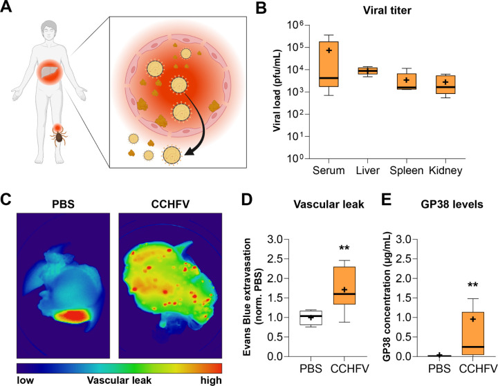
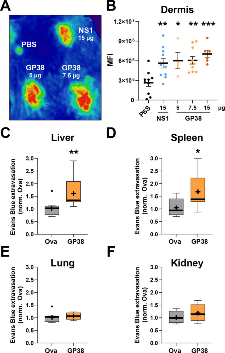
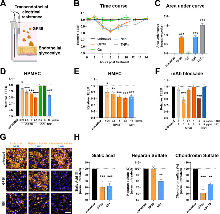
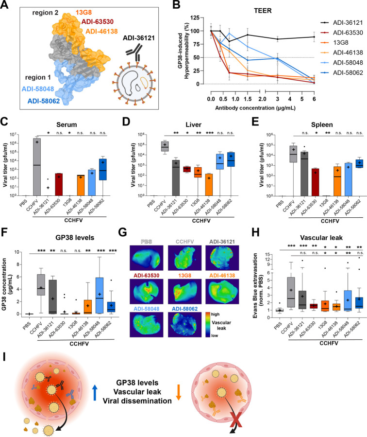
Crimean-Congo hemorrhagic fever virus (CCHFV) is a priority pathogen transmitted by tick bites, with no vaccines or specific therapeutics approved to date. Severe disease manifestations include hemorrhage, endothelial dysfunction, and multiorgan failure. Infected cells secrete the viral glycoprotein GP38, whose extracellular function is presently unknown. GP38 is considered an important target for vaccine and therapeutic design as GP38-specific antibodies can protect against severe disease in animal models, albeit through a currently unknown mechanism of action. Here, we show that GP38 induces endothelial barrier dysfunction in vitro, and that CCHFV infection, and GP38 alone, can trigger vascular leak in a mouse model. Protective antibodies that recognize specific antigenic sites on GP38, but not a protective neutralizing antibody binding the structural protein Gc, potently inhibit endothelial hyperpermeability in vitro and vascular leak in vivo during CCHFV infection. This work uncovers a function of the secreted viral protein GP38 as a viral toxin in CCHFV pathogenesis and elucidates the mode of action of non-neutralizing GP38-specific antibodies.


Figures

Figure 1.
Figure 1.. CCHFV infection leads to viral dissemination, vascular leak, and circulation of GP38.
(A) Transmission of CCHFV by tick bites is followed by virus amplification in the bloodstream, endothelial dysfunction, and liver pathology. (B) Viral load in tissues (liver, spleen, kidney) and serum from mice infected with CCHFV and treated with anti-IFNAR mAb MAR1-5A3 three days post-infection (n=5–10). (C) Representative images of leak of tracer dye Dextran-680 in the liver of PBS-treated or CCHFV-infected mice. (D) Extravasation of tracer dye Evans Blue dye into the liver of PBS-treated or CCHFV-infected mice relative to PBS control animals (n=5–10). (E) Concentration of GP38 in the serum of PBS-treated or CCHFV-infected mice determined by quantitative sandwich ELISA (n=5–14). Data shown as a Tukey distribution with the median as a bar and mean as +. Statistical comparisons were performed by Mann-Whitney test with **, p < 0.01.
Figure 2.
Figure 2.. CCHFV GP38 causes vascular leak independently of virus infection.
(A) Leak of the tracer dye Dextran-680 in the mouse dorsal dermis was measured after intradermal injection of CCHFV GP38 with the indicated controls (negative, PBS; positive dengue virus NS1) into the shaved backs of mice. One representative scan is shown. (B) Quantification of (A) as mean fluorescence intensity (MFI). (C-F) Mice were intravenously injected with 0.1 mg of GP38 or ovalbumin (Ova) and after two days, mice were intravenously injected with Evans Blue dye before organ harvest. Relative levels of Evans Blue dye extracted from the liver (C), spleen (D), lung (E) and kidney (F) compared to the ovalbumin control are shown as a Tukey distribution with the median as a bar and mean as + (n=8). Statistical comparisons were performed by Ordinary one-way ANOVA with Dunnett’s multiple comparison’s test (A) or Mann-Whitney tests (C-D) with *, p < 0.05; **, p < 0.01; ***, p < 0.001.
Figure 3.
Figure 3.. CCHFV GP38 triggers endothelial hyperpermeability and endothelial glycocalyx layer component disruption.
(A) Endothelial barrier dysfunction can be assessed by measuring transendothelial electrical resistance (TEER) in a Transwell system and staining of endothelial glycocalyx layer (EGL) components. (B) Relative TEER of human pulmonary microvascular endothelial cells (HPMEC) treated with GP38 or PBS and measured over a 24-hour time-course (n=3). (C) Quantification of (B) by area under the curve of the negative peaks compared to the untreated control (n=3). (D-F) Relative TEER of a monolayer of HPMEC (D, F) and human dermal microvascular endothelial cells (HMEC) (E) at 6 hours post-treatment (hpt) with GP38 complexed or not with mAb (n≥2), normalized to the untreated control. (G-H) HPMEC were stained for EGL components sialic acid, heparan sulfate, and chondroitin sulfate 6 hours after GP38 or dengue virus NS1 treatment. Representative images are shown in (G) (scale bar = 50 µm), and MFI was quantified and normalized to the untreated control in (H) (n≥3). Statistical comparisons were performed by Ordinary one-way ANOVA with Holm-Sidak test with *, p < 0.05; **, p < 0.01; ***, p < 0.001.
Figure 4.
Figure 4.. GP38-specific, non-neutralizing antibodies protect against endothelial barrier dysfunction and vascular leak.
(A) Surface representation of GP38 from CCHFV strain IbAr10200 (PDB ID: 6VKF) colored by GP38-specific mAbs to antigenic region 1 (blue) and antigenic region 2 (orange). Schematic of virion with Gc-binding mAb (black). (B) HPMEC were treated with GP38 complexed with varying amounts of the indicated mAb, and TEER was measured after six hours incubation. The area under the curve of the negative peaks was calculated and normalized to 100% for GP38 in the absence of mAbs (n=4). (C-E) Viral load in the serum (C), liver (D) and spleen (E) of mice treated with an anti-IFNAR mAb MAR1-5A3 three days after PBS treatment or CCHFV infection in the presence or absence of GP38 mAb treatment (n≥5). (F) Concentration of GP38 in the serum of PBS-treated or CCHFV-infected mice determined by quantitative sandwich ELISA (n≥8). (G) Vascular leak visualized by Dextran-680 in the liver of CCHFV-infected mice treated with and GP38-specific mAbs as indicated. One representative image is shown. (H) Quantification of extravasated tracer dye Evans Blue in the liver of CCHFV-infected and GP38 mAb-treated mice (n≥13). (I) Visualization of proposed mode of action of protective, non-neutralizing GP38-specific mAbs in preventing GP38-mediated vascular leak. Data shown as a Tukey distribution with the median as a bar and mean as +. Statistical comparisons were performed by Kruskal-Wallis test with an uncorrected Dunn’s test with n.s., non-significant, p > 0.05; *, p < 0.05; **, p < 0.01; ***, p < 0.001.

Similar articles

References

    1. Hawman D. W., Feldmann H., Crimean-Congo haemorrhagic fever virus. Nat. Rev. Microbiol. 21, 463–477 (2023). - PMC - PubMed
    1. Ergö Ö., Crimean-Congo haemorrhagic fever. Lancet Infect. Dis. 6, 203–214 (2006). - PMC - PubMed
    1. Swanepoel R., Gill D. E., Shepherd A. J., Leman P. A., Mynhardt J. H., Harvey S., The Clinical Pathology of Crimean-Congo Hemorrhagic Fever. Rev. Infect. Dis. 11, 794–800 (1989). - PubMed
    1. Welch S. R., Ritter J. M., McElroy A. K., Harmon J. R., Coleman-McCray J. A. D., Scholte F. E. M., Kobinger G. P., Bergeron EÉ ., Zaki S. R., Nichol S. T., Spengler J. R., Spiropoulou C. F., Fluorescent Crimean-Congo hemorrhagic fever virus illuminates tissue tropism patterns and identifies early mononuclear phagocytic cell targets in IFNAR-/- mice. PLoS Pathog. 15, 1–23 (2019). - PMC - PubMed
    1. Ergonul O., Celikbas A., Baykam N., Eren S., Dokuzoguz B., Analysis of risk-factors among patients with Crimean-Congo haemorrhagic fever virus infection: severity criteria revisited. Clin. Microbiol. Infect. 12, 551–554 (2006). - PubMed

Publication types