[Preprint]. 2024 May 20:2024.05.20.594972.
doi: 10.1101/2024.05.20.594972.

A PARP2-specific active site α-helix melts to permit DNA damage-induced enzymatic activation

Affiliations

A PARP2-specific active site α-helix melts to permit DNA damage-induced enzymatic activation

Emily S Smith-Pillet et al. bioRxiv. .

Update in

Abstract

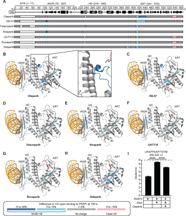
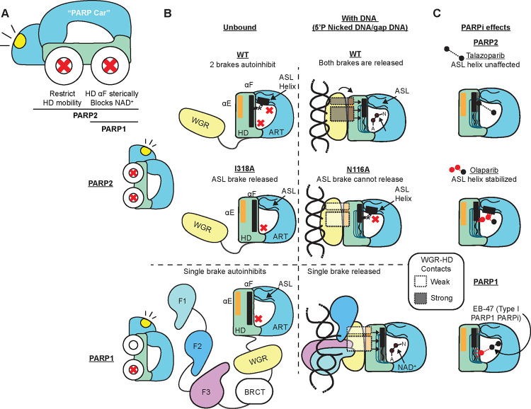
PARP1 and PARP2 recognize DNA breaks immediately upon their formation, generate a burst of local PARylation to signal their location, and are co-targeted by all current FDA-approved forms of PARP inhibitors (PARPi) used in the cancer clinic. Recent evidence indicates that the same PARPi molecules impact PARP2 differently from PARP1, raising the possibility that allosteric activation may also differ. We find that unlike for PARP1, destabilization of the autoinhibitory domain of PARP2 is insufficient for DNA damage-induced catalytic activation. Rather, PARP2 activation requires further unfolding of an active site α-helix absent in PARP1. Only one clinical PARPi, Olaparib, stabilizes the PARP2 active site α-helix, representing a structural feature with the potential to discriminate small molecule inhibitors. Collectively, our findings reveal unanticipated differences in local structure and changes in activation-coupled backbone dynamics between PARP1 and PARP2.


Conflict of interest statement

Declarations of interest B.E.B., J.M.P., and T.T.T. are co-founders of Hysplex, Inc. with interests in PARP inhibitor development. B.E.B., J.M.P., and T.T.T. are co-inventors on a provisional patent application filed by UPenn that is related to this work. B.E.B is on the scientific advisory board of Denovicon Therapeutics.

Figures

Figure 1:
Figure 1:. PARP2 exhibits local HX protection and deprotection from DNA break induced activation not observed in PARP1.
(A) Domain architecture of PARP1 and PARP2 and the percent sequence identity between each shared domain is displayed. (B) HX difference plot between WT PARP2 and WT PARP2 complexed with 5’P nicked DNA at 100 s. Horizontal bar represent PARP2 peptides. White gaps represent the missing coverage. The color key of HX differences is shown next to panel A. (C) Consensus HX data from panel B mapped on the cryo-EM structure of PARP2 complexed with DNA and HPF1 (PDB 6X0L) with HPF1 removed for clarity. (D) Consensus HX data of PARP1 upon binding to DNA mapped on the crystal structure of PARP1 on DNA damage (PDB 4DQY: F3, WGR, HD, ART) combined with the NMR structure of PARP1 on DNA damage (PDB 2N8A: F1, F2, DNA). (E) HX at 100 s of representative peptides in the C-terminus of αE of PARP2 or PARP1 with the indicated condition (DNA and/or EB-47). PARP1 specific residues are shown in parenthesis. Each bar represents the average value from three replicates, with the error bars representing the SD. * and **** indicate differences with a P-value <0.05 or <0.0001, based on a two-sided t-test performed between triplicates of the indicated conditions. The maximum number of exchangeable deuterons (maxD) is indicated by a dotted line. (F) HX at 100 s of the representative peptide in the ASL of PARP2 or PARP1 with or without DNA damage is shown. *** and ns indicate differences with a P-value <0.001 or >0.05, respectively. (G) PARP1 and PARP2 structures used in panel C and D, highlighting differences between the PARP1 and PARP2 ASL during DNA engagement.
Figure 2:
Figure 2:. Extended contacts between the WGR β-sheet and the HD confer ASL Helix destabilization upon PARP2 engaging a DNA break.
(A) HX difference plot between N116A PARP2 with and without 5’P nicked DNA at 10 s. The consensus behavior at each N116A PARP2 residue is displayed in a horizontal bar below the secondary structure annotation, along with a horizontal bar representing WT PARP2 data for comparison. (B-E) HX at 10 s of the indicated representative peptides in the HD αB helix (B), HD C-terminus of αF helix (C), HD C-terminus of the αE helix (D), or ASL (E) for the indicated conditions. *, **, ***, and **** indicate differences with a P-value <0.05, <0.01 <0.001 or <0.0001, respectively, based on a two-sided t-test performed between triplicate samples of the indicated conditions. (F) Consensus HX data from N116A PARP2 with or without 5’P nicked DNA in panel A mapped on the cryo-EM structure of PARP2 complexed with DNA and HPF1 (PDB 6X0L; displayed as shown in Fig. 1C). The position of N116 is indicated with an *. (G) Consensus HX data from WT PARP2 upon binding to 5’P nicked DNA in panel A mapped on the cryo-EM structure of PARP2/DNA/HPF1 complex (PDB 6X0L; displayed as shown in Fig. 1C).
Figure 3:
Figure 3:. The PARP2 active site loop forms an α-helix upon stabilization through contacting the HD.
(A) The 3D structures with the HD (αF helix) contacting the ART, excluding the HD, or with the HD distant from the ASL are shown. (B-C) Sequence alignment of ASL with α-helical residues overlayed in green (B) or sequence alignment of HD αF (C) of PARP1 and PARP2 in humans and in selected orthologs. (D) Crystal structures in corresponding orientations highlighting the unique contacts between ASL and αF of PARP2 (PDB 3KJD) and PARP1 (PDB 3GN7) are shown. V762 PARP1 model (middle) was visualized using PDB 3GN7 and the Pymol mutagenesis wizard.
Figure 4:
Figure 4:. Contacts between the HD and the ASL Helix maintain PARP2 autoinhibition until their separation via DNA damage engagement.
(A) HX difference plots between WT PARP2 and I318A at 100 s. Note that both WT and I318A are measured in the absence of DNA. (B) HX at 100 s of the representative peptide of ASL in WT PARP2 and I318A. (C) HX at 100 s of a representative peptide in an unchanging region of the ART in WT PARP2 and I318A. (D) Catalytic activity of WT PARP2, I318A PARP2, and ΔHD PARP2 (1 μM) in the presence of NAD+ (500 μM) was measured using an SDS-PAGE automodification assay.
Figure 5:
Figure 5:. Olaparib and EB-47 stabilize the ASL of PARP2.
(A) Consensus HX between PARP2 with 5’P nicked DNA and PARP2 complexed with 5’P nicked DNA and the indicated PARPi at 100 s. (B-H) Consensus HX data from Panel A mapped on the cryo-EM structure of PARP2/DNA/HPF1 (PDB 6X0L; displayed as shown in Fig. 1C). (I) HX at 100 s of the representative ASL peptide of PARP2 with the indicated conditions is shown. **** indicates a difference with a P-value <0.0001, based on a two-sided t-test performed between triplicate samples of the indicated conditions.
Figure 6:
Figure 6:. PARP2 undergoes an additional forward allosteric pathway during DNA dependent activation that is distinguished from PARP1.
(A) Analogy of PARP1 and PARP2 to a “PARP car”. Models of PARP1 and PARP2 comparing autoinhibition (B) and PARPi response within the CAT (C).

Similar articles

References

    1. Pandey N., and Black B.E. (2021). Rapid detection and signaling of DNA damage by PARP-1. Trends Biochem Sci 46, 744–757. 10.1016/j.tibs.2021.01.014. - DOI - PMC - PubMed
    1. Ray Chaudhuri A., and Nussenzweig A. (2017). The multifaceted roles of PARP1 in DNA repair and chromatin remodelling. Nat Rev Mol Cell Biol 18, 610–621. 10.1038/nrm.2017.53. - DOI - PMC - PubMed
    1. Satoh M.S., and Lindahl T. (1992). Role of poly(ADP-ribose) formation in DNA repair. Nature 356, 356–358. 10.1038/356356a0. - DOI - PubMed
    1. Bonfiglio J.J., Fontana P., Zhang Q., Colby T., Gibbs-Seymour I., Atanassov I., Bartlett E., Zaja R., Ahel I., and Matic I. (2017). Serine ADP-ribosylation depends on HPF1. Mol Cell 65, 932–940.e6. 10.1016/j.molcel.2017.01.003. - DOI - PMC - PubMed
    1. Palazzo L., Leidecker O., Prokhorova E., Dauben H., Matic I., and Ahel I. (2018). Serine is the major residue for ADP-ribosylation upon DNA damage. eLife 7, e34334. 10.7554/eLife.34334. - DOI - PMC - PubMed

Publication types