[Preprint]. 2024 May 22:rs.3.rs-4307848.
doi: 10.21203/rs.3.rs-4307848/v1.

VCP regulates early tau seed amplification via specific cofactors

Affiliations

VCP regulates early tau seed amplification via specific cofactors

Sushobhna Batra et al. Res Sq. .

Update in

  • VCP regulates early tau seed amplification via specific cofactors.
    Batra S, Vaquer-Alicea J, Valdez C, Taylor SP, Manon VA, Vega AR, Kashmer OM, Kolay S, Lemoff A, Cairns NJ, White CL 3rd, Diamond MI. Batra S, et al. Mol Neurodegener. 2025 Jan 7;20(1):2. doi: 10.1186/s13024-024-00783-z. Mol Neurodegener. 2025. PMID: 39773263 Free PMC article.

Abstract

Background: Neurodegenerative tauopathies may progress based on seeding by pathological tau assemblies, whereby an aggregate is released from one cell, gains entry to an adjacent or connected cell, and serves as a specific template for its own replication in the cytoplasm. In vitro seeding reactions typically take days, yet seeding into the complex cytoplasmic milieu happens within hours, implicating a machinery with unknown players that controls this process in the acute phase.

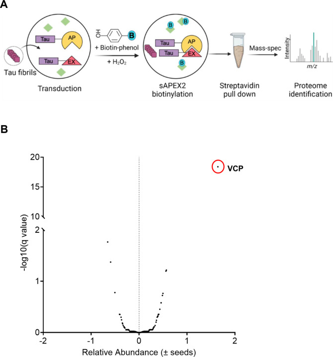
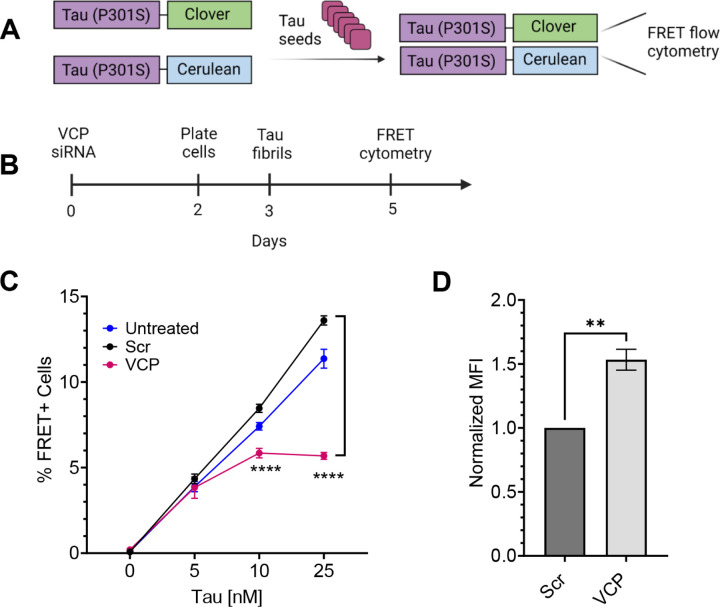
Methods: We used proximity labeling to identify factors that control seed amplification within 5h of seed exposure. We fused split-APEX2 to the C-terminus of tau repeat domain (RD) to reconstitute peroxidase activity 5h after seeded intracellular tau aggregation. Valosin containing protein (VCP/p97) was the top hit. VCP harbors dominant mutations that underlie two neurodegenerative diseases, multisystem proteinopathy and vacuolar tauopathy, but its mechanistic role is unclear. We used immortalized cells and human neurons to study the effects of VCP on tau seeding. We exposed cells to fibrils or brain homogenates in cell culture media and measured effects on uptake and induction of intracellular tau aggregation following various genetic and chemical manipulations of VCP.

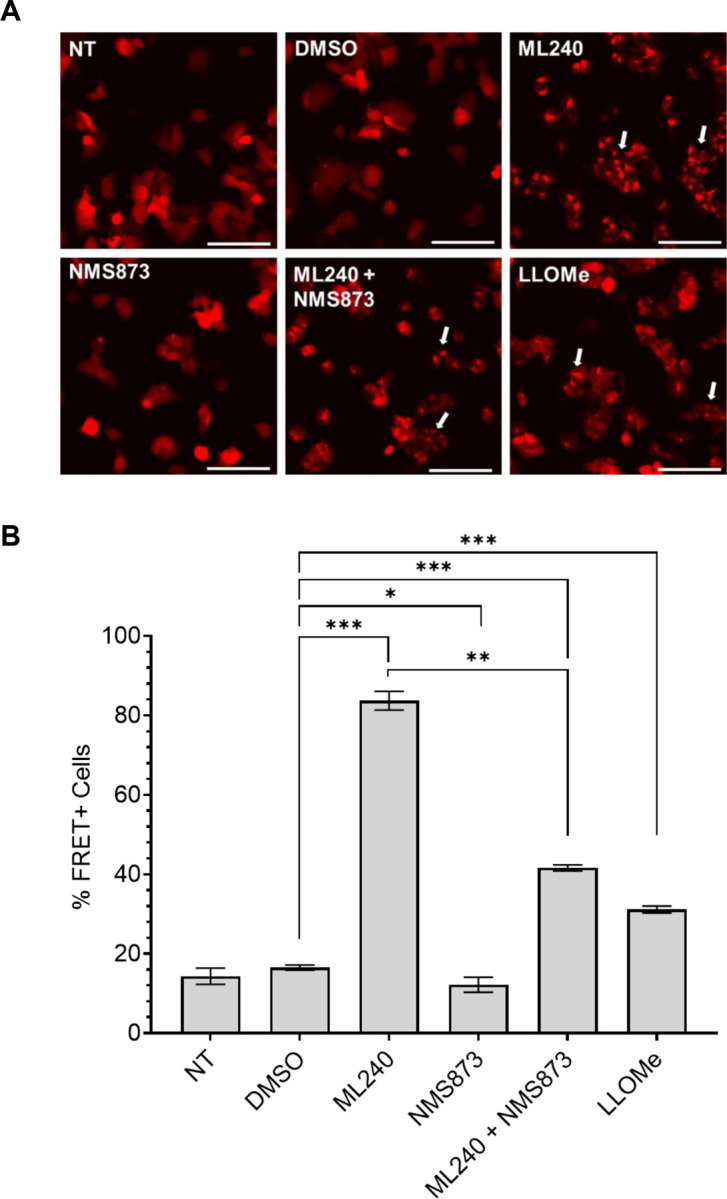
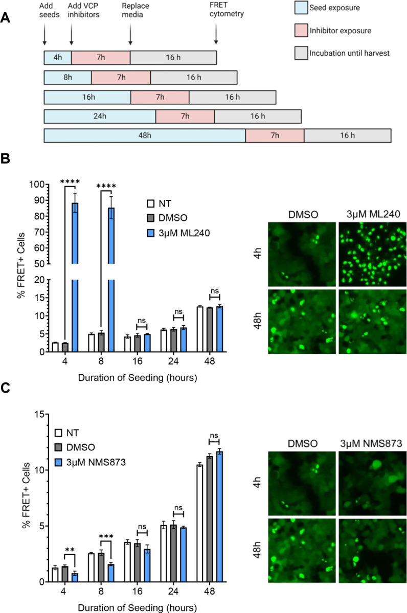
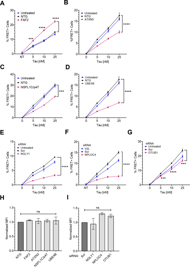
Results: VCP knockdown reduced tau seeding. Chemical inhibitors had opposing effects on aggregation in HEK293T tau biosensor cells and human neurons alike: ML-240 increased seeding efficiency, whereas NMS-873 decreased it. The inhibitors were effective only when administered within 8h of seed exposure, indicating a role for VCP early in seed processing. We screened 30 VCP co-factors in HEK293T biosensor cells by genetic knockout or knockdown. Reduction of ATXN3, NSFL1C, UBE4B, NGLY1, and OTUB1 decreased tau seeding, as did NPLOC4, which also uniquely increased soluble tau levels. By contrast, reduction of FAF2 increased tau seeding.

Conclusions: Divergent effects on tau seeding of chemical inhibitors and cofactor reduction indicate that VCP regulates this process. This is consistent with a dedicated cytoplasmic processing complex based on VCP that directs seeds acutely towards degradation vs. amplification.

Keywords: APEX2; Cofactors; Disaggregase; Seeding; Tau; VCP; p97.


Conflict of interest statement

Competing interests The authors declare no competing interests.

Figures

Figure 1
Figure 1. VCP identified by proximity labeling from tau aggregation.
(A)Schematic of the TMT-MS study performed for proteomics. (B) VCP was identified as the most significant hit enriched in the early tau aggregation proteome. Unpaired t- test, P value < 0.000001.
Figure 2
Figure 2. Reduction of VCP inhibits tau seeding.
(A) The tau biosensor seeding assay is based on exposure of cells to exogenous tau seeds which trigger intracellular aggregation of tau RD (P301S)-cerulean/cloverthat is detected by FRET. (B) Timeline of siRNA treatment to knockdown (KD) VCP in biosensors. (C) KD of VCP reduced tau seeding. Error bars represent S.D. Graph represents n=3 independent experiments. P value **** < 0.0001; One-Way ANOVA with a 95% confidence interval. (D) VCP KD increased uptake of tau fibrils labeled with AF-647, measured by flow cytometry. Error bars represent S.E.M (n=3). P value ** 0.0029; Unpaired t-test with a 95% confidence interval.
Figure 3
Figure 3. Acute exposure of inhibitors differentially impacts tau aggregation.
(A) Timeline of experiment: 1h exposure of tau biosensor cells to inhibitors, followed by 4h of 25nM tau, before washout. (B) ML-240 dose-dependently increased tau seeding. P values: ns = 0.4353, **** < 0.0001(C) NMS-873 dose-dependently decreased tau seeding. Error bars represent S.D. Representative data of n=3 independent experiments. P values: ** 0.0030, **** < 0.0001 (D) Proteasome inhibitor MG132 increased tau seeding. P values: ns = 0.8492, * 0.0415, ** 0.0024, **** < 0.0001 (E) Fluorescence microscopy confirmed the effects of VCP and proteasome inhibition on tau seeding. (F) Compound treatment did not change tau-AF-647 uptake as measured by flow cytometry. Error bars represent S.E.M (n=3). P values: ns = 0.9960, 0.9992, 0.1742, in order of bars on the graph. One-Way ANOVA with a 95% confidence interval.
Figure 4
Figure 4. ML-240 induces Gal3 puncta formation.
v2L biosensors overexpressing galectin-3-mRuby3 were treated with different compounds for 1h prior to addition of tau fibrils and media replacement after 4h. (A) Both ML-240 (3μM) and LLOMe (1mM) induced Gal3 puncta with no effects of NMS-873 (3μM). Co-treatment of ML-240 and NMS-873 also induced Gal3 puncta. Representative images of n=3 independent experiments taken before media replacement (5h of compound alone incubation). (B) ML-240 increased tau seeding ~5x; LLOMe induced seeding by ~2x. NMS-873 suppressed the tau seeding enhanced by ML-240. Representative data of n=3 independent experiments. Error bars represent S.D. P values: **** 0.005, * 0.0331, *** 0.0007, ** 0.0017, *** 0.0001 in order of the bars on the graph. One-Way ANOVA with a 95% confidence interval. Scale bar = 100mm.
Figure 5
Figure 5. VCP inhibition impacts tau seedingearly in the process.
(A) Timeline of compound and tau treatments at different time points in the seeding process. (B) ML-240 increased tau aggregation ~16 to 25-fold, but only when administered <8h after seed exposure. Representative images (20x magnification) are shown in the right panel. P values: **** < 0.0001, ns (16hr) = 0.5892, ns (24hr) = 0.4340, ns (48hr) = 0.3569 (C) NMS-873 decreased tau seeding by ~50%, but only when administered <8h after seed exposure. Representative images (20x magnification) are shown in the right panel. P values: ** 0.0039, *** 0.0004, ns (16hr) = 0.0788, ns (24hr) = 0.8695, ns (48hr) = 0.0547. Error bars represent S.D. Representative data of n=3 independent experiments. One-Way ANOVA with a 95% confidence interval.
Figure 6
Figure 6. VCP inhibitors differentially impact tau seeding in human neurons.
(A) Differentiated iPSC-derived human neurons were transduced with tau RD(P301S)-clover/ruby lentivirus for 48h followed by seeding in the presence or absence of 100nM VCP inhibitors. (B) ML-240 enhanced seeded tau aggregation in neuronal biosensors whereas (C)NMS-873 suppressed tau seeding. Error bars represent S.D. Representative data for n=3 independent experiments. Each dot represents an image taken per condition, with 4 different points captured per well, for a total of 5 wells per condition. P value: **** < 0.0001; Paired t-test with a95% confidence interval.
Figure 7
Figure 7. ML-240 enhances seeding by human brain lysates.
(A) ML-240 increased seeding by AD and CBD brain samples into tau biosensors. (B) ML-240 increased seeding by FTLD type A brain lysate onto TDP-43 biosensors. Error bars represent S.D. Representative data for n =3 independent experiments. (C) Representative microscopy images showing effects of ML-240 on tauopathy lysates and FTLD seeding as observed under the YFP channel. Error bars represent S.D. Representative data of n=3 independent experiments. P value: **** < 0.0001, ** 0.0088; One-Way ANOVA with a 95% confidence interval.
Figure 8
Figure 8. VCP cofactors differentially regulate tau seeding.
VCP cofactors were either knocked out via CRISPR/Cas9 (A-D) or knocked down via siRNA (E-G) in v2L biosensors prior to exposure to increasing amounts of tau fibrils. (A) Knockout of FAF2 increased tau seeding whereas knockout of (B) ATXN3, (C) NSFL1C, and (D) UBE4B reduced tau seeding. P values: FAF2 (*** 0.0001, **** < 0.0001); ATXN3 (**** <0.0001); NSFL1C (*** 0.0002); UBE4B (**** < 0.0001). (E) Knockdown of NGLY1, (F)NPLOC4, and (G) OTUB1, decreased tau seeding. P values: NGLY1(**** < 0.0001); NPLOC4 (**** < 0.0001); OTUB1 (*** 0.0004, **** < 0.0001, *** 0.0001). Graphs are representative of n= 3 independent experiments. Error bars represent S.D. (H) Cofactor KO did not affect tau uptake. P values: ns = 0.9819, 0.9988, 0.9956, 0.9928, in order of bars on the graph. (I) Cofactor KD did not affect tau uptake. P values: ns = 0.9795, 0.1856, 0.3928, in order of bars on the graph. Graphs are representative of n= 3 independent experiments. Error bars represent S.E.M. One-Way ANOVA with a 95% confidence interval.
Figure 9
Figure 9. VCP regulation of tau seeding.
VCP regulates endolysosome integrity, which governs the amount of tau seeds escaping into the cytoplasm. VCP then acts on tau seeds that enter the cytoplasm either to promote degradation or amplification. Disaggregase activity of VCP removes monomer for degradation. This might occur at the end of filaments, which would decrease seeding, or from within, which could fragment fibrils and promote seeding. Chemical inhibitors and cofactors bias the process towards different processing paths.

References

    1. Lee VM, Goedert M, Trojanowski JQ. Neurodegenerative tauopathies. Annu Rev Neurosci. 2001;24:1121–59. - PubMed
    1. Braak H, Braak E. Neuropathological stageing of Alzheimer-related changes. Acta Neuropathol. 1991;82(4):239–59. - PubMed
    1. Seeley WW, Crawford RK, Zhou J, Miller BL, Greicius MD. Neurodegenerative diseases target large-scale human brain networks. Neuron. 2009. Apr 16;62(1):42–52. - PMC - PubMed
    1. Hoenig MC, Bischof GN, Seemiller J, Hammes J, Kukolja J, Onur ÖA, et al. Networks of tau distribution in Alzheimer’s disease. Brain. 2018. Feb 1;141(2):568–81. - PubMed
    1. Ramirez DMO, Whitesell JD, Bhagwat N, Thomas TL, Ajay AD, Nawaby A, et al. Endogenous pathology in tauopathy mice progresses via brain networks [Internet]. bioRxiv; 2023. [cited 2023 Jul 19]. p. 2023.05.23.541792. Available from: https://www.biorxiv.org/content/10.1101/2023.05.23.541792v1 - DOI

Publication types