[Preprint]. 2024 May 22:2024.05.21.595181.
doi: 10.1101/2024.05.21.595181.

Astrocytic RIPK3 exerts protective anti-inflammatory activity during viral encephalitis via induction of serpin protease inhibitors

Affiliations

Astrocytic RIPK3 exerts protective anti-inflammatory activity during viral encephalitis via induction of serpin protease inhibitors

Marissa Lindman et al. bioRxiv. .

Update in

Abstract

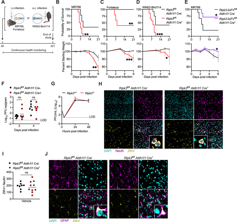
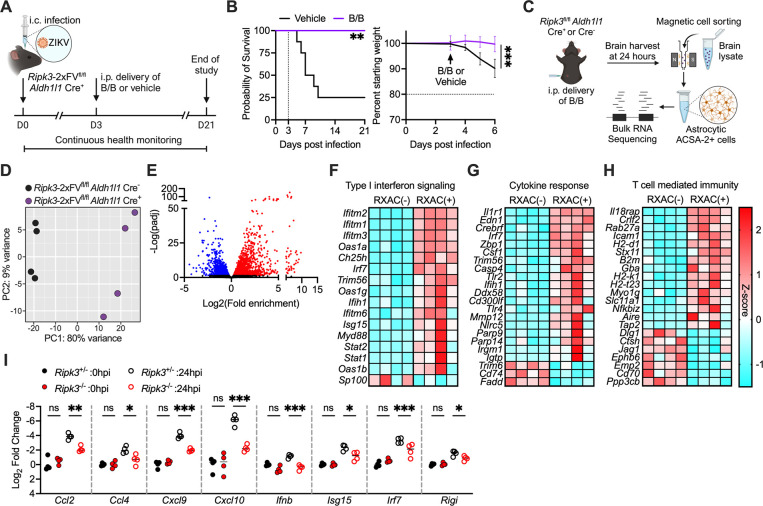
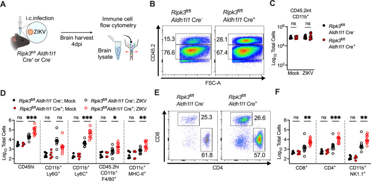
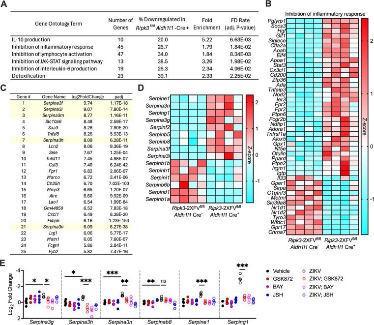
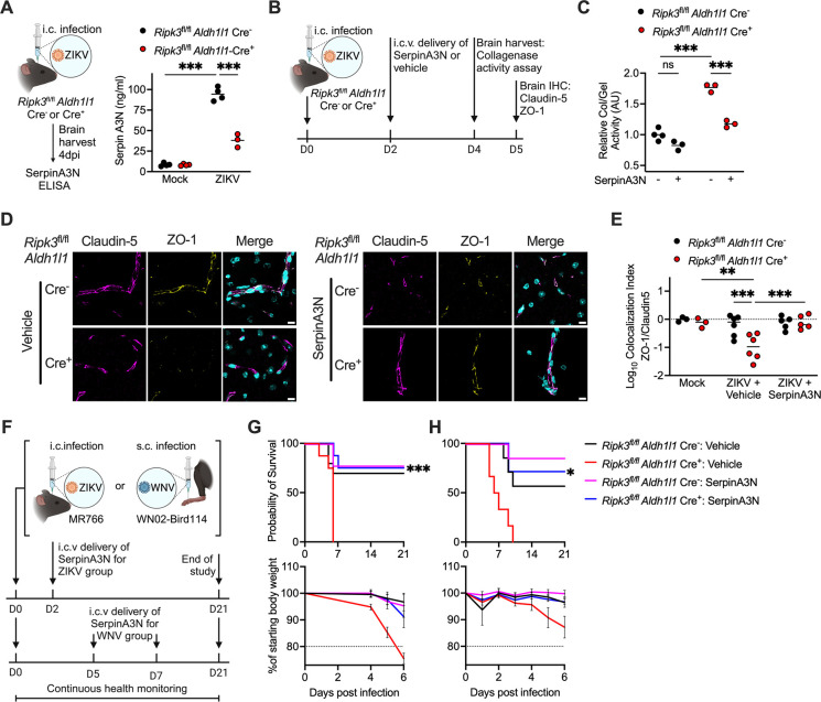
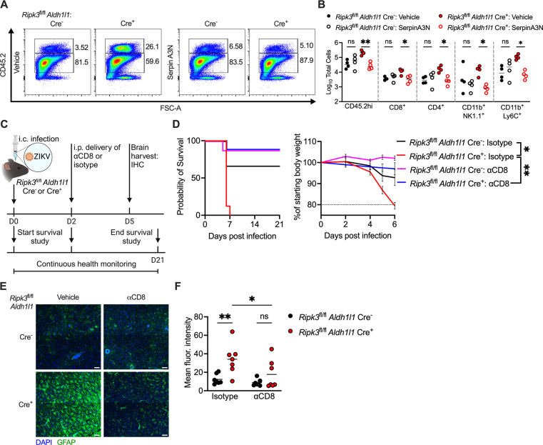
Flaviviruses pose a significant threat to public health due to their ability to infect the central nervous system (CNS) and cause severe neurologic disease. Astrocytes play a crucial role in the pathogenesis of flavivirus encephalitis through their maintenance of blood-brain barrier (BBB) integrity and their modulation of immune cell recruitment and activation within the CNS. We have previously shown that receptor interacting protein kinase-3 (RIPK3) is a central coordinator of neuroinflammation during CNS viral infection, a function that occurs independently of its canonical function in inducing necroptotic cell death. To date, however, roles for necroptosis-independent RIPK3 signaling in astrocytes are poorly understood. Here, we use mouse genetic tools to induce astrocyte-specific deletion, overexpression, and chemogenetic activation of RIPK3 to demonstrate an unexpected anti-inflammatory function for astrocytic RIPK3. RIPK3 activation in astrocytes was required for host survival in multiple models of flavivirus encephalitis, where it restricted neuropathogenesis by limiting immune cell recruitment to the CNS. Transcriptomic analysis revealed that, despite inducing a traditional pro-inflammatory transcriptional program, astrocytic RIPK3 paradoxically promoted neuroprotection through the upregulation of serpins, endogenous protease inhibitors with broad immunomodulatory activity. Notably, intracerebroventricular administration of SerpinA3N in infected mice preserved BBB integrity, reduced leukocyte infiltration, and improved survival outcomes in mice lacking astrocytic RIPK3. These findings highlight a previously unappreciated role for astrocytic RIPK3 in suppressing pathologic neuroinflammation and suggests new therapeutic targets for the treatment of flavivirus encephalitis.


Conflict of interest statement

Declaration of Interests The authors declare no competing interests.

Figures

Figure 1:
Figure 1:. Astrocytic RIPK3 restricts flavivirus pathogenesis but not cell-intrinsic viral replication
(A) Schematic illustrating survival studies in which mice underwent either intracranial (i.c.) ZIKV or subcutaneous (s.c.) WNV infections. (B-E) Survival and weight measurements in mice of indicated genotypes following i.c. ZIKV-MR766 infection (B), i.c. ZIKV-Fortaleza infection (C) infection, s.c. WNV-WN02-Bird114 infection (D), or i.c. ZIKV-MR766 infection (E). n=5–14 mice/group. (F) qRT-PCR analysis of ZIKV-MR766 genome copies in whole brain homogenates derived from mice of indicated genotypes, two or four days following i.c. ZIKV-MR766 infection. Data are expressed as plaque forming unit equivalents (PFU eq)/gram of brain tissue. (G) Multistep growth curve analysis of ZIKV-MR766 replication in primary cortical astrocytes derived from mice of indicated genotypes. Viral titers were assessed via plaque assay. N=4 replicates/group. (H) Immunohistochemical (IHC) staining of NeuN (magenta), DAPI (cyan), and pan-flavivirus E protein (yellow) staining in cortical brain tissue derived from mice of indicated genotypes five days following i.c. ZIKV-MR766 infection. Images are 2×2 tiled composites at 20x magnification. Scale bar = 20μm. (I) Quantification of cells exhibiting colocalization of NeuN and pan-flavivirus E protein in cortical brain tissue derived from mice described in (H). (J) IHC staining of GFAP (magenta), DAPI (cyan), and pan-flavivirus E protein (yellow) in mice as described in (H). Scale bar = 20μm. Images are at 40x magnification. *p<0.05, **p < 0.01, ***p < 0.001. Error bars represent SEM.
Figure 2:
Figure 2:. Activation of astrocytic RIPK3 drives proinflammatory gene expression
(A) Schematic illustrating survival studies in which mice expressing a chemogenetically activatable form of RIPK3 (Ripk3–2xFV) received intraperitoneal (i.p.) administration of homodimerizer ligand (B/B) three days following high dose (103 PFU) intracranial (i.c.) ZIKV-MR766 infection. (B) Survival and weight measurements in Ripk3–2xFVfl/fl Aldh1l1 Cre+ mice treated as described in (A). (C) Schematic illustrating magnetic activated cell sorting (MACS) of ACSA-2+ astrocytes derived from mice of indicated genotypes 24 hours following i.p. administration of B/B. (D-E) Principal component analysis (PCA) (D) and volcano plot depicting significant differentially expressed genes (E) in bulk RNA sequencing data derived from ACSA-2+ astrocytes as described in (C). Transcripts with significant differential expression (>1.5-fold change, adj. P < 0.05) are highlighted. Downregulated transcripts are shown in blue and upregulated transcripts are shown in red. (F-H) Heatmap depicting significant differentially expressed genes within indicated gene ontology terms. (I) qRT-PCR analysis of indicated inflammatory chemokines and interferon stimulated genes in primary astrocytes derived from mice of indicated genotypes 0 or 24 hours following ZIKV-MR766 infection. *p<0.05, **p < 0.01, ***p < 0.001. Error bars represent SEM.
Figure 3:
Figure 3:. Astrocytic RIPK3 suppresses CNS leukocyte infiltration during flavivirus encephalitis
(A) Schematic illustrating flow cytometric analysis of CNS leukocytes derived from mice of indicated genotypes four days following intracranial (i.c.) ZIKV-MR766 infection. (B) Representative flow cytometry plots depicting resident (CD45.2int) or infiltrating (CD45.2hi) CNS leukocytes derived from mice infected with ZIKV-MR766 as described in (A). (C) Total numbers of CD45int CD11b+ resident microglia in brain tissue derived from mice of indicated genotypes following ZIKV-MR766 infection. (D) Total numbers of indicated myeloid cell populations in brain tissue derived from mice of indicated genotypes following ZIKV-MR766 infection. (E) Representative flow cytometry plots depicting CD4+ T cells and CD8+ T cells derived as described in (A). (F) Total numbers of CD8+ T cells, CD4+ T cells, and NK1.1+ natural killer cells in brain tissue derived from mice of indicated genotypes following ZIKV-MR766 infection. *p<0.05, **p < 0.01, ***p < 0.001.
Figure 4:
Figure 4:. Astrocytic RIPK3 promotes a complex immunologic transcriptional program, including multiple Serpin protease inhibitors
(A) Gene ontology enrichment analysis of terms related to anti-inflammatory signaling and immunoregulation, derived from RNAseq analysis comparing ACSA-2+ astrocytes sorted from Ripk3–2xFVfl/fl Aldh1l1 Cre+ mice compared to astrocytes sorted from Cre littermate controls 24 hours following B/B treatment. (B) Heatmap depicting significant differentially expressed genes within indicated gene ontology term. (C) Top 25 differentially expressed genes induced by chemogenetic RIPK3 activation in ACSA-2+ astrocytes. (D) Heatmap depicting all significantly differentially expressed serpin genes induced by chemogenetic RIPK3 activation in astrocytes. (E) qRT-PCR analysis of indicated serpin genes in primary astrocytes derived from WT mice. Astrocytes were pretreated for two hours with indicated inhibitors then infected with ZIKV-MR766. Gene expression was measured 24 hours post infection. *p<0.05, **p < 0.01, ***p < 0.001.
Figure 5:
Figure 5:. SerpinA3N is neuroprotective in models of flavivirus encephalitis
(A) SerpinA3N concentrations (measured via ELISA) in whole brain homogenates derived from mice of indicated genotypes four days following intracranial (i.c.) ZIKV-MR766 infection. (B) Schematic illustrating timeline of i.c. ZIKV-MR766 infection and i.c.v. treatment with recombinant SerpinA3N, prior to various endpoint readouts. (C) Collagenase/gelatinase activity assay in whole brain homogenates derived from mice of indicated genotypes following ZIKV-MR766 infection and SerpinA3N treatment, as described in (B). (D) Immunohistochemical (IHC) staining of Claudin5 (magenta), Zo-1 (yellow), and DAPI (cyan) in cortical brain tissue of mice of indicated genotypes following ZIKV-MR766 infection and SerpinA3N treatment, as described in (B). Scale bar = 10μm. Images are at 63x magnification. (E) Colocalization index of Claudin5 and Zo-1 staining in cortical brain sections described in (D). (F) Schematic illustrating survival studies in which mice received i.c. ZIKV-MR766 infection or subcutaneous (s.c.) WNVWN02-Bird114 infection, followed by i.c.v. administration of recombinant SerpinA3N at indicated intervals following infection. (G-H) Survival and weight measurements in mice of indicated genotypes following ZIKV-MR766 infection (G) or WNV-WN02-Bird114 infection (H) with or without i.c.v. SerpinA3N treatment, as described in (F). n=6–10 mice/group. *p<0.05, **p < 0.01, ***p < 0.001. Error bars represent SEM.
Figure 6:
Figure 6:. SerpinA3N suppresses deleterious neuroinflammation during flavivirus encephalitis
(A) Representative flow cytometry plots depicting resident (CD45.2int) or infiltrating (CD45.2hi) CNS leukocytes derived from mice of indicated genotypes infected with ZIKV-MR766 with or without i.c.v. treatment with SerpinA3N two days following infection. Flow cytometry was performed four days following infection. (B) Total numbers of indicated leukocyte populations in brain tissue derived from mice described in (A). (C) Schematic illustrating survival study and immunohistochemical (IHC) analysis in which mice received intraperitoneal administration of an anti-CD8 neutralizing antibody (αCD8) two days following intracranial (i.c.) ZIKV-MR766 infection. (D) Survival and weight measurements in mice of indicated genotypes following ZIKV-MR766 infection with or without depletion of CD8+ T cells, as described in (C). (E) IHC analysis of GFAP (green) and DAPI (blue) in cortical brain tissue derived from mice of indicated genotypes treated as described in (C). Images are 2×2 tiled composites at 20x magnification. Scale bar = 50μm. (F) Mean fluorescence intensity of GFAP staining in cortical brain tissue derived from mice of indicated genotypes and treated as described in (C). *p<0.05, **p < 0.01. Error bars represent SEM.

Similar articles

References

    1. Espinal M. A. et al. , Emerging and Reemerging Aedes-Transmitted Arbovirus Infections in the Region of the Americas: Implications for Health Policy. Am J Public Health 109, 387–392 (2019). - PMC - PubMed
    1. Lazear H. M., Stringer E. M., de Silva A. M., The Emerging Zika Virus Epidemic in the Americas: Research Priorities. JAMA 315, 1945–1946 (2016). - PubMed
    1. Asad H., Carpenter D. O., Effects of climate change on the spread of zika virus: a public health threat. Rev Environ Health 33, 31–42 (2018). - PubMed
    1. Musso D., Ko A. I., Baud D., Zika Virus Infection - After the Pandemic. N Engl J Med 381, 1444–1457 (2019). - PubMed
    1. Pardy R. D., Richer M. J., Zika Virus Pathogenesis: From Early Case Reports to Epidemics. Viruses 11, (2019). - PMC - PubMed

Publication types