[Preprint]. 2024 May 25:2024.05.24.595760.
doi: 10.1101/2024.05.24.595760.

Differential TLR-ERK1/2 activity promotes viral ssRNA and dsRNA mimic-induced dysregulated immunity in macrophages

Affiliations

Differential TLR-ERK1/2 activity promotes viral ssRNA and dsRNA mimic-induced dysregulated immunity in macrophages

Rakshya Shrestha et al. bioRxiv. .

Update in

Abstract

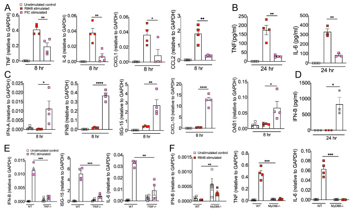
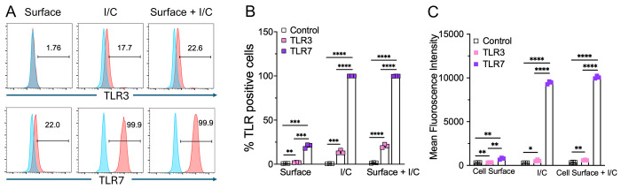
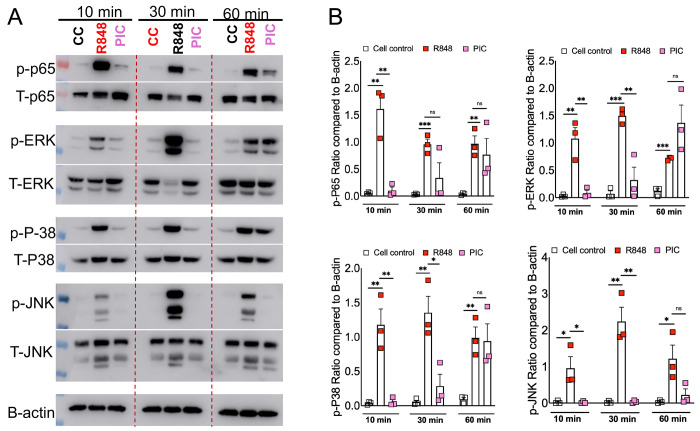
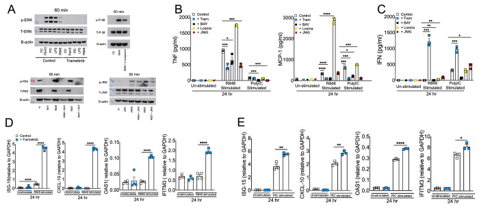
RNA virus induced excessive inflammation and impaired antiviral interferon (IFN-I) responses are associated with severe disease. This innate immune response, also referred to as 'dysregulated immunity,' is caused by viral single-stranded RNA (ssRNA) and double-stranded-RNA (dsRNA) mediated exuberant inflammation and viral protein-induced IFN antagonism. However, key host factors and the underlying mechanism driving viral RNA-mediated dysregulated immunity are poorly defined. Here, using viral ssRNA and dsRNA mimics, which activate toll-like receptor 7 (TLR7) and TLR3, respectively, we evaluated the role of viral RNAs in causing dysregulated immunity. We show that murine bone marrow-derived macrophages (BMDMs) stimulated with TLR3 and TLR7 agonists induce differential inflammatory and antiviral cytokine response. TLR7 activation triggered a robust inflammatory cytokine/chemokine induction compared to TLR3 activation, whereas TLR3 stimulation induced significantly increased IFN/IFN stimulated gene (ISG) response relative to TLR7 activation. To define the mechanistic basis for dysregulated immunity, we examined cell-surface and endosomal TLR levels and downstream mitogen-activated protein kinase (MAPK) and nuclear factor kappa B (NF-kB) activation. We identified a significantly higher cell-surface and endosomal TLR7 expression compared to TLR3, which further correlated with early and robust MAPK (pERK1/2 and p-P38) and NF-kB activation in TLR7-stimulated macrophages. Furthermore, blocking EKR1/2, p38, and NF-kB activity reduced TLR3/7-induced inflammatory cytokine/chemokine levels, whereas only ERK1/2 inhibition enhanced viral RNA-mimic-induced IFN/ISG responses. Collectively, our results illustrate that high cell surface and endosomal TLR7 expression and robust ERK1/2 activation drive viral ssRNA mimic-induced excessive inflammatory and reduced IFN/ISG responses, and blocking ERK1/2 activity would mitigate viral-RNA/TLR-induced dysregulated immunity.

Keywords: ERK1/2; SARS-COV-2; TLRs; inflammation; interferon; macrophages.


Figures

Fig 1:
Fig 1:. ssRNA/TLR7 stimulation induces robust inflammatory cytokine/chemokine responses whereas dsRNA mimics are potent inducers of antiviral IFN and ISG responses.
Murine BMDMs were stimulated with viral RNA mimics; R848 (ssRNA mimic) and poly I:C (dsRNA mimic) each with 10 ug/ml. mRNA (A) and protein levels (B) of inflammatory cytokines and chemokines and mRNA (C) and protein levels (D) of interferons and ISGs were assessed 8 & 24 hours after stimulation respectively. WT and TRIF−/− BMDMs (E) and WT and MyD88−/− BMDMs (F) were stimulated with viral RNA mimics; poly I:C (dsRNA mimic) and R848 (ssRNA mimic) each with 10 ug/ml respectively. mRNA levels of interferons and inflammatory cytokines were assessed 8 hours post-stimulation. Data are representative of 3 independent experiments. Statistical significance was determined using Student’s t-test with *P < .05, **P < .01, **P < .001 and ****P < .0001.
Fig 2:
Fig 2:. TLR3 and TLR7 expression in mouse bone marrow macrophages.
Expression of cell surface and intracellular TLR3 and TLR7 in naive mouse bone marrow macrophages was analyzed by flow cytometry (FACS) using standard protocols (A). Scatter plot graphs show the percentage and Mean fluorescent intensity of TLR3 and TLR7 positive cells (B and C). Data are representative of 3 independent experiments. Statistical significance was determined using Student’s t test with *P < .05,**P < .01, **P < .001 and ****P < .0001.
Fig 3:
Fig 3:. RNA mimic stimulation induced differential phosphorylation of MAPK and NF-kB signaling.
Murine bone marrow macrophages were treated with ssRNA mimic (R848) and dsRNA mimic (PIC) for 10, 30, and 60 mins. Cell lysates were collected and western blot was performed using mAbs against phosphorylated MAPK (ERK1/2, JNK and p-38) and NF-kB (p-65) (A) and relative quantification of protein levels by densitometry analysis (B). Data are representative of 3 independent experiments (A) or pooled from 3 independent experiments (B). Statistical significance was determined using Student’s t-test with *P < .05 , **P < .01, **P < .001 and ****P < .0001.
Fig 4:
Fig 4:. Effect of blocking MAPK and NF-kB signaling in viral RNA mimic induced inflammatory and antiviral cytokine response.
Murine BMDMs were treated with each specific inhibitor of NF-kB and MAPK pathways for 2 hrs followed by stimulation with R848 and or Poly I:C each with 10 ug/ml. Western blot showing the inhibitory effect of Trametinib, Losmapimod, BAY, and JNKi on p-ERK, p-P38, p-P65, and p-JNK respectively (A) Protein levels of cytokines and interferons (B and C) and mRNA levels of ISGs (D and E) were assessed at 24 hours post-stimulation. Data are representative of 2-3 independent experiments (B-E). Statistical significance was determined using Student’s t-test with *P < .05 , **P < .01, **P < .001 and ****P < .0001.

Similar articles

References

    1. Woolhouse MEJ, Gowtage-Sequeria S. Host Range and Emerging and Reemerging Pathogens. Emerg Infect Dis. 2005;11(12):1842–1847. doi:10.3201/eid1112.050997 - DOI - PMC - PubMed
    1. Taylor LH, Latham SM, Woolhouse ME. Risk factors for human disease emergence. Philos Trans R Soc B Biol Sci. 2001;356(1411):983–989. doi:10.1098/rstb.2001.0888 - DOI - PMC - PubMed
    1. Li W, Shi Z, Yu M, et al. Bats Are Natural Reservoirs of SARS-Like Coronaviruses. Science. 2005;310(5748):676–679. doi:10.1126/science.1118391 - DOI - PubMed
    1. Lau SKP, Woo PCY, Li KSM, et al. Severe acute respiratory syndrome coronavirus-like virus in Chinese horseshoe bats. Proc Natl Acad Sci. 2005;102(39):14040–14045. doi:10.1073/pnas.0506735102 - DOI - PMC - PubMed
    1. Wang LF, Eaton BT. Bats, Civets and the Emergence of SARS. In: Childs JE, Mackenzie JS, Richt JA, eds. Wildlife and Emerging Zoonotic Diseases: The Biology, Circumstances and Consequences of Cross-Species Transmission. Springer; 2007:325–344. doi:10.1007/978-3-540-70962-6_13 - DOI

Publication types