[Preprint]. 2024 May 25:2024.05.25.595886.
doi: 10.1101/2024.05.25.595886.

Co-dependent formation of the Toxoplasma gondii sub-pellicular microtubules and inner membrane skeleton

Affiliations

Co-dependent formation of the Toxoplasma gondii sub-pellicular microtubules and inner membrane skeleton

Klemens Engelberg et al. bioRxiv. .

Update in

Abstract

One of the defining features of apicomplexan parasites is their cytoskeleton composed of alveolar vesicles, known as the inner membrane complex (IMC) undergirded by intermediate-like filament network and an array of subpellicular microtubules (SPMTs). In Toxoplasma gondii, this specialized cytoskeleton is involved in all aspects of the disease-causing lytic cycle, and notably acting as a scaffold for parasite offspring in the internal budding process. Despite advances in our understanding of the architecture and molecular composition, insights pertaining to the coordinated assembly of the scaffold are still largely elusive. Here, T. gondii tachyzoites were dissected by advanced, iterative expansion microscopy (pan-ExM) revealing new insights into the very early sequential formation steps of the tubulin scaffold. A comparative study of the related parasite Sarcocystis neurona revealed that different MT bundling organizations of the nascent SPMTs correlate with the number of central and basal alveolar vesicles. In absence of a so far identified MT nucleation mechanism, we genetically dissected T. gondii γ-tubulin and γ-tubulin complex protein 4 (GCP4). While γ-tubulin depletion abolished the formation of the tubulin scaffold, a set of MTs still formed that suggests SPMTs are nucleated at the outer core of the centrosome. Depletion of GCP4 interfered with the correct assembly of SPMTs into the forming daughter buds, further indicating that the parasite utilizes the γ-tubulin complex in tubulin scaffold formation .

Keywords: APR; IMC; SFA; Sarcocystis; Toxoplasma; alveoli; microtubules; γ-tubulin; γTuRC.


Figures

Fig 1.
Fig 1.. Early daughter cytoskeleton assembly displays five-fold symmetry, alternating between nSPMT rafts and the foundation of longitudinal alveolar sutures
A. Schematic representation of T. gondii cytoskeleton assembly. Stage 1-7 represent advancing steps in daughter cytoskeleton scaffold assembly. Modified from [21]. B. Original observation of BCC0 bud initiation as five distinct puncta, with five-fold symmetry (puncta marked by arrowheads). C. Pan-ExM of three different parasites progressing through mitosis and early bud assembly. Asterisks mark mitotic spindle; arrows mark the apical end of the mother’s cytoskeleton (conoid); arrowheads mark the BCC0-5xV5 signals deposited before nSPMT assembly in five foci around the centrioles, which subsequently accumulates between the nSPMTs rafts
Fig 2.
Fig 2.. The nSPMTs display a five-fold symmetrical organization early in the division, in connection with the IMC and the basal complex
A. Pan-ExM of early budding parasites co-stained with HsCentrin2 (Cen) and α-tubulin (MAb 12G10) reveals that the centrioles are connected by a centrin fiber. SPMT formation starts during mitotic anaphase (left panel; mitotic spindle between the centrosomes). Five-fold symmetrical rafts of nSPMTs assemble during telophase (middle and right panels), which transitions from a flat organization (middle panel) into a domed orientation (right panel). Arrows: mitotic spindles; asterisks: nSPMT rafts. B. Pan-ExM snapshots of sequential stages of conoid (open arrowheads) and nSPMT formation. nSPMT rafts are numbered. Arrows mark pairs of short MTs that are being added to the growing scaffold. Note that the ICMT pair is visible within the conoid in the middle panel. Ce: centrioles. C. Transmission electron microscopy of early daughter budding. Cross section through daughter buds wherein nSPMT rafts are visible with IMC membrane sheets on top. Arrowheads mark individual nSPMTs; arrows mark breaks in the membrane sheets of the nascent IMC alveoli. A: apicoplast, Ce: centriole; G: Golgi apparatus. D. Pan-ExM of the MT cytoskeleton co-stained with MORN1, highlighting how the nascent basal complex interfaces with the early scaffold (nSPTMs). Ce: centrioles; asterisks mark mitotic spindle; white arrowhead mark the nSPMT rafts; yellow arrowheads mark the discontinuity of the nascent basal complex assembled close to the (+)-end of nSPMTs. E. Pan-ExM reveals the differential susceptibility of microtubules to Oryzalin treatment. As previously observed [60, 61] division is not halted by this treatment, but cytoskeleton organization is substantially altered. After oryzalin treatment, IMC32-5xV5 forms plaques (asterisks) around (single) centrioles, contrary to its localization in discrete foci (arrowheads) between nSPMT rafts in untreated controls. Ce: centrioles. arrows: mitotic spindle;
Fig 3.
Fig 3.. Spindle and SPMTs in Sarcocystis neurona endopolygeny
A. Representative stages of endopolygeny imaged by single expansion. 1: A just invaded, single merozoite; 2. Early schizont undergoing the first mitosis; 32-64: late schizont going through the last round of mitosis (32 spindles) coupled to budding 64 merozoites – the first outlines of the nSPMTs are visible at the ends of each spindle; 64 budding: later budding schizont with the nSPMTs well defined. Asterisks mark the apical end (conoid) of the mother’s cytoskeleton, which is visible till the very end; arrows mark mitotic spindles; arrowhead marks the nucleolus. All panels are from the same expansion experiment and are shown on the same scale. B. Single expansion imaging of the S. neurona IMC. The IMC is composed of an apical cap and 3x11 alveolar sheets (note that only the front of the schizont is displayed here). The sutures between the sheets are marked by stable overexpression of YFP-TgIMC15 [63]. Note the SPMTs (arrowhead) only extend along the apical cap in this mid-stage schizont. Arrows mark the mitotic spindles. C. Pan-ExM of S. neurona daughter budding stages co-stained with α-tubulin and MORN1. α-tubulin marks the spindle, centrioles and SPMTs. MORN1 highlights the centrocone and the basal complex. Ce: centriole; arrows mark mitotic spindle; yellow arrowheads mark the 11 pairs of nSPMTs; asterisks: centrocone. D. Pan-ExM of S. neurona schizonts, zoomed in on the spindles. MTs of unknown identity emanate from the centrioles into the cytoplasm during mitosis. Two different parasites are shown: left panels undergoing the first round of mitosis (two spindles); right panels undergoing third round of mitosis (eight spindles). Ce: centriole; arrows mark overlapping mitotic spindles; yellow arrowheads mark the MTs of unknown identity; white arrowheads mark the nucleolus.
Fig. 4.
Fig. 4.. γ-tubulin is essential for cell division
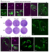
A. Pan-ExM of parasites expressing γ-tubulin endogenously tagged at the C-terminus with mAID-5xV5 (γ-tub-cKD), co-stained with V5 and α-tubulin antisera. Asterisks: the spindle or spindle pole; arrow: horseshoe shape at the nAPR; open arrowhead: γ-tubulin at the ICMT; yellow arrowhead: γ-tubulin pairs of different length protruding from the centriole (Ce). B. Kinetics of γ-tub-cKD depletion upon IAA addition visualized by western blot. α-tubulin (MAb 12G10) is used as loading control. The mAID system allows fast protein degradation. C. Seven-day plaque assay of γ-tubulin depletion demonstrates its essentiality for the lytic cycle. D. Pan-ExM of 2 hrs γ-tubulin depleted parasites stained for α-tubulin and protein density marker NHS ester conjugated to ATTO488. Mother MT populations appear normal whereas two daughter MT populations are visible: one (arrows) connected to round structures (putatively centrioles), and the other (arrowheads) presents as a long bundle of MT. E. Pan-ExM of 2 hrs γ-tubulin depleted parasites co-stained for α-tubulin and HsCentrin2 (centrosomes). A variable number of unstructured microtubules emanates from typically one side of each centriole, which are connected by Centrin. Numbered boxes are expanded in panels on the right. F. Quantification of panel D for the number of MTs emanating from centrioles. The majority of centrioles, roughly 30% is connected to 2 MTs, the remainder is a mixture of 0-5 MTs. 145 centrioles from 39 parasites were quantified. G. Depletion of γ-tubulin for 24 hrs imaged by pan-ExM reveals long unstructured MTs without any daughter cytoskeletons within an unstructured mother cell. NHS-ATTO488 reports on protein density.
Fig 5.
Fig 5.. Identity of microtubule populations in γ-tubulin depleted parasites
A. Pan-ExM of control and 2 hrs γ-tubulin depleted parasites stained with α-tubulin and IMC6 antisera. Arrows: bundled MT, not coated with IMC6; open arrowheads: centrioles from which IMC6-coated MTs are emanating. B. Pan-ExM of control and 2 hrs γ-tubulin depleted parasites stained for α-tubulin and polyglutamylated (polyE) tubulin antisera. Arrows: bundled MT, not marked with polyglutamylated tubulin; open arrowheads: centrioles from which polyglutamylated tubulin-coated MTs are emanating. C. Pan-ExM of control and 2 hrs γ-tubulin depleted parasites stained for α-tubulin and MORN1 antisera. Yellow-1 labeled panels shows a very early budding cytoskeleton, with only a few nSPMTs (arrow) of the scaffold surrounded by the nascent basal complexes (nBC). MORN1 also highlights the centrocone (blue arrow) in the nuclear envelope, where the spindle reaches into the nucleus. Yellow-2 labeled panels show early budding parasites with dome shaped nSPMTs coated with the nBC close to the nSPMT (+)-ends. Blue arrows: centrocone. After γ-tubulin depletion (2 hrs IAA), MORN1 localizes in a semi-circle to the ‘centriolar” MTs and partially coats them. MORN1 also localizes to the middle of the bundled MT, highlighting the centrocone. mBC: mother parasite’s BC; asterisk: mitotic spindle; open arrowheads: centrioles; yellow arrowheads: bundled MTs coated with MORN1, marking the mitotic spindle embedded in the nuclear envelope. Dotted-line boxed areas are 2-fold enlarged. Yellow numbers indicate corresponding panels.
Fig 6.
Fig 6.. γ-tubulin depletion causes deviant daughter IMC buds
A, B. Airyscan superresolution images after 2 hrs of γ-tubulin depletion using two different co-stains: A. ISP1 (apical cap) and IMC3 (mother and daughter IMC); B. ISP1 (apical cap) and HsCentrin2 (centrosomes). Arrows mark unstructured IMC3, whereas ISP1 appears normally clustered around the centrosomes. DAPI highlights DNA. C. Airyscan superresolution images of 2 hrs γ-tubulin depleted parasites. No interfereance with initiation of daughter budding is observed (arrows in ‘bud initiation’ panel) but the morphology of the daughter buds is severely affected: daughters appear disrupted and no cone-shape bud is assembled when γ-tubulin is depleted (arrows in ‘late buds’ panel). D. Quantification of panel C for incidence of budding and bud shapes upon 2 hrs of γ-tubulin depletion. n=3 biological replicates, 100 vacuoles each, error bars denote SEM. Data points indicate averages of individual replicates. One-way ANOVA with Tukey’s HSD. ns: not significant; ** p = 0.01; *** p = 0.001; **** p = 0.0001.
Figure 7.
Figure 7.. SFA2 and GCP4 depletion strongly affects bud formation
A. SFA2-mAID5xV5 (SFA2-cKD) localization throughout mitosis and early daughter bud assembly Ce: centriole; asterisks: mitotic spindle; arrows: SFA2 ring-like structure aligning with the conoid. Arrowhead: SFA2 signal apical to nAPR and conoid; yellow arrowhead mark the broken fiber at mid-budding. Imaged by pan-ExM. B. Seven-day plaque assays of SFA2 and GCP4 depleted parasites using the mAID system. Parent line is RHΔKu80-Tir1. C. Pan-ExM of 2 hr SFA2 depleted parasites. Mitosis progresses normally, but single daughter buds appear at a high frequency. The single buds display a normal nSPMT organization. Ce: centrioles; asterisks: mitotic spindle; arrowheads: apical end of the single daughter bud. D. GCP4-mAID-5xV5 (GCP4-cKD) localizes to the spindle poles (asterisks) as well as in between the centrioles, and to the forming scaffold. Imaged by pan-ExM. E. Pan-ExM of 2 hr GCP4 depleted parasites. Mitosis progresses normally, and two daughter buds are formed, however, the organization and number of nSPMTs in each sheet is irregular (number of nSPMTs per sheet indicated by numericals). Ce: centrioles; asterisks: spindle poles.
Figure 8.
Figure 8.. Model of daughter scaffold formation in T. gondii.
Summarizing schematic: The daughter scaffold is initiated close to the centrioles (Ce) and relies on two factors; γ-tubulin (magenta) for the nucleation and/or stabilization/anchoring of tubulin components, and as previously reported on the SFA fiber [19] (black). The basal complex (MORN1 [31], red), the alveoli (not shown), and IMC components (e.g. BCC0 [21] and IMC32 [23], blue) align with the earliest observable tubulin scaffold structure. SPMTs are nucleated close to the centrioles, presumably in the pericentriolar material of the outer centrosomal core, and subsequently added to the forming APR (highlighted by γ-tubulin/magenta). Closure of the circular conoid and nAPR, in concert with nSPMT addition, establishes the scaffold’s planar configuration. At this point the basal complex assumes ring shape. Most abundantly, four rafts of four nSPMTs and one raft of six nSPMTs (this study and [56]) support the alveoli, which are being added to the growing scaffold (not shown), The apical cap (ISP1 [67], yellow) is formed by a so far incompletely understood process, but presumably establishes the cone shape of the scaffold, which then growths in basal direction. At this point γ-tubulin no longer localizes to the APR (blue), which is then stabilized by other factors (e.g. KinA/APR1 [42] and/or AC9/10 [22]).

Similar articles

References

    1. Gubbels M.J., Keroack C.D., Dangoudoubiyam S., Worliczek H.L., Paul A.S., Bauwens C., Elsworth B., Engelberg K., Howe D.K., Coppens I., and Duraisingh M.T., 2020. Fussing About Fission: Defining Variety Among Mainstream and Exotic Apicomplexan Cell Division Modes. Front Cell Infect Microbiol, 10: 269. - PMC - PubMed
    1. Harding C.R. and Frischknecht F., 2020. The Riveting Cellular Structures of Apicomplexan Parasites. Trends Parasitol, 36: 979–991. - PubMed
    1. Harding C.R. and Meissner M., 2014. The inner membrane complex through development of Toxoplasma gondii and Plasmodium. Cell Microbiol, 16: 632–41. - PMC - PubMed
    1. Gubbels M.J. and Morrissette N.S., The Toxoplasma cytoskeleton: structures, proteins, and processes, in Toxoplasma gondii: the model apicomplexan - perspective and methods, Weiss L.M. and Kim K., Editors. 2020, Academic Press: London, IK. 743–788.
    1. Goodenough U., Roth R., Kariyawasam T., He A., and Lee J.H., 2018. Epiplasts: Membrane Skeletons and Epiplastin Proteins in Euglenids, Glaucophytes, Cryptophytes, Ciliates, Dinoflagellates, and Apicomplexans. MBio, 9: e02020–18. - PMC - PubMed

Publication types