

Rare adrenal incidentaloma: ganglioneuroma
- PMID: 38826854
- PMCID: PMC11140512
- DOI: 10.1093/jscr/rjae352
Rare adrenal incidentaloma: ganglioneuroma
Abstract
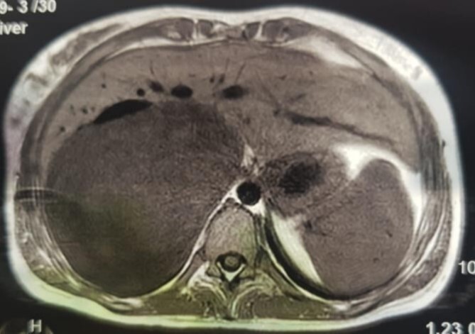
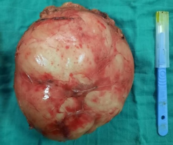
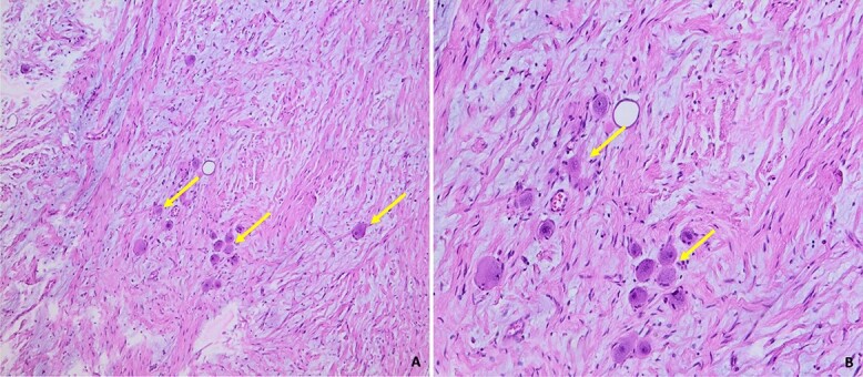
Ganglioneuroma (GN) is a rare, benign neurogenic tumor that develops from sympathetic ganglion cells. It occurs mainly in the retroperitoneal region. Adrenal localization is rare. We report a case of adrenal ganglioneuroma in a 22-year-old woman with no previous history of the disease. The tumor was discovered incidentally on an entero scan ordered as part of the etiological assessment for chronic diarrhea. The diagnosis was confirmed by pathological examination.
Keywords: ganglioneuroma; oncology; retroperitoneal tumor; urology.
Published by Oxford University Press and JSCR Publishing Ltd. © The Author(s) 2024.
Conflict of interest statement
None declared. The examination of the patient was conducted in accordance with the Declaration of Helsinki Principles. Written informed consent was obtained from the patient for publication of this article.
Figures


References
-
- Adraoui J, El Jai SR, Chehab F, et al. Ganglioneurome rétropéritonéal. Journal Marocain d’Urologie 2008;10:34–6.
-
- Sarf I, el Mejjad A, Badre L, et al. Le ganglioneurome rétropéritonéal géant. Prog Urol 2003;13:502–5. - PubMed
Publication types
LinkOut - more resources
Full Text Sources
