

Rare Encounter: Giant Hemangiopericytoma of Thigh in a Young Male
- PMID: 38826872
- PMCID: PMC11143947
- DOI: 10.7759/cureus.59514
Rare Encounter: Giant Hemangiopericytoma of Thigh in a Young Male
Abstract
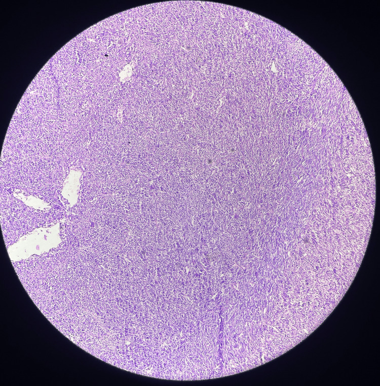
A rare tumor called hemangiopericytoma develops from the pericytes, the cells that surround blood vessels. They frequently grow slowly and might be asymptomatic initially. Although they can develop anywhere in the body, these tumors are most frequently found in the head, pelvis, and legs. This uncommon tumor originates in soft tissues like fat, muscles, tendons, nerves, blood vessels, and other fibrous tissues. The tumor in adolescence can be benign or malignant; it frequently develops in the bones but has the potential to metastasize to the lungs. Imaging tests, such as MRIs or CT scans, are commonly used in diagnosis to determine the location and size of the tumor. We present a case of a 23-year-old male who complained of swelling in his left thigh that had persisted for two years. He underwent multiple biopsies which were inconclusive until wide local excision of the swelling was done. On histopathology, the excised tumor was suggestive of hemangiopericytoma. The patient was advised of radiotherapy for completion of the treatment.
Keywords: hemangiopericytoma; myofibromatosis; pericytes; soft tissue tumor; thigh; thigh swelling.
Copyright © 2024, Bhargava et al.
Conflict of interest statement
The authors have declared that no competing interests exist.
Figures


Similar articles
-
[Hemangiopericytoma (extrapleural solitary fibrous tumour). Exemplified and discussed on the basis of two cases].Mund Kiefer Gesichtschir. 2005 Nov;9(6):404-8. doi: 10.1007/s10006-005-0649-x. Mund Kiefer Gesichtschir. 2005. PMID: 16220316 German.
-
Giant Leiomyosarcoma Arising in Posterior Thigh: Management of a Rare Case.Cureus. 2020 Aug 30;12(8):e10146. doi: 10.7759/cureus.10146. Cureus. 2020. PMID: 33014644 Free PMC article.
-
Giant hemangiopericytoma of the neck: a case report.Kulak Burun Bogaz Ihtis Derg. 2002 Jul-Aug;9(4):297-300. Kulak Burun Bogaz Ihtis Derg. 2002. PMID: 12422087
-
[Solitary fibrous hemangiopericytoma of atypical location: importance of immunohistochemical study].Cir Cir. 2014 May-Jun;82(3):323-7. Cir Cir. 2014. PMID: 25238475 Review. Spanish.
-
CD34-positive infantile myofibromatosis: Case report and review of hemangiopericytoma-like pattern tumors.J Dermatol. 2016 Sep;43(9):1088-91. doi: 10.1111/1346-8138.13400. Epub 2016 Apr 14. J Dermatol. 2016. PMID: 27074874 Review.
References
-
- Haemangiopericytoma of left thigh: a rare case report. Mane K, Khan S, Singhania S, Singh PK, Gudhe M, Nikose S. https://jorjournal.com/haemangiopericytoma-of-left-thigh-a-rare-case-rep... J Orthop Rehabil. 2015;1:28–30.
-
- Hemangiopericitoma de muslo: a propósito de un caso . Cascallo JC, Salvadó NM, Fontanet MC, Mullerat AMC. https://www.elsevier.es/index.php?p=revista&pRevista=pdf-simple&pii=9002.... Angiologia. 1993;45:103–106. - PubMed
-
- Brain microvascular pericytes in health and disease. Dalkara T, Gursoy-Ozdemir Y, Yemisci M. Acta Neuropathol. 2011;122:1–9. - PubMed
Publication types
LinkOut - more resources
Full Text Sources