Case Reports
. 2024 May 23;19(8):3372-3375.
doi: 10.1016/j.radcr.2024.04.080. eCollection 2024 Aug.

Spigelian-cryptorchidism syndrome: Lesson based on a case report

Affiliations
Case Reports

Spigelian-cryptorchidism syndrome: Lesson based on a case report

Renato Farina et al. Radiol Case Rep. .

Abstract

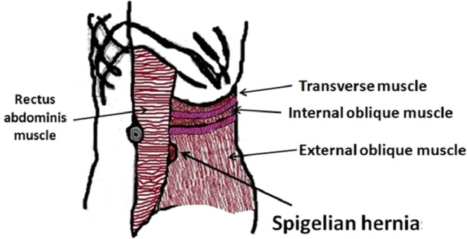
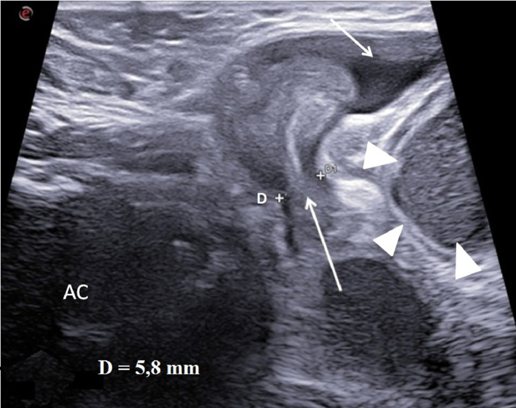
The Spigelian hernia is a abdominal wall hernia that originates from a discontinuity of the Spigelian fascia located lateral to the rectus abdominis muscle. It can be acquired in adults or congenital in newborns. In very rare cases in male it can be associated with cryptorchidism, in which case it is known as "Spigellian-Cryptorchidism Syndrome". It can be clinically highlighted with abdominal swelling wall along the semilunar line and intestinal obstruction. The diagnosis, as in all pediatric emergencies, must be timely and the method of choice is ultrasound which allows a rapid localization of the hernia breach and herniated structures. The treatment of choice is surgical with herniopexy and repositioning of the testicle into the scrotal sac, or orchipessy in cases of testicular necrosis. We describe ultrasound characteristics of Spigellian-cryptorchidism syndrome presenting with acute intestinal obstruction in a newborn.

Keywords: Congenital anomalies; Criptorchidism; Spigelian hernia; Ultrasound.


Figures

Fig 1
Fig. 1
Spigelian hernia. Diagram showing the anatomical relationships of the main structures involved in this pathology.
Fig 2
Fig. 2
Abdominal wall ultrasound. In this scan the width of the hernia breach (D) (long arrow) were measured. Fluid collection (short arrow). Ileal dilatation upstream of the hernia (arrow heads). Abdominal cavity (AC).
Fig 3
Fig. 3
Abdominal wall ultrasound. The scan performed with linear probe in the abdominal wall swelling highlighted a breach of abdominal wall with colon (long arrows) and testicle (short arrow) involvement. Intestinal fluid (IL). Intestinal caliper (C). Abdominal Cavity (AC).

References

    1. Jones BC, Hutson JM. The syndrome of Spigelian hernia and cryptorchidism: a review of paediatric literature. J Pediatr Surg. 2015;50(2):325–330. doi: 10.1016/j.jpedsurg.2014.10.059. - DOI - PubMed
    1. Mittal T, Kumar V, Khullar R, et al. Diagnosis and management of Spigelian hernia: a review of literature and our experience. J Minim Access Surg. 2008;4:95–98. doi: 10.4103/0972-9941.45204. - DOI - PMC - PubMed
    1. Sanae A, Kaoutar I, Hind S, et al. Complicated abdominal wall hernia mimicking an abdominal wall abscess: a case report. Radiol Case Rep. 2021;16(6):1451–1453. doi: 10.1016/j.radcr.2021.03.037. - DOI - PMC - PubMed
    1. Durham MM, Ricketts RR. Congenital spigelian hernias and cryptorchidism. J Pediatr Surg. 2006;41:1814–1817. doi: 10.1016/j.jpedsurg.2006.06.043. - DOI - PubMed
    1. Levy G., Nagar H., Blachar A., et al. Pre-operative sonographic diagnosis of incarcerated neonatal spigelian hernia containing the testis. Pediatr Radiol. 2003;33:407–409. doi: 10.1007/s00247-003-0879-8. - DOI - PubMed

Publication types

LinkOut - more resources