

Exploring varied radiologic appearance in pulmonary embolism with CT pulmonary angiography: Case series with literature review
- PMID: 38827043
- PMCID: PMC11143776
- DOI: 10.1016/j.radcr.2024.04.081
Exploring varied radiologic appearance in pulmonary embolism with CT pulmonary angiography: Case series with literature review
Abstract
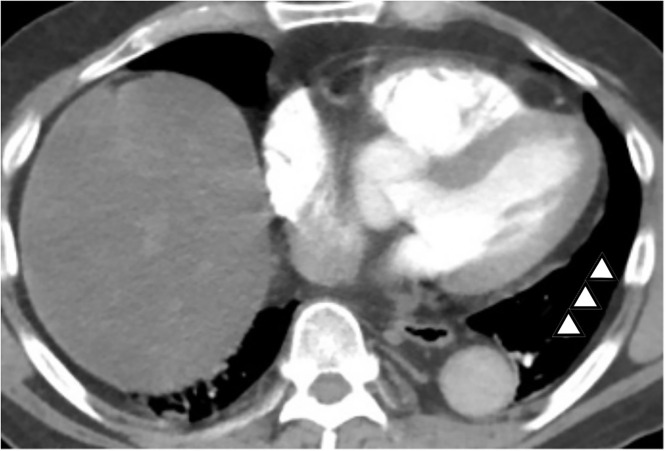
Pulmonary embolism (PE) is a life-threatening condition caused by a sudden blockage of pulmonary arteries. Nonspecific and extremely variable clinical presentation frequently leads to undetected cases, making computed tomography pulmonary angiography (CTPA) hold a crucial role in the diagnosis of PE. This case series presents numerous types and findings of PE in CTPA among patients with different initial presentations followed by a literature review. We presented 3 cases with different initial presentations such as dyspnea with wheezing, productive cough, and hematemesis. All patients were consequently evaluated for D-dimer due to suspicion of PE from cardiac ultrasonography, electrocardiography (ECG), and persistent symptoms. Large to subsegmental PE can be found with various secondary findings such as pleural effusion and Hampton's hump. All patient's conditions were improved after anticoagulant treatment. This case series highlights the significance of CTPA as an imaging modality in the diagnosis of PE, as well as the necessity of evaluating the main to subsegmental pulmonary artery through an in-depth understanding of the images that can be assessed on CTPA.
Keywords: Case report; Computed tomography pulmonary angiography; High clinical suspicion; Pulmonary embolism; Radiologic findings.
© 2024 The Authors. Published by Elsevier Inc. on behalf of University of Washington.
Figures


Similar articles
-
Hampton's Hump-A Rare Radiological Feature in Patients with Pulmonary Embolism in a Single-Center Study.J Clin Med. 2025 Mar 12;14(6):1900. doi: 10.3390/jcm14061900. J Clin Med. 2025. PMID: 40142708 Free PMC article.
-
Multislice computed tomography angiography as an imaging modality of choice in patients with suspicion of pulmonary embolism - own experiences and modern imaging techniques.Adv Clin Exp Med. 2013 Sep-Oct;22(5):705-13. Adv Clin Exp Med. 2013. PMID: 24285456
-
Application of CT pulmonary angiography and echocardiography in acute pulmonary embolism: A cross-sectional study.Health Sci Rep. 2023 Sep 4;6(9):e1546. doi: 10.1002/hsr2.1546. eCollection 2023 Sep. Health Sci Rep. 2023. PMID: 37670847 Free PMC article.
-
Peripheral pulmonary embolism on multidetector CT pulmonary angiography.JBR-BTR. 2007 Mar-Apr;90(2):100-8. JBR-BTR. 2007. PMID: 17555069 Review.
-
Symptomatic subsegmental pulmonary embolism: what is the next step?J Thromb Haemost. 2012 Aug;10(8):1486-90. doi: 10.1111/j.1538-7836.2012.04804.x. J Thromb Haemost. 2012. PMID: 22672341 Review.
References
Publication types
LinkOut - more resources
Full Text Sources
