. 2024 May 4;32(2):101263.
doi: 10.1016/j.omtm.2024.101263. eCollection 2024 Jun 13.

Prenatal AAV9-GFP administration in fetal lambs results in transduction of female germ cells and maternal exposure to virus

Affiliations

Prenatal AAV9-GFP administration in fetal lambs results in transduction of female germ cells and maternal exposure to virus

Beltran Borges et al. Mol Ther Methods Clin Dev. .

Abstract

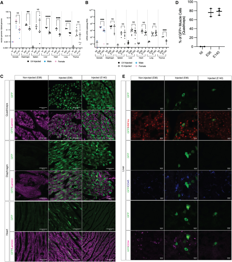
Prenatal somatic cell gene therapy (PSCGT) could potentially treat severe, early-onset genetic disorders such as spinal muscular atrophy (SMA) or muscular dystrophy. Given the approval of adeno-associated virus serotype 9 (AAV9) vectors in infants with SMA by the U.S. Food and Drug Administration, we tested the safety and biodistribution of AAV9-GFP (clinical-grade and dose) in fetal lambs to understand safety and efficacy after umbilical vein or intracranial injection on embryonic day 75 (E75) . Umbilical vein injection led to widespread biodistribution of vector genomes in all examined lamb tissues and in maternal uteruses at harvest (E96 or E140; term = E150). There was robust GFP expression in brain, spinal cord, dorsal root ganglia (DRGs), without DRG toxicity and excellent transduction of diaphragm and quadriceps muscles. However, we found evidence of systemic toxicity (fetal growth restriction) and maternal exposure to the viral vector (transient elevation of total bilirubin and a trend toward elevation in anti-AAV9 antibodies). There were no antibodies against GFP in ewes or lambs. Analysis of fetal gonads demonstrated GFP expression in female (but not male) germ cells, with low levels of integration-specific reads, without integration in select proto-oncogenes. These results suggest potential therapeutic benefit of AAV9 PSCGT for neuromuscular disorders, but warrant caution for exposure of female germ cells.

Keywords: adeno-associated virus; germ-cell transduction; prenatal somatic cell gene therapy; spinal muscular atrophy.


Conflict of interest statement

G.G., S.P., T.D.R., and F.O. are employees and stockholders of Novartis. C.J.S. receives grant support from Roche Ltd., Biogen, and Actio Bio and has served as a paid advisor, consultant, and/or speaker to Biogen, Roche/Genentech, and Novartis; these arrangements have been reviewed and approved by the Johns Hopkins University in accordance with its conflict-of-interest policies. T.C.M. receives grant funding from Novartis, BioMarin, and Biogen and is on the SAB of Acrigen; these arrangements have been reviewed and approved by UCSF in accordance with its conflict of interest policies.

Figures

None
Graphical abstract
Figure 1
Figure 1
Experimental design and survival after in utero injection of scAAV9-GFP in fetal lambs (A) Experimental design. Fetal lambs were injected on E75 with 1 × 1014 vector genomes/kg of sc-AAV9-GFP and harvested at E96 ± 1 day or E140 ± 3 days (term = E150). Image created with www.biorender.com. (B) Representative pictures of an umbilical vein injection (left) or an ultrasound-guided IC injection (right). (C) Survival to harvest (non-injected [NI, n = 13]; umbilical vein [UV, n = 13]; IC [n = 4]). (D and E). Harvest weights at E96 (D) or at E140 (E) based on litter size and experimental group. Each dot represents one fetus; symbols indicate total number of fetuses in each litter. Center lines and whiskers indicate mean and SEM, respectively. ∗p < 0.05 by one-way ANOVA with Tukey’s multiple comparison test (n = 6 for NI and n = 5 for umbilical vein at E96). No significant differences were seen between groups at the E140 time point by one-way ANOVA with Tukey’s multiple comparison test.
Figure 2
Figure 2
Biodistribution of scAAV9-GFP in the CNS after prenatal administration (A) Vector genome copies per diploid genome (vg/dg) in various CNS tissues after prenatal umbilical vein injection and harvest at E96 or E140. Each dot represents one fetus. Center lines and whiskers indicate mean and SEM, respectively. ∗p < 0.05 and ∗∗p < 0.01 by parametric, unpaired, two-tailed t tests (n = 5 for E96 and n = 6 for E140). LSC, lumbar spinal cord; TSC, thoracic spinal cord. (B) GFP mRNA copies/μg RNA in various CNS tissues of umbilical vein-injected (filled circles) and IC-injected (open circles) fetuses harvested at E96 and E140. Each dot represents one fetus. Center lines and whiskers indicate mean and SEM, respectively. No significant differences were seen between time points by parametric, unpaired, two-tailed t tests. (C) Representative images of GFP expression (green) and NeuN (pink) in cervical (top row), thoracic (middle row), and lumbar spinal cord (bottom row) in the indicated experimental groups (original magnification ×63). (D) Representative images of GFP expression (green) and Tuj1 (red) in lumbar DRG in indicated experimental groups (original magnification ×63). NI, non-injected; UV, umbilical vein.
Figure 3
Figure 3
Biodistribution of scAAV9-GFP in peripheral tissues after prenatal administration (A) Vector genome copies per diploid genome (vg/dg) in various peripheral tissues after umbilical vein injection and harvest at E96 or E140. Each dot represents one fetus. Blue dots represent male fetuses; pink dots represent female fetuses. Center lines and whiskers indicate mean and SEM, respectively. ∗p < 0.05, ∗∗p < 0.01, and ∗∗∗∗p < 0.0001 by parametric, unpaired, two-tailed t tests (n = 4 for E96 and n = 6 for E140). (B) GFP mRNA copies/μg RNA in various peripheral tissues of umbilical vein-injected (filled circles) and IC-injected (open circles) fetuses harvested at E96 and E140. Each dot represents one fetus. Blue dots represent male fetuses. Pink dots represent female fetuses. Center lines and whiskers indicate mean and SEM, respectively. No significant differences were seen between time points by parametric, unpaired, two-tailed t tests. (C) Representative images of GFP expression in quadriceps (top row), diaphragm (middle row), and heart (bottom row) after umbilical vein injection and harvest at the indicated time points (original magnification ×20). Pink, laminin. (D) Quantification of GFP+ myocytes in quadriceps after prenatal umbilical vein injection. Each dot represents one fetus. Center lines and whiskers indicate mean and range, respectively. No significant differences were seen between time points by Mann-Whitney test. (E) Representative images of GFP expression in various cell types in fetal liver after umbilical vein injection. Hepatocytes (HNF-4α, top); leukocytes (CD45, middle), and hematopoietic stem cells (CD34, bottom) (original magnification ×63). NI, non-injected; UV, umbilical vein.
Figure 4
Figure 4
Detection of scAAV9-GFP in fetal gonads after prenatal administration Representative images of GFP expression by cells in gonads after prenatal umbilical vein injection in testes (A) or ovaries (B) of fetal lambs at the indicated time points (original magnification ×20; last column, original magnification ×63). Co-localization of GFP with the germ cell marker DDx4 (pink) was not seen in testes, but was detected in ovaries.
Figure 5
Figure 5
Detection of Anti-AAV9 IgM and IgG antibodies in maternal ewes and lambs after prenatal exposure to scAAV9-GFP (A and B) Anti-AAV9 IgM (A) and IgG (B) endpoint titers prior to prenatal injections (predose) and at the time of harvest in ewes whose fetuses underwent umbilical vein injection (filled circles) or IC injection (open circles) of scAAV9-GFP. Each dot represents one ewe. Center lines and whiskers indicate mean and SEM, respectively. (C and D) Anti-AAV9 IgM (C) or IgG (D) endpoint titers at the time of harvest in non-injected fetuses (open triangles) and fetuses that underwent umbilical vein injection (filled circles) and IC injection (open circles). Each dot represents one fetus. Center lines and whiskers indicate mean and SEM, respectively. p > 0.05 between time points by Wilcoxon signed rank test in both IgG and IgM of fetuses and ewes. (E) Paired maternal-fetal anti-AAV9 IgG endpoint titers at the time of harvest after umbilical vein injection (filled circles) or IC injection (open circles) of scAAV9-GFP. (F) Anti-GFP IgM and IgG endpoint titers at the specified times of harvest in fetuses who underwent umbilical vein injection (filled circles) and IC injection (open circles). E96 = 3 weeks after injection; E140 = 9 weeks after injection.
Figure 6
Figure 6
Serum chemistries and relevant histology analysis in ewes and lambs after prenatal exposure to scAAV9-GFP (A) Maternal ewe blood tests obtained serially at the indicated time points. Each circle represents one ewe; red circles denote ewes with a fetal demise. Lines represents the lower and upper limit of normal. Center lines and whiskers indicate mean and SEM, respectively. ∗p < 0.05 by parametric, paired, two-tailed t tests. (n = 7 for E75, n = 6 for E76 and n = 9 for E96 for ewes with umbilical vein-injected fetuses.) (B) Fetal lamb blood tests with and without in utero injection of scAAV9-GFP at indicated harvest time points. Open circles represent IC-injected fetuses. Each dot represents one fetus. Center lines and whiskers indicate mean and SEM, respectively. ∗∗p < 0.01 by parametric, unpaired, two-tailed t tests. (n = 6 for NI and n = 5 for injected at the E96 harvest.) (C) Representative hematoxylin and eosin images of grades I and II lesions in kidney (top row) and liver (bottom row) in the indicated groups (original magnification ×40). Kidney and liver grade I lesions show intracellular pigment accumulation (black arrows); kidney grade II lesions show tubular epithelial necrosis (asterisks); liver grade II lesions show bile canalicular stasis (red arrows). (D and E) Kidney (D) and liver (E) lesion scores for injected and uninjected fetuses at E96 and E140. Each dot represents one fetus. Open circles represent IC-injected fetuses. ∗p < 0.05 by nonparametric, Wilcoxon signed rank test (n = 4 for NI and n = 6 for injected at E96).
Figure 7
Figure 7
Analysis of genomic integration of scAAV9-GFP in gonadal tissues (A) Schematic outline of shearing extension primer tag selection ligation mediated PCR (S-EPTS/LM-PCR) to amplify AAV vector-genomic fusion sequences. Image created with www.biorender.com (B) Details of total sorted reads, IS-specific reads, ISs and genes with associated ISs in each replicate. (C) Distribution of AAV vector ISs in each chromosome (Chr); different color codes refer to unique samples as indicated in the legend. (D) IS location with respect to transcription sites.

References

    1. Herzeg A., Almeida-Porada G., Charo R.A., David A.L., Gonzalez-Velez J., Gupta N., Lapteva L., Lianoglou B., Peranteau W., Porada C., et al. Prenatal Somatic Cell Gene Therapies: Charting a Path Toward Clinical Applications (Proceedings of the CERSI-FDA Meeting) J. Clin. Pharmacol. 2022;62:S36–S52. doi: 10.1002/jcph.2127. - DOI - PMC - PubMed
    1. Almeida-Porada G., Atala A., Porada C.D. In utero stem cell transplantation and gene therapy: rationale, history, and recent advances toward clinical application. Mol. Ther. Methods Clin. Dev. 2016;5 doi: 10.1038/mtm.2016.20. - DOI - PMC - PubMed
    1. Schwab M.E., MacKenzie T.C. Prenatal Gene Therapy. Clin. Obstet. Gynecol. 2021;64:876–885. doi: 10.1097/GRF.0000000000000655. - DOI - PubMed
    1. Shen J.S., Meng X.L., Maeda H., Ohashi T., Eto Y. Widespread gene transduction to the central nervous system by adenovirus in utero: implication for prenatal gene therapy to brain involvement of lysosomal storage disease. J. Gene Med. 2004;6:1206–1215. doi: 10.1002/jgm.630. - DOI - PubMed
    1. Chan J.K.Y., Gil-Farina I., Johana N., Rosales C., Tan Y.W., Ceiler J., McIntosh J., Ogden B., Waddington S.N., Schmidt M., et al. Therapeutic expression of human clotting factors IX and X following adeno-associated viral vector-mediated intrauterine gene transfer in early-gestation fetal macaques. FASEB J. 2019;33:3954–3967. doi: 10.1096/fj.201801391R. - DOI - PMC - PubMed