

Clinical Characteristics and Progression of Pachychoroid and Conventional Geographic Atrophy
- PMID: 38827489
- PMCID: PMC11143896
- DOI: 10.1016/j.xops.2024.100528
Clinical Characteristics and Progression of Pachychoroid and Conventional Geographic Atrophy
Abstract
Purpose: To elucidate the clinical characteristics and progression rates of pachychoroid and conventional geographic atrophy (GA).
Design: Retrospective, multicenter, observational study.
Participants: A total of 173 eyes from 173 patients (38 eyes with pachychoroid GA and 135 with conventional GA) from 6 university hospitals in Japan were included. All patients were Japanese, aged ≥50 years and with fundus autofluorescence images having analyzable image quality. A total of 101 eyes (22 with pachychoroid GA and 79 with conventional GA) were included in the follow-up group.
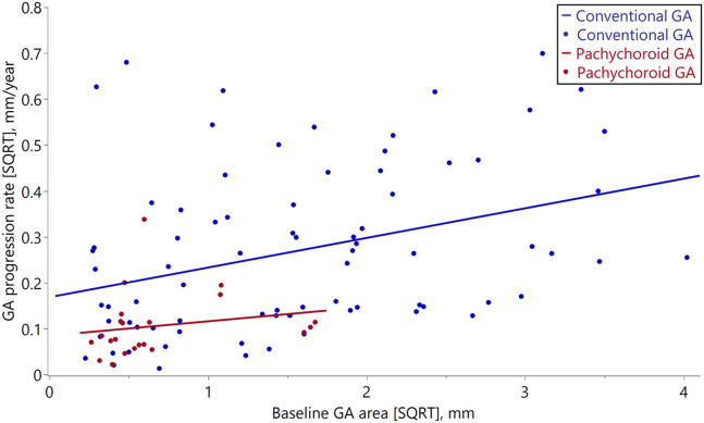
Methods: The studied eyes were classified as having pachychoroid or conventional GA; the former was diagnosed if the eye had features of pachychoroid and no drusen. The GA area was semiautomatically measured on fundus autofluorescence images, and the GA progression rate was calculated for the follow-up group. Multivariable linear regression analysis was used to determine whether the rate of GA progression was associated with GA subtype.
Main outcome measures: Clinical characteristics and progression rates of pachychoroid and conventional GA.
Results: The pachychoroid GA group was significantly younger (70.3 vs. 78.7 years; P < 0.001), more male-dominant (89.5 vs. 55.6%; P < 0.001), and had better best-corrected visual acuity (0.15 vs. 0.40 in logarithm of the minimum angle of resolution; P = 0.002), thicker choroid (312.4 vs. 161.6 μm; P < 0.001), higher rate of unifocal GA type (94.7 vs. 49.6%; P < 0.001), and smaller GA area (0.59 vs. 3.76 mm2;P < 0.001) than the conventional GA group. In the follow-up group, the mean GA progression rate (square-root transformation) was significantly lower in the pachychoroid GA group than in the conventional GA group (0.11 vs. 0.27 mm/year; P < 0.001).
Conclusions: Demographic and ocular characteristics differed between GA subtypes. The progression rate of pachychoroid GA, adjusted for age and baseline GA area, was significantly lower than that of conventional GA. Japanese patients with conventional GA showed characteristics and progression rates similar to those in White populations. Some characteristics of GA in Japanese population differ from those in Waucasian populations, which may be due to the inclusion of pachychoroid GA.
Financial disclosures: Proprietary or commercial disclosure may be found in the Footnotes and Disclosures at the end of this article.
Keywords: Age-related macular degeneration; Geographic atrophy; Japanese; Pachychoroid geographic atrophy; Subtype.
© 2024 by the American Academy of Ophthalmology.
Figures


References
-
- Flaxman S.R., Bourne R.R.A., Resnikoff S., et al. Global causes of blindness and distance vision impairment 1990-2020: a systematic review and meta-analysis. Lancet Glob Health. 2017;5:e1221–e1234. - PubMed
-
- Rim T.H., Kawasaki R., Tham Y.C., et al. Prevalence and pattern of geographic atrophy in asia: the Asian eye Epidemiology consortium. Ophthalmology. 2020;127:1371–1381. - PubMed
-
- Rudnicka A.R., Jarrar Z., Wormald R., et al. Age and gender variations in age-related macular degeneration prevalence in populations of European ancestry: a meta-analysis. Ophthalmology. 2012;119:571–580. - PubMed
-
- Liao D.S., Grossi F.V., El Mehdi D., et al. Complement C3 inhibitor Pegcetacoplan for geographic atrophy secondary to age-related macular degeneration: a randomized phase 2 trial. Ophthalmology. 2020;127:186–195. - PubMed
LinkOut - more resources
Full Text Sources
