

Antimicrobial Susceptibility Testing for Colistin: Extended Application of Novel Quantitative and Morphologic Assay Using Scanning Electron Microscopy
- PMID: 38827502
- PMCID: PMC11144066
- DOI: 10.1155/2024/8917136
Antimicrobial Susceptibility Testing for Colistin: Extended Application of Novel Quantitative and Morphologic Assay Using Scanning Electron Microscopy
Abstract
Background: Colistin (Polymyxin E) has reemerged in the treatment of MDR Gram-negative infections. Traditional Colistin AST methods have long turnaround times and are cumbersome for routine use. We present a SEM-AST technique enabling rapid detection of Colistin resistance through direct observation of morphological and quantitative changes in bacteria exposed to Colistin.
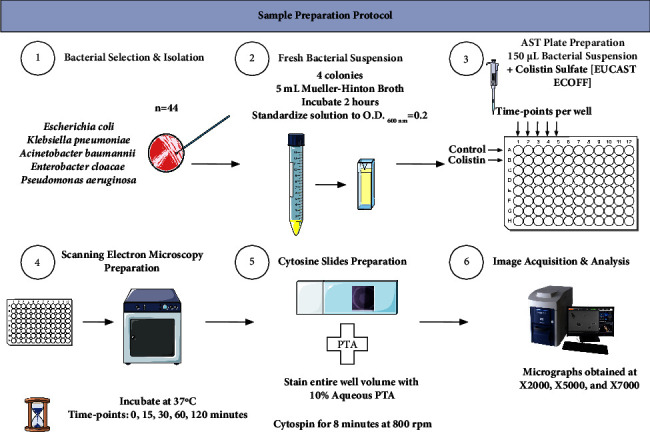
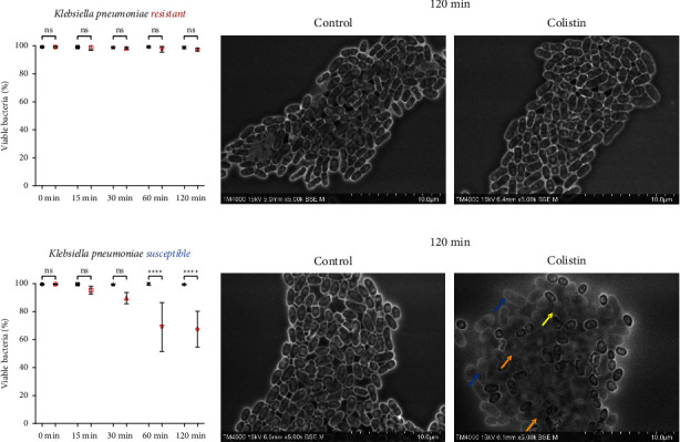
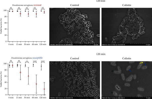
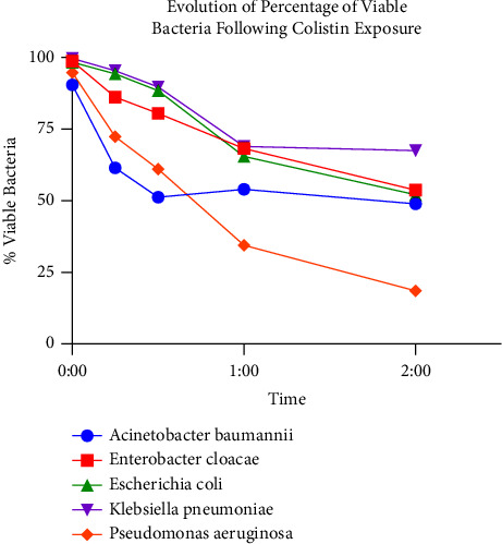
Methods: Forty-four Gram-negative reference organisms were chosen based on their Colistin susceptibility profiles. Bacterial suspensions of ∼107 CFU/mL were exposed to Colistin at EUCAST-ECOFF, with controls not exposed, incubated at 37°C, and then sampled at 0, 15, 30, 60, and 120 minutes. Phosphotungstic Acid (PTA) staining was applied, followed by SEM imaging using Hitachi TM4000PlusII-Tabletop-SEM at ×2000, ×5000 and ×7000 magnifications. Bacterial viability analysis was performed for all conditions by quantifying viable and dead organisms based on PTA-staining and morphologic changes.
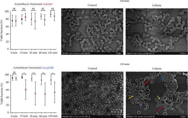
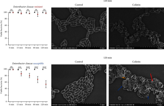
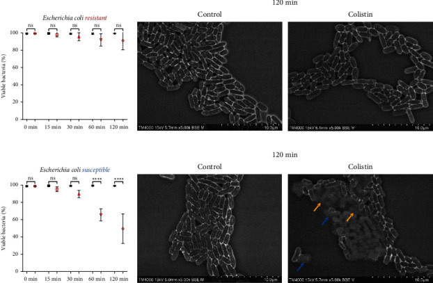
Results: We identified a significant drop in the percentage of viable organisms starting 30 minutes after exposure in susceptible strains, as compared to nonsignificant changes in resistant strains across all tested organisms. The killing effect of Colistin was best observed after 120 minutes of incubation with the antibiotic, with significant changes in morphologic features, including bacterial inflation, fusion, and lysis, observed as early as 30 minutes. Our observation matched the results of the gold standard-based broth microdilution method.
Conclusions: We provide an extended application of the proof of concept for the utilization of the SEM-AST assay for Colistin for a number of clinically relevant bacterial species, providing a rapid and reliable susceptibility profile for a critical antibiotic.
Copyright © 2024 Omar Zmerli et al.
Conflict of interest statement
The authors would like to declare that DR was a consultant in microbiology for Hitachi High-Tech Corporation from March 2018 until March 2021. YO is employed by Hitachi High-Tech Corporation. AH and EM are employed by Hitachi, Ltd. Personal fees for GH, SB, and JBK are paid through a collaborative contract from the Hitachi High-Tech Corporation. OZ and RI declare no relevant competing interests. A patent application for this methodology is pending (PCT/FR2021/052468).
Figures
Similar articles
-
[Evaluation of the BD Phoenix100 System and Colistin Broth Disk Elution Method for Antimicrobial Susceptibility Testing of Colistin Against Gram-negative Bacteria].Mikrobiyol Bul. 2019 Jul;53(3):254-261. doi: 10.5578/mb.68066. Mikrobiyol Bul. 2019. PMID: 31414627 Turkish.
-
Rapid microbial viability assay using scanning electron microscopy: a proof-of-concept using Phosphotungstic acid staining.Comput Struct Biotechnol J. 2023 Jul 11;21:3627-3638. doi: 10.1016/j.csbj.2023.07.010. eCollection 2023. Comput Struct Biotechnol J. 2023. PMID: 37501704 Free PMC article.
-
[Evaluation of the Two Commercial Methods Based on Broth Microdilution Against Reference Microdilution Method for Klebsiella pneumoniae Isolates].Mikrobiyol Bul. 2020 Oct;54(4):606-612. doi: 10.5578/mb.69828. Mikrobiyol Bul. 2020. PMID: 33107289 Turkish.
-
Ultra-rapid flow cytometry assay for colistin MIC determination in Enterobacterales, Pseudomonas aeruginosa and Acinetobacter baumannii.Clin Microbiol Infect. 2020 Nov;26(11):1559.e1-1559.e4. doi: 10.1016/j.cmi.2020.08.019. Epub 2020 Aug 21. Clin Microbiol Infect. 2020. PMID: 32835792
-
Antimicrobial Susceptibility Testing for Polymyxins: Challenges, Issues, and Recommendations.J Clin Microbiol. 2019 Mar 28;57(4):e01390-18. doi: 10.1128/JCM.01390-18. Print 2019 Apr. J Clin Microbiol. 2019. PMID: 30541939 Free PMC article. Review.
References
LinkOut - more resources
Full Text Sources
