

A Case of Squamous Cell Carcinoma Arising in Disseminated Superficial Porokeratosis
- PMID: 38827628
- PMCID: PMC11144422
- DOI: 10.2147/CCID.S463569
A Case of Squamous Cell Carcinoma Arising in Disseminated Superficial Porokeratosis
Abstract
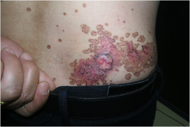
Porokeratosis (PK), characterized by keratotic lesions with an atrophic center and a prominent peripheral ridge, with a typical histological hallmark, namely, the cornoid lamella, has two forms: disseminated and localized. While PK often converts into squamous cell carcinoma (SCC), conversion from disseminated superficial porokeratosis (DSP) alone is rarely reported except for one case in which DSP and LP coexisted and converted to SCC. Here, we report the case of a patient with SCC converted from DSP alone, presenting with coin-sized macules on the bottom right of his waist that developed into an ulcer at the center. The patient underwent radiation therapy, which effectively treated the SCC but did not resolve the PK. This article highlights regular follow-up and undergo comprehensive diagnosis, both of which are beneficial to enable early detection and management of DSP that has converted to into SCC; in addition, standardized medical treatment may help improve the treatment therapeutic effect of in similar diseases.
Keywords: disseminated superficial porokeratosis; porokeratosis; squamous cell carcinoma.
© 2024 Zheng et al.
Conflict of interest statement
The authors declare that they have no conflicts of interest in this work.
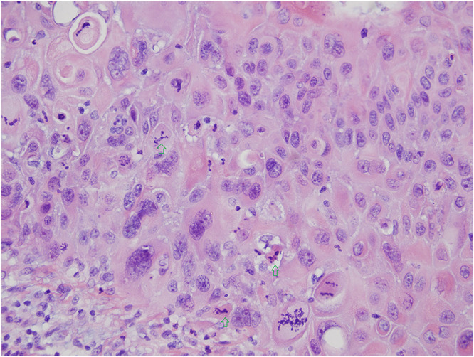
Figures
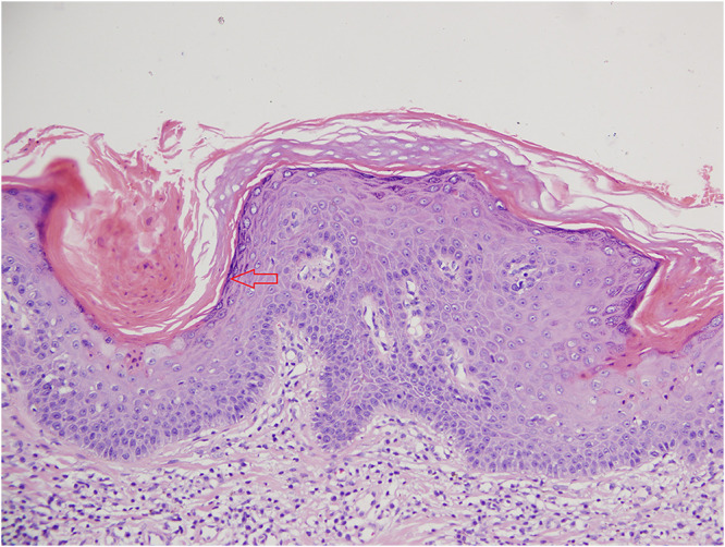
 .
.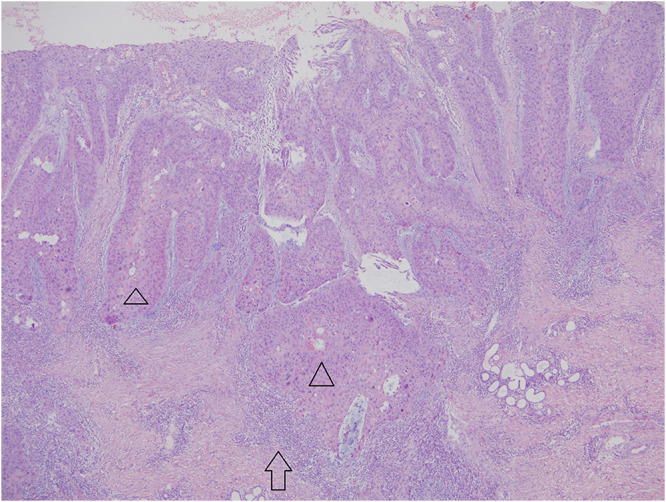
 . Inflammatory cell infiltration:
  .
.
 .
.Similar articles
-
Generalized type 2 segmental disseminated superficial actinic porokeratosis coexisted with multiple cutaneous squamous cell carcinomas: Analysis of two cases.Indian J Pathol Microbiol. 2020 Oct-Dec;63(4):634-636. doi: 10.4103/IJPM.IJPM_987_19. Indian J Pathol Microbiol. 2020. PMID: 33154323
-
Case Report on Rare Clinical Variant of Porokeratosis: Disseminated Superficial Porokeratosis.J Cutan Aesthet Surg. 2020 Apr-Jun;13(2):145-148. doi: 10.4103/JCAS.JCAS_35_19. J Cutan Aesthet Surg. 2020. PMID: 32792775 Free PMC article.
-
A case of inflammatory disseminated superficial porokeratosis in a colon cancer patient.Ann Dermatol. 2009 May;21(2):150-3. doi: 10.5021/ad.2009.21.2.150. Epub 2009 May 31. Ann Dermatol. 2009. PMID: 20523774 Free PMC article.
-
Type 2 segmental manifestation of disseminated superficial actinic porokeratosis in a 7-year-old girl.Eur J Dermatol. 2009 Jan-Feb;19(1):25-8. doi: 10.1684/ejd.2008.0567. Epub 2008 Dec 5. Eur J Dermatol. 2009. PMID: 19059828 Review.
-
Exclusive facial porokeratosis: histopathologically showing follicular cornoid lamellae.J Dermatol. 2011 Nov;38(11):1072-1075. doi: 10.1111/j.1346-8138.2011.01260.x. Epub 2011 Sep 20. J Dermatol. 2011. PMID: 21933257 Review.
References
Publication types
LinkOut - more resources
Full Text Sources
Research Materials
Miscellaneous
