. 2024 May 17:15:1345046.
doi: 10.3389/fimmu.2024.1345046. eCollection 2024.

IFNγ at the early stage induced after cryo-thermal therapy maintains CD4+ Th1-prone differentiation, leading to long-term antitumor immunity

Affiliations

IFNγ at the early stage induced after cryo-thermal therapy maintains CD4+ Th1-prone differentiation, leading to long-term antitumor immunity

Junjun Wang et al. Front Immunol. .

Abstract

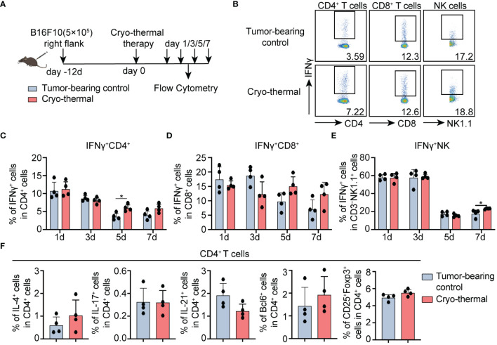
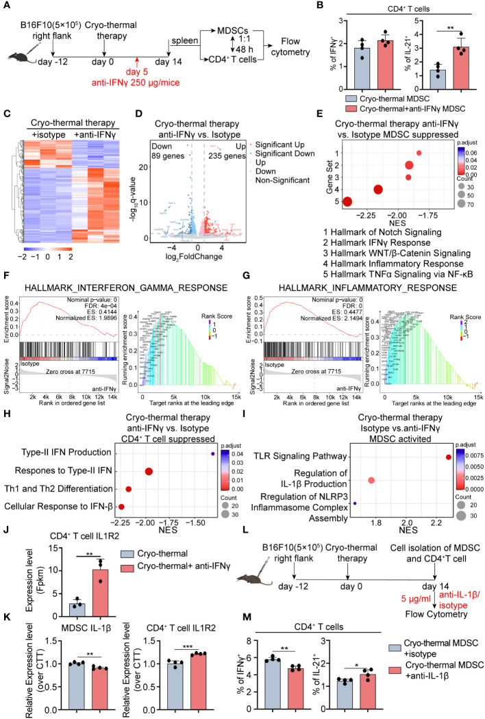
Introduction: Recently, more and more research illustrated the importance of inducing CD4+ T helper type (Th)-1 dominant immunity for the success of tumor immunotherapy. Our prior studies revealed the crucial role of CD4+ Th1 cells in orchestrating systemic and durable antitumor immunity, which contributes to the satisfactory outcomes of the novel cryo-thermal therapy in the B16F10 tumor model. However, the mechanism for maintaining the cryo-thermal therapy-mediated durable CD4+ Th1-dominant response remains uncovered. Additionally, cryo-thermal-induced early-stage CD4+ Th1-dominant T cell response showed a correlation with the favorable prognosis in patients with colorectal cancer liver metastasis (CRCLM). We hypothesized that CD4+ Th1-dominant differentiation induced during the early stage post cryo-thermal therapy would affect the balance of CD4+ subsets at the late phase.

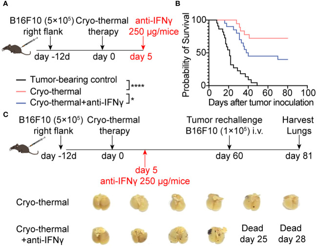
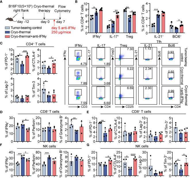
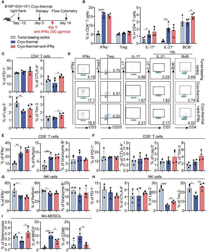
Methods: To understand the role of interferon (IFN)-γ, the major effector of Th1 subsets, in maintaining long-term CD4+ Th1-prone polarization, B16F10 melanoma model was established in this study and a monoclonal antibody was used at the early stage post cryo-thermal therapy for interferon (IFN)-γ signaling blockade, and the influence on the phenotypic and functional change of immune cells was evaluated.

Results: IFNγ at the early stage after cryo-thermal therapy maintained long-lasting CD4+ Th1-prone immunity by directly controlling Th17, Tfh, and Tregs polarization, leading to the hyperactivation of Myeloid-derived suppressor cells (MDSCs) represented by abundant interleukin (IL)-1β generation, and thereby further amplifying Th1 response.

Discussion: Our finding emphasized the key role of early-phase IFNγ abundance post cryo-thermal therapy, which could be a biomarker for better prognosis after cryo-thermal therapy.

Keywords: CD4+ Th1 cells; cryo-thermal therapy; interferon-γ; interleukin-1β; myeloid-derived suppressor cells.


Conflict of interest statement

The authors declare that the research was conducted in the absence of any commercial or financial relationships that could be construed as a potential conflict of interest.

Figures

Figure 1
Figure 1
Phenotype of CD4+ T cells, CD8+ T cells, and NK cells after cryo-thermal therapy. (A) Scheme of study design. Briefly, CD4+ T cells, CD8+ T cells, and NK cells from tumor-bearing mice and cryo-thermal treated mice were detected by flow cytometry at indicated times. (B–E) The representative figure of gating strategy (B), the expression of IFNγ in (C) CD4+ T cells, (D) CD8+ T cells, (E) and NK cells from tumor-bearing control (gray) and cryo-thermal therapy (red). (F) The subsets of CD4+ T cells on day 5 after cryo-thermal therapy. Experiments were independently repeated at least three times. *p < 0.05. n=4 for each group.
Figure 2
Figure 2
IFNγ at the early stage after cryo-thermal therapy mediated antitumor immune memory and led to a better prognosis. (A) Scheme of the experiment design. (B) Kaplan–Meier survival curve of tumor-bearing control, cryo-thermal therapy, or cryo-thermal therapy with IFNγ neutralization. The survival curves were compared using log-rank tests. *p < 0.05. ****p < 0.0001. n=22 for each group. (C) Scheme of tumor rechallenge design. The treated mice were injected with 1×10⁵ B16F10 cells intravenously on day 60 after cryo-thermal therapy. The lungs were collected on day 21 after the rechallenge. n=6 for the cryo-thermal group. n=4 for the cryo-thermal + anti-IFNγ group for two mice that died before tumor rechallenge.
Figure 3
Figure 3
Phenotype of CD4+ T cells, CD8+ T cells, and NK cells after IFNγ in vivo neutralization at the early stage. (A) Scheme of study design. 250 µg anti-IFNγ antibodies was injected i.p. on day 5 after cryo-thermal therapy, the phenotype of immune cells was detected by flow cytometry on day 7 post cryo-thermal therapy. (B, C) The subsets (B) and the immune checkpoints (C) of CD4+ T cells. (D, E) The cytotoxic molecules (D) and the immune checkpoints (E) of CD8+ T cells. (F, G) The cytotoxic molecules (F) and the immune checkpoints (G) of NK cells. Experiments were independently repeated at least three times. *p < 0.05, **p < 0.01, ***p < 0.001, ****p < 0.0001. n=4 for each group.
Figure 4
Figure 4
Phenotype of CD4+ T cells, CD8+ T cells, and NK cells after IFNγ neutralization at the late stage in vivo. (A) Scheme of study design. Briefly, 250 µg anti-IFNγ antibodies was injected i.p. on day 5 after cryo-thermal therapy, the phenotype of immune cells was detected by flow cytometry on day 14 post cryo-thermal therapy. (B–D) The subsets (B, D) and the immune checkpoints (C) of CD4+ T cells. (E, F) The cytotoxic molecules (E) and the immune checkpoints (F) of CD8+ T cells. (G, H) The cytotoxic molecules (G) and the immune checkpoints (H) of NK cells. (I) The proportion of MDSCs and the expression levels of MHCII and CD86 on MDSCs. Experiments were independently repeated at least three times. *p < 0.05, **p < 0.01, ***p < 0.001, ****p < 0.0001. n=4 for each group.
Figure 5
Figure 5
Transcriptomic profile of MDSCs and IL-1β secreted by MDSCs are involved in maintaining CD4+ Th1-dominated differentiation. (A) Scheme of in vitro design. MDSCs from cryo-thermal therapy and cryo-thermal therapy with IFNγ neutralization on day 14 were isolated by MASC and cocultured with CD4+ T cells from tumor-bearing control (0d). After 48 hours, the subsets (B) of CD4+ T cells were analyzed by flow cytometry. MDSCs and CD4+ T cells were isolated by MACS and analyzed by RNA-seq. (C) Heatmap of differentially expressed genes. (D) Volcano plot of gene expression change of MDSCs from cryo-thermal therapy injected with anti-IFNγ antibody over cryo-thermal treated therapy. (E) GSEA analysis showing significantly suppressed gene sets of MDSCs cryo-thermal with IFNγ neutralization mice. (F) Individual GSEA enrichment plot for the hallmark inflammation response gene set (left), GSEA dot lot (right). (G) Individual GSEA enrichment plot for the hallmark IFNγ response gene set (left), GSEA dot plot (right). n=3 for each group in RNA-seq. (H) GSEA analysis showing significantly suppressed gene sets of CD4+ T cells cryo-thermal with IFNγ neutralization mice. (I) GSEA analysis showing significantly activated gene sets of MDSCs from cryo-thermal therapy. (J) The expression level of IL-1R2 on CD4+ T cells from RNA-seq. (K) The expression levels of IL-1β in MDSCs and IL-1R2 on CD4+ T cells from cryo-thermal therapy and cryo-thermal therapy with IFNγ neutralization were detected by RT-PCR. (L) Scheme of in vitro design. MDSCs from the cryo-thermal therapy on day 14 were isolated by MASC and cocultured with CD4+ T cells from cryo-thermal therapy (14d) in the presence of 5 µg/mL isotype or the anti-IL-1β antibody. Forty-eight hours, the subsets (M) of CD4+ T cells were analyzed by flow cytometry. In both in vitro experiments, the corresponding serum was administered to both groups to mimic the in vivo environment, and an anti-CD3 agonist (1 µg/mL) was added to stimulate the activation of CD4+ T cells. Experiments were independently repeated at least three times. *p < 0.05, **p < 0.01, ***p < 0.001. n=4 for each group in vitro experiments.

Similar articles

Cited by

  • CD4+ T cells in antitumor immunity.
    Montauti E, Oh DY, Fong L. Montauti E, et al. Trends Cancer. 2024 Oct;10(10):969-985. doi: 10.1016/j.trecan.2024.07.009. Epub 2024 Sep 5. Trends Cancer. 2024. PMID: 39242276 Review.

References

    1. Basu A, Ramamoorthi G, Albert G, Gallen C, Beyer A, Snyder C, et al. . Differentiation and regulation of T(H) cells: A balancing act for cancer immunotherapy. Front Immunol. (2021) 12:669474. doi: 10.3389/fimmu.2021.669474 - DOI - PMC - PubMed
    1. Peng D, Kryczek I, Nagarsheth N, Zhao L, Wei S, Wang W, et al. . Epigenetic silencing of TH1-type chemokines shapes tumour immunity and immunotherapy. Nature. (2015) 527:249–53. doi: 10.1038/nature15520 - DOI - PMC - PubMed
    1. Sun Z, Chen F, Meng F, Wei J, Liu B. MHC class II restricted neoantigen: A promising target in tumor immunotherapy. Cancer Lett. (2017) 392:17–25. doi: 10.1016/j.canlet.2016.12.039 - DOI - PubMed
    1. Kondo T, Nakazawa H, Ito F, Hashimoto Y, Osaka Y, Futatsuyama K, et al. . Favorable prognosis of renal cell carcinoma with increased expression of chemokines associated with a Th1-type immune response. Cancer Sci. (2006) 97:780–6. doi: 10.1111/j.1349-7006.2006.00231.x - DOI - PMC - PubMed
    1. Zhu J, Zhang Y, Zhang A, He K, Liu P, Xu LX. Cryo-thermal therapy elicits potent anti-tumor immunity by inducing extracellular Hsp70-dependent MDSC differentiation. Sci Rep. (2016) 6:27136. doi: 10.1038/srep27136 - DOI - PMC - PubMed

Substances