

Identification of Diagnostic Genes of Aortic Stenosis That Progresses from Aortic Valve Sclerosis
- PMID: 38828052
- PMCID: PMC11144011
- DOI: 10.2147/JIR.S453100
Identification of Diagnostic Genes of Aortic Stenosis That Progresses from Aortic Valve Sclerosis
Abstract
Background: Aortic valve sclerosis (AVS) is a pathological state that can progress to aortic stenosis (AS), which is a high-mortality valvular disease. However, effective medical therapies are not available to prevent this progression. This study aimed to explore potential biomarkers of AVS-AS advancement.
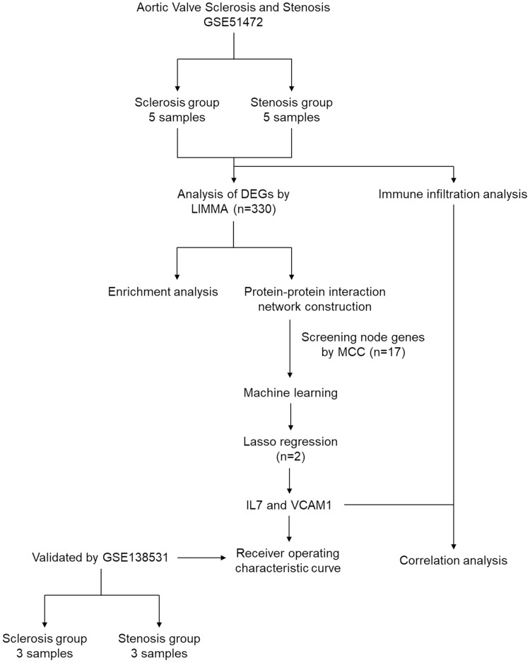
Methods: A microarray dataset and an RNA-sequencing dataset were obtained from the Gene Expression Omnibus (GEO) database. Differentially expressed genes (DEGs) were screened from AS and AVS samples. Functional enrichment analysis, protein-protein interaction (PPI) network construction, and machine learning model construction were conducted to identify diagnostic genes. A receiver operating characteristic (ROC) curve was generated to evaluate diagnostic value. Immune cell infiltration was then used to analyze differences in immune cell proportion between tissues. Finally, immunohistochemistry was applied to further verify protein concentration of diagnostic factors.
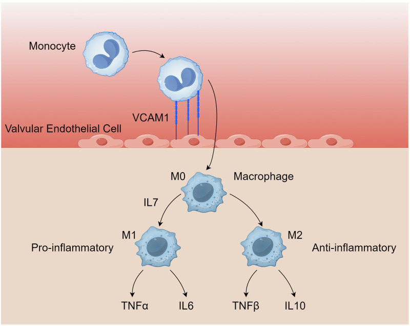
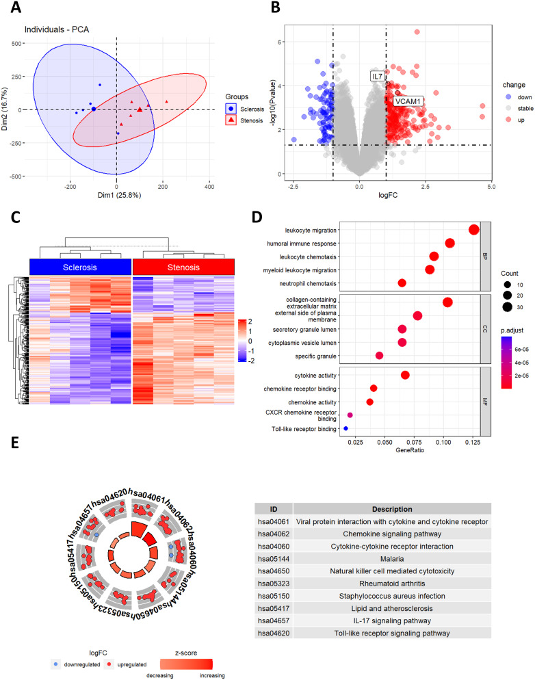
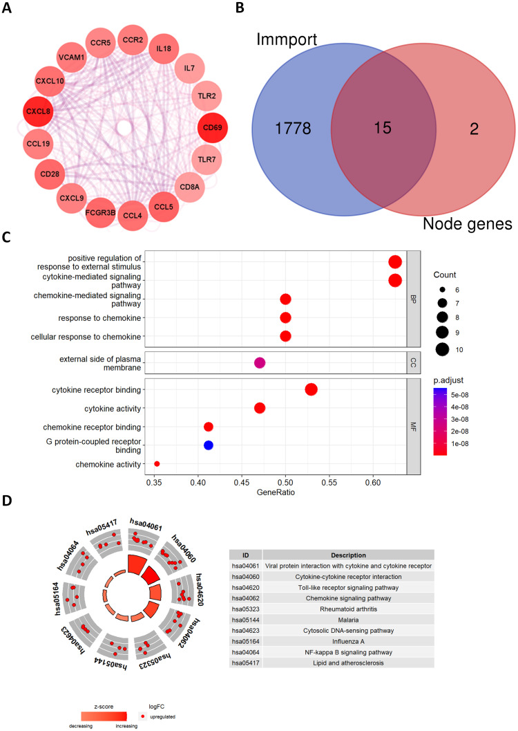
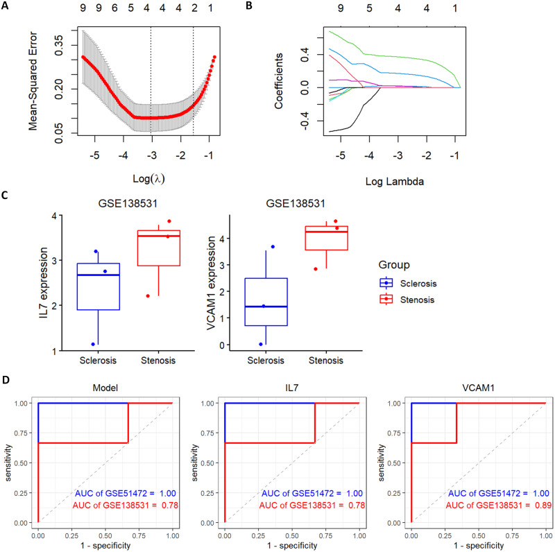
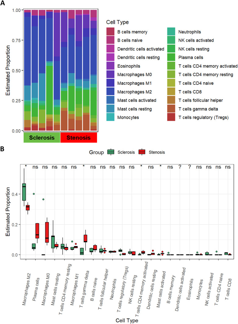
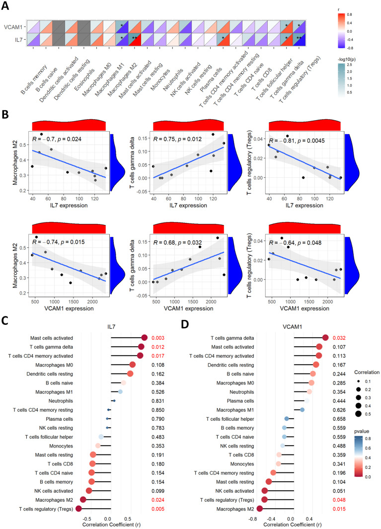
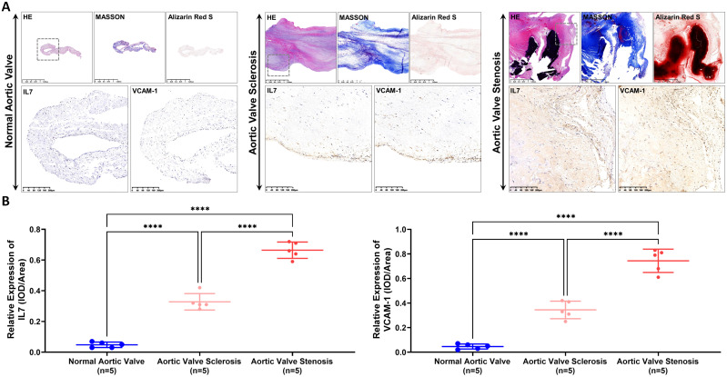
Results: A total of 330 DEGs were identified, including 92 downregulated and 238 upregulated genes. The top 5% of DEGs (n = 17) were screened following construction of a PPI network. IL-7 and VCAM-1 were identified as the most significant candidate genes via least absolute shrinkage and selection operator (LASSO) regression. The diagnostic value of the model and each gene were above 0.75. Proportion of anti-inflammatory M2 macrophages was lower, but the fraction of pro-inflammatory gamma-delta T cells was elevated in AS samples. Finally, levels of IL-7 and VCAM-1 were validated to be higher in AS tissue than in AVS tissue using immunohistochemistry.
Conclusion: IL-7 and VCAM-1 were identified as biomarkers during the disease progression. This is the first study to analyze gene expression differences between AVS and AS and could open novel sights for future studies on alleviating or preventing the disease progression.
Keywords: aortic stenosis; aortic valve sclerosis; diagnostic genes; immune infiltration; immunohistochemistry; machine learning.
© 2024 Yu et al.
Conflict of interest statement
The authors report no conflicts of interest in this work.
Figures
Similar articles
-
Potential biomarkers and immune cell infiltration involved in aortic valve calcification identified through integrated bioinformatics analysis.Front Physiol. 2022 Dec 15;13:944551. doi: 10.3389/fphys.2022.944551. eCollection 2022. Front Physiol. 2022. PMID: 36589450 Free PMC article.
-
Identification of Immune-Associated Genes in Diagnosing Aortic Valve Calcification With Metabolic Syndrome by Integrated Bioinformatics Analysis and Machine Learning.Front Immunol. 2022 Jul 4;13:937886. doi: 10.3389/fimmu.2022.937886. eCollection 2022. Front Immunol. 2022. PMID: 35865542 Free PMC article.
-
Identification of pyroptosis-associated genes with diagnostic value in calcific aortic valve disease.Front Cardiovasc Med. 2024 Jan 25;11:1340199. doi: 10.3389/fcvm.2024.1340199. eCollection 2024. Front Cardiovasc Med. 2024. PMID: 38333413 Free PMC article.
-
Identifying the hub gene and immune infiltration of Parkinson's disease using bioinformatical methods.Brain Res. 2022 Jun 15;1785:147879. doi: 10.1016/j.brainres.2022.147879. Epub 2022 Mar 10. Brain Res. 2022. PMID: 35278479
-
Integrated identification of key immune related genes and patterns of immune infiltration in calcified aortic valvular disease: A network based meta-analysis.Front Genet. 2022 Sep 21;13:971808. doi: 10.3389/fgene.2022.971808. eCollection 2022. Front Genet. 2022. PMID: 36212153 Free PMC article.
References
-
- Otto CM, Nishimura RA, Bonow RO, et al. 2020 ACC/AHA guideline for the management of patients with valvular heart disease: executive summary: a report of the American college of cardiology/American heart association joint committee on clinical practice guidelines. Circulation. 2021;143(5):e35–e71. doi:10.1161/cir.0000000000000932 - DOI - PubMed
LinkOut - more resources
Full Text Sources
Research Materials
Miscellaneous
