. 2024 Jun;14(6):2554-2566.
doi: 10.1016/j.apsb.2024.03.007. Epub 2024 Mar 10.

Pharmacokinetic enhancement of oncolytic virus M1 by inhibiting JAK‒STAT pathway

Affiliations

Pharmacokinetic enhancement of oncolytic virus M1 by inhibiting JAK‒STAT pathway

Jingyi Tan et al. Acta Pharm Sin B. 2024 Jun.

Abstract

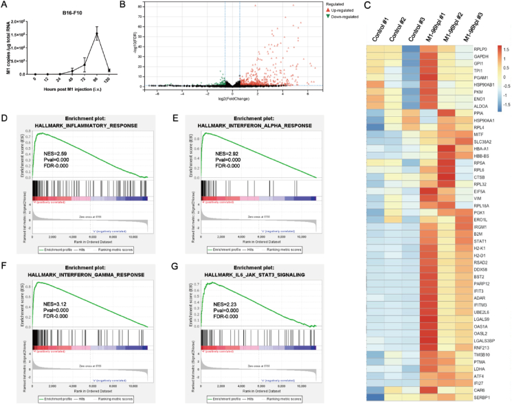
Oncolytic viruses (OVs), a group of replication-competent viruses that can selectively infect and kill cancer cells while leaving healthy cells intact, are emerging as promising living anticancer agents. Unlike traditional drugs composed of non-replicating compounds or biomolecules, the replicative nature of viruses confer unique pharmacokinetic properties that require further studies. Despite some pharmacokinetics studies of OVs, mechanistic insights into the connection between OV pharmacokinetics and antitumor efficacy remain vague. Here, we characterized the pharmacokinetic profile of oncolytic virus M1 (OVM) in immunocompetent mouse tumor models and identified the JAK‒STAT pathway as a key modulator of OVM pharmacokinetics. By suppressing the JAK‒STAT pathway, early OVM pharmacokinetics are ameliorated, leading to enhanced tumor-specific viral accumulation, increased AUC and Cmax, and improved antitumor efficacy. Rather than compromising antitumor immunity after JAK‒STAT inhibition, the improved pharmacokinetics of OVM promotes T cell recruitment and activation in the tumor microenvironment, providing an optimal opportunity for the therapeutic outcome of immune checkpoint blockade, such as anti-PD-L1. Taken together, this study advances our understanding of the pharmacokinetic-pharmacodynamic relationship in OV therapy.

Keywords: Anticancer; JAK‒STAT; Oncolytic virus; Pharmacokinetics.


Conflict of interest statement

The authors declare no conflicts of interest.

Figures

Image 1
Graphical abstract
Figure 1
Figure 1
Pharmacokinetic profiles of OVM in syngeneic tumor mouse models (the OVM injection dose was 3 × 106 PFU). (A, C, E, G, I) The change of the OVM RNA copies in tumor and normal organs over time, n = 3. (B, D, F, H, J) Main parameters of pharmacokinetics in tumor is shown, n = 3.
Figure 2
Figure 2
Peak intratumoral OVM level accompanies with activation of inflammatory response. (A) C57BL/6 mice were implanted subcutaneously in the right flank with B16-F10 cells and treated intravenously with OVM, the OVM injection dose was 1 × 105 PFU (n = 5). The tumors were harvested at 0 and 96 h after OVM administration, the total RNA was extracted, and RNA sequencing was then performed. (B) Volcano plot in tumor tissue is shown. (C) Gene enrichment heatmap in tumor tissue. (D–G) GSEA results for the inflammatory response (D), IFN-α response (E), IFN-γ response (F), and JAK‒STAT signaling (G) are shown.
Figure 3
Figure 3
Inhibiting JAK‒STAT pathway increases intratumoral Cmax and AUC of OVM (3 × 106 PFU, single dose). (A–D) Effects of two concentrations of MCC950 (A), amlexanox (B), fludarabine (C), and ruxolitinib (D) on the replication of OVM in tumor, n = 3. (E and F) Western blot image (left) of JAK‒STAT pathway and IRF-1, IRF-9 expression and quantitative statistics (right), n = 3. (G–H) IFN-α (G) and IFN-β (H) mRNA level of B16-F10 tumor with OVM alone or combined with ruxolitinib administration, n = 5. (I) Western blot image (left) of OVM protein expression and quantitative statistics (right), n = 4. (J–M) Pharmacokinetic profiles of OVM combined neutralizing or depleting antibodies in B16-F10 models, n = 3. (N–O) OVM replication curve and AUC in B16-F10 tumor (N) or Pan02 tumor (O) when OVM alone or combined with ruxolitinib administration, n = 5. (P) Copy number of OVM in different tissues when OVM alone or combined with ruxolitinib administration at 96 hpi, n = 3. (Q) OVM RNA copy number in normal and tumor tissues at 21 days post-treatment initiation, n = 3. hpi, h post infection. AUC, area under curve. Data are reported as the mean ± SD. ns, no statistical difference. ∗P < 0.05; ∗∗P < 0.01; ∗∗∗P < 0.001; ∗∗∗∗P < 0.0001.
Figure 4
Figure 4
Inhibiting JAK‒STAT pathway improves the therapeutic efficacy of OVM in immunocompetent mice (6 × 105 PFU/dose/day, five daily injections). (A, E) C57BL/6 mice were implanted subcutaneously in the right flank with B16-F10 (A) or Pan02 (E) cells on day 0 and treated intravenously with control or OVM once per day on Days 6–10 (A) or Days 8–12 (E). Ruxolitinib was treated 3 times. (B, F) Tumor growth curves in B16-F10 (C) or Pan02 (F) tumor-bearing mice are shown, n = 6. (C, G) Survival curves in B16-F10 (C) or Pan02 (G) tumor-bearing mice are shown, n = 10. (D, H) Body weight changes in different treatment groups of B16-F10 (D) and Pan02 (H) tumor-bearing mouse models, n = 6. (I) H&E staining of heart, liver, spleen, lung, kidney, and intestine of B16-F10 tumor-bearing mice 21 days post treatment with OVM alone or OVM combined with ruxolitinib. Scale bar, 100 μm ns, no statistical difference; ∗P < 0.05; ∗∗P < 0.01; ∗∗∗P < 0.001; ∗∗∗∗P < 0.0001.
Figure 5
Figure 5
Inhibiting JAK‒STAT pathway improves the therapeutic efficacy of OVM in mouse model implanted with human melanoma cells (6 × 105 PFU/dose/day, five daily injections). (A) OVM copies in A375 human melanoma model tumor, n = 4. (B and C) Tumor growth curve and T/C (%) in A375 tumor-bearing mice are shown, n = 6. (E) A375 tumor-bearing mouse weight curve is shown, n = 6. (D) A375 tumors and quantitative statistics are shown. Group Ruxolitinib + M1 had 1/6 tumor regression, n = 6. T/C (%) = TRTV/CRTV × 100; TRTV, RTV in treatment group; CRTV, RTV in isotype control group; RTV: relative tumor volume; RTV = Vt/V0; Vt, tumor volume after treatment; V0, tumor volume before treatment. ns, no statistical difference; ∗P < 0.05; ∗∗P < 0.01; ∗∗∗P < 0.001; ∗∗∗∗P < 0.0001.
Figure 6
Figure 6
Inhibiting JAK‒STAT pathway boosts the OVM-induced intratumoral T cell infiltration and activation. (A–C) The infiltration of CD45+ CD3+ T cells(A), CD8+ T cell (B), and CD4+ T cells (C) in B16-F10 model tumor was detected by flow cytometry, n = 4. (D–F) The infiltration of CD45+ CD3+ T cells (D), CD8+ T cell (E), and CD4+ T cells (F) in spleen of B16-F10 model, n = 4. (G and H) The mRNA level of IFN-I in B16-F10 model tumor after 36 h (G) or 120 h (H) post OVM alone or combined ruxolitinib administration, n = 3. (I) The mRNA level of Granzyme B and IFNγ in B16-F10 model tumor after 36 h (I) or 120 h (J) post OVM alone or combined ruxolitinib administration, n = 3. ns, no statistical difference; ∗P < 0.05; ∗∗P < 0.01; ∗∗∗P < 0.001; ∗∗∗∗P < 0.0001.
Figure 7
Figure 7
PD-L1 antibody enhances the anti-tumor efficacy of OVM and ruxolitinib combination therapy (6 × 105 PFU/dose/day, five daily injections). (A) The mRNA level of PD-L1 in B16-F10 tumor-bearing model after OVM alone or combined ruxolitinib administration, n = 3. (B) C57BL/6 mice were implanted subcutaneously in the right flank with B16-F10 cells on day 0 and treated intravenously with control (n = 10) or OVM (n = 10) once per day on Days 6–10. Ruxolitinib and PD-L1 antibody were respectively treated 3 times. (C–E) Tumor growth curve (C, n = 6), weight of mice (D, n = 6), survival curve of tumor-bearing mice (E, n = 10) in each group are shown. ns, no statistical difference; ∗P < 0.05; ∗∗P < 0.01; ∗∗∗P < 0.001; ∗∗∗∗P < 0.0001.

Similar articles

Cited by

References

    1. Macedo N., Miller D.M., Haq R., Kaufman H.L. Clinical landscape of oncolytic virus research in 2020. J Immunother Cancer. 2020;8 - PMC - PubMed
    1. Wang W., Liu S., Dai P., Yang N., Wang Y., Giese R.A., et al. Elucidating mechanisms of antitumor immunity mediated by live oncolytic vaccinia and heat-inactivated vaccinia. J Immunother Cancer. 2021;9 - PMC - PubMed
    1. Hemminki O., Dos Santos J.M., Hemminki A. Oncolytic viruses for cancer immunotherapy. J Hematol Oncol. 2020;13:84. - PMC - PubMed
    1. Pol J., Kroemer G., Galluzzi L. First oncolytic virus approved for melanoma immunotherapy. OncoImmunology. 2016;5 - PMC - PubMed
    1. Corrigan P.A., Beaulieu C., Patel R.B., Lowe D.K. Talimogene Laherparepvec: an oncolytic virus therapy for melanoma. Ann Pharmacother. 2017;51:675–681. - PubMed