. 2024 Jun;14(6):2567-2580.
doi: 10.1016/j.apsb.2024.02.023. Epub 2024 Mar 3.

Carrimycin inhibits coronavirus replication by decreasing the efficiency of programmed -1 ribosomal frameshifting through directly binding to the RNA pseudoknot of viral frameshift-stimulatory element

Affiliations

Carrimycin inhibits coronavirus replication by decreasing the efficiency of programmed -1 ribosomal frameshifting through directly binding to the RNA pseudoknot of viral frameshift-stimulatory element

Hongying Li et al. Acta Pharm Sin B. 2024 Jun.

Abstract

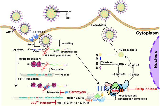
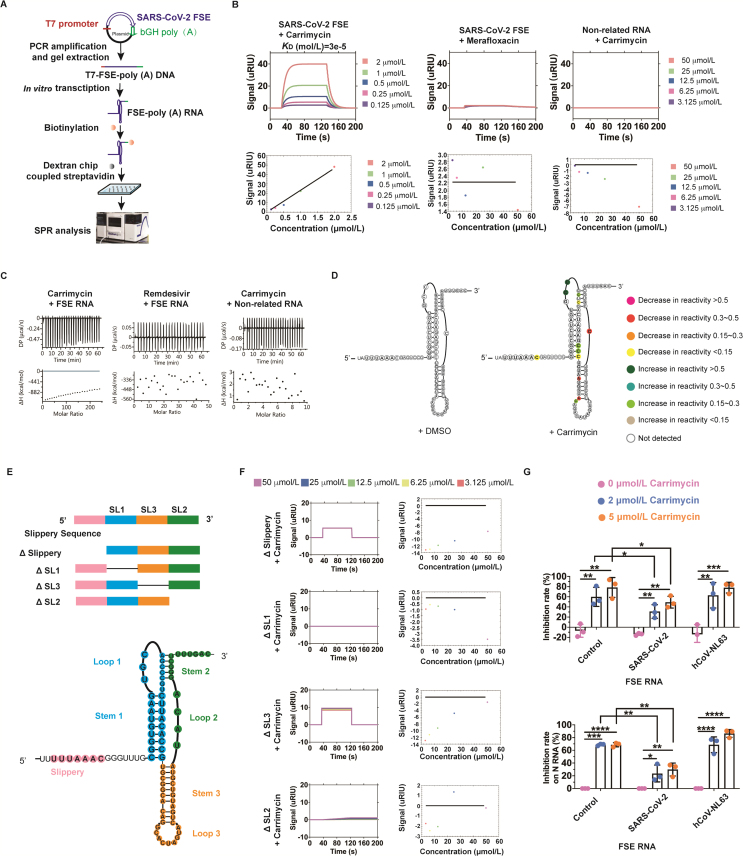
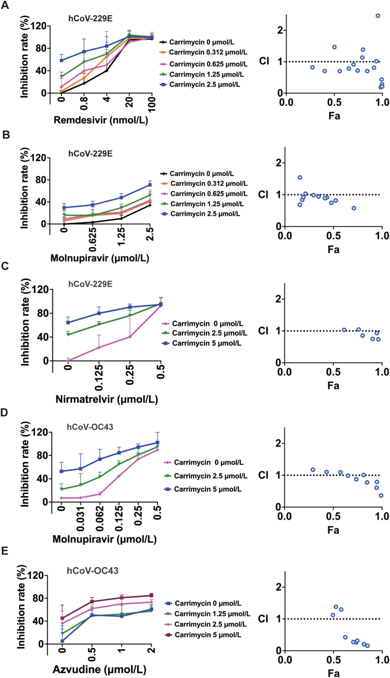
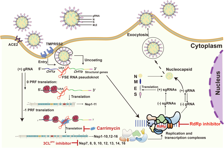
The pandemic of SARS-CoV-2 worldwide with successive emerging variants urgently calls for small-molecule oral drugs with broad-spectrum antiviral activity. Here, we show that carrimycin, a new macrolide antibiotic in the clinic and an antiviral candidate for SARS-CoV-2 in phase III trials, decreases the efficiency of programmed -1 ribosomal frameshifting of coronaviruses and thus impedes viral replication in a broad-spectrum fashion. Carrimycin binds directly to the coronaviral frameshift-stimulatory element (FSE) RNA pseudoknot, interrupting the viral protein translation switch from ORF1a to ORF1b and thereby reducing the level of the core components of the viral replication and transcription complexes. Combined carrimycin with known viral replicase inhibitors yielded a synergistic inhibitory effect on coronaviruses. Because the FSE mechanism is essential in all coronaviruses, carrimycin could be a new broad-spectrum antiviral drug for human coronaviruses by directly targeting the conserved coronaviral FSE RNA. This finding may open a new direction in antiviral drug discovery for coronavirus variants.

Keywords: Antiviral agent; Broad-spectrum antiviral activity; Carrimycin; Coronavirus; Programmed –1 ribosomal frameshifting; RNA pseudoknot; RNA target; Synergistic inhibitory effect.


Conflict of interest statement

The authors declare no conflicts of interest.

Figures

Image 1
Graphical abstract
Figure 1
Figure 1
Carrimycin inhibits hCoV replication in cells. (A) The chemical structure of carrimycin. (B) Carrimycin inhibited hCoV-229E replication under a different multiplicity of infection (MOI) detected by CCK-8 assay at 72 h of drug treatment. Remdesivir (RDV 0.02 μmol/L) as a positive control. (C) Carrimycin inhibited hCoV-229E (MOI = 0.03) at the RNA levels quantified by qRT-PCR at 24 h of drug treatment. (D) Carrimycin inhibited the SARS-CoV-2 Omicron strain in Vero-E6 cells (CPE assay). (E) Anti-coronaviral activity of macrolide antibiotics. Viral dsRNA (green) and cell nuclei (blue) in Huh7 cells infected with hCoV-229E and in H460 cells infected with hCoV-OC43 visualized by immunofluorescent staining assay at 24 h of drug treatment. Scale bar: 100 μm. The experiments were carried out three times. P values were calculated using Student's t-test (mean ± SD, n = 3). ∗P < 0.05, ∗∗P < 0.01, ∗∗∗P < 0.001 vs. virus control. CC50, 50% cytotoxic concentration; EC50, 50% effective concentration; TCID50, 50% tissue culture infective dose.
Figure 2
Figure 2
Carrimycin interrupts the viral protein translation switch from ORF1a to ORF1b. (A) Huh7 cells were infected with hCoV-229E (MOI = 5.6) and treated simultaneously with carrimycin (5 μmol/L). Viral dsRNA and cell nuclei were visualized by immunofluorescent staining assay, the ratio of dsRNA/nuclei fluorescent intensity was calculated by Image J, and intracellular RNAs were quantified by qRT-PCR at the indicated hours post-infection (hpi). Remdesivir (0.05 μmol/L) as a positive control. (B) Carrimycin did not inhibit the activities of viral replicases. SARS-CoV-2 3CLpro and RdRp activities were detected using sandwich-like FP and fluorometric assay, respectively. PF-07321332 or C646 as an individual positive control. Data are shown as mean ± SD (n = 3). (C) Proteomics sequencing. Huh7 cells were infected with hCoV-229E (MOI = 0.88) and treated with 5 μmol/L carrimycin. After 24 or 36 h, cell lysates were subjected to tandem mass tag (TMT) proteomic sequencing. The protein ratio of RdRp/3CLpro was calculated. (D) Ribosome profiling sequencing. Ribosome profiling sequencing (Ribo-seq) read counts within the genome of hCoV-229E gp1 (ORF1ab) in Huh7 cells infected with hCoV-229E (MOI = 0.88) and treated with carrimycin for 24 h. The relative translation ratios of ORF1b-to ORF1a-translated proteins and RdRp to 3CLpro were calculated. P values were calculated using Student's t-test (mean ± SD, n = 3). ∗P < 0.05, ∗∗P < 0.01, and ns, P > 0.05 vs. control.
Figure 3
Figure 3
Carrimycin decreases the efficiency of –1 PRF of coronaviral RNAs. (A) The principle of the dual fluorescent reporter system showed the translational outcome, and the fluorescent density of mCherry and EGFP was visualized. Scale bar: 200 μm. (B) Carrimycin reduced the efficacy of –1 PRF of SARS-CoV-2 FSE detected by the fluorescent intensity at 24 h of drug treatment in HEK293T, Huh7, and Huh7.5 cells. The efficiency of –1 PRF was quantified by the ratio of EGFP to mCherry fluorescence intensity described in Method. (C) Inhibition on –1 PRF of SARS-CoV-2 by carrimycin in a rabbit reticulocyte lysate translation system. The efficiency of –1 PRF was quantified by the protein ratio of mCherry-EGFP to mCherry. (D) Carrimycin reduced the efficacy of –1 PRF of coronaviral FSEs in Huh7 cells. P values were calculated using Student's t-test (mean ± SD, n = 3). ∗P < 0.05, ∗∗P < 0.01, ∗∗∗P < 0.001, and ∗∗∗∗P < 0.0001 vs. control. –1 PRF, programmed –1 ribosomal frameshifting.
Figure 4
Figure 4
Carrimycin binds directly to viral FSE RNA pseudoknots. (A) Methodology for detecting affinity between the compound and FSE RNA. (B) Affinity between the compound and SARS-CoV-2 FSE RNA. (C) Interaction between carrimycin and FSE RNA detected by ITC. Up panel: power required to maintain the temperature of the RNA solution (baseline-corrected). Down panel: integrated heats of interaction plotted against the molar ratio of ligand over RNA and fitted to a single binding site model (MicroCal PEAQ-ITC Analysis Software 1.1.0). (D) DMS-Map upon carrimycin binding in Huh7 cells infected with hCoV-229E. FSE RNA pseudoknot structure (left) and average changes (n = 2) in DMS reactivity upon carrimycin binding (right). Colored nucleotides represent a decrease or increase in reactivity. (E) The typical structure of SARS-CoV-2 FSE RNA pseudoknot and truncated mutations of FSE RNAs. (F) The affinity between carrimycin and FSE RNA with truncated mutations. (G) Exogenous SARS-CoV-2 FSE RNA reduced the antiviral activity of carrimycin against hCoV-229E in Huh7 cells detected with CCK-8 staining assay (up) and qRT-PCR (down) methods (n = 3). P values were calculated using Student's t-test (mean ± SD). ∗P < 0.05, ∗∗P < 0.01, ∗∗∗P < 0.001, ∗∗∗∗P < 0.0001 and ns, P > 0.05 vs. control.
Figure 5
Figure 5
Carrimycin synergistically enhances the antiviral activities of viral replicase inhibitors. (A–C) Carrimycin in combination with remdesivir (A), molnupiravir (B), or nirmatrelvir (C) synergistically inhibited hCoV-229E (MOI = 0.05) replication in Huh7 cells. (D) Carrimycin in combination with molnupiravir synergistically inhibited hCoV-OC43 (MOI = 1) replication. Cells were infected with the virus and treated simultaneously with compounds. At 72 h, the cell viability was detected by CCK-8 assay, and antiviral activities were calculated. Data are shown as mean ± SD (n = 3). (E) Carrimycin plus azvudine synergistically inhibited hCoV-OC43 using qRT-PCR (RdRp RNA). The combination index (CI) was calculated by the Chou-Talalay method using CompuSyn version 1.0, where CI > 1 indicates antagonism, CI = 1 indicates addition, and CI < 1 suggests synergy between the two drugs. Fa, inhibition of test compounds combination.
Figure 6
Figure 6
Overview of the mechanism of action and role of carrimycin against hCoV. Coronaviruses apply the –1 PRF mechanism to switch complete protein translation from ORF1a to ORF1b, while carrimycin binds directly to the coronaviral FSE RNA pseudoknot to stop the switch procedure and thus reduces the amount of viral encoded polyprotein, which is hydrolyzed by 3CLpro into core components of viral replication and transcription complexes. Since carrimycin, 3CLpro inhibitor, and RdRp inhibitor interrupt viral replication at different stages of the viral life cycle, the combination of carrimycin and them produces a synergistic inhibitory effect on coronaviruses.

Similar articles

Cited by

References

    1. Kung Y.A., Lee K.M., Chiang H.J., Huang S.Y., Wu C.J., Shih S.R. Molecular virology of SARS-CoV-2 and related coronaviruses. Microbiol Mol Biol Rev. 2022;86 - PMC - PubMed
    1. Food and Drug Administration . 2023. Coronavirus (COVID-19)|Drugs.https://www.fda.gov/drugs/emergency-preparedness-drugs/coronavirus-covid... Available from:
    1. Syed Y.Y. Molnupiravir: first approval. Drugs. 2022;82:455–460. - PMC - PubMed
    1. Zhang J.L., Li Y.H., Wang L.L., Liu H.Q., Lu S.Y., Liu Y., et al. Azvudine is a thymus-homing anti-SARS-CoV-2 drug effective in treating COVID-19 patients. Signal Transduct Targeted Ther. 2021;6:414. - PMC - PubMed
    1. Cao Z.J., Gao W.Y., Bao H., Fenng H.Y., Mei S.Y., Chen P.Z., et al. VV116 versus nirmatrelvir–ritonavir for oral treatment of Covid-19. N Engl J Med. 2023;388:406–417. - PMC - PubMed

LinkOut - more resources