. 2024 May 27:19:4779-4801.
doi: 10.2147/IJN.S452548. eCollection 2024.

Dual-mRNA Delivery Using Tumor Cell Lysate-Based Multifunctional Nanoparticles as an Efficient Colon Cancer Immunogene Therapy

Affiliations

Dual-mRNA Delivery Using Tumor Cell Lysate-Based Multifunctional Nanoparticles as an Efficient Colon Cancer Immunogene Therapy

Kaiyu Wang et al. Int J Nanomedicine. .

Abstract

Background: Messenger RNA (mRNA)-based immunogene therapy holds significant promise as an emerging tumor therapy approach. However, the delivery efficiency of existing mRNA methods and their effectiveness in stimulating anti-tumor immune responses require further enhancement. Tumor cell lysates containing tumor-specific antigens and biomarkers can trigger a stronger immune response to tumors. In addition, strategies involving multiple gene therapies offer potential optimization paths for tumor gene treatments.

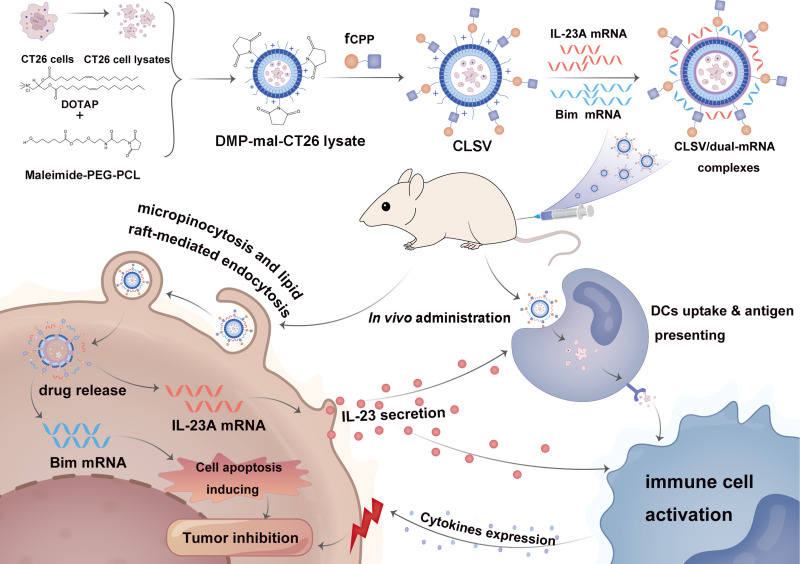
Methods: Based on the previously developed ideal mRNA delivery system called DOTAP-mPEG-PCL (DMP), which was formed through the self-assembly of 1.2-dioleoyl-3-trimethylammonium-propane (DOTAP) and methoxypoly (ethylene glycol)-b-poly (ε-caprolactone) (mPEG-PCL), we introduced a fused cell-penetrating peptide (fCPP) into the framework and encapsulated tumor cell lysates to form a novel nanovector, termed CLSV system (CLS: CT26 tumor cell lysate, V: nanovector). This system served a dual purpose of facilitating the delivery of two mRNAs and enhancing tumor immunogene therapy through tumor cell lysates.

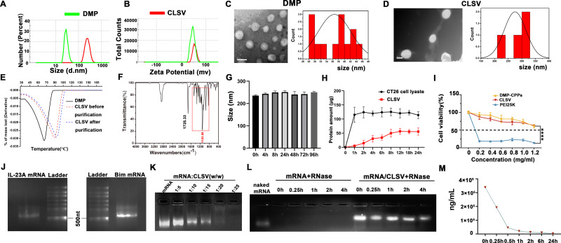
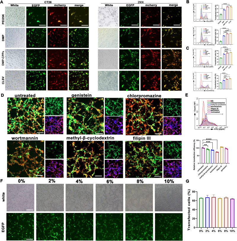
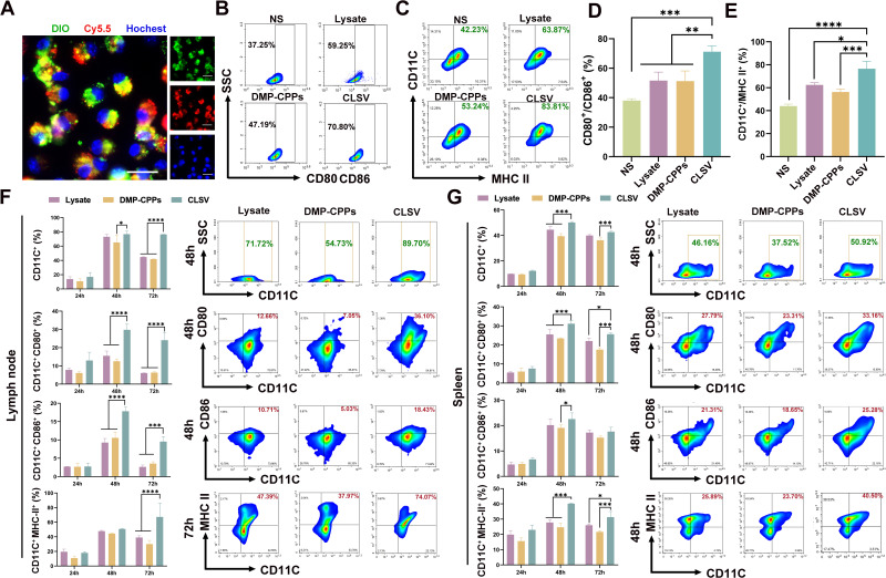
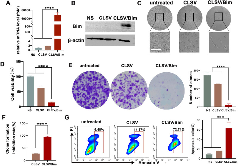
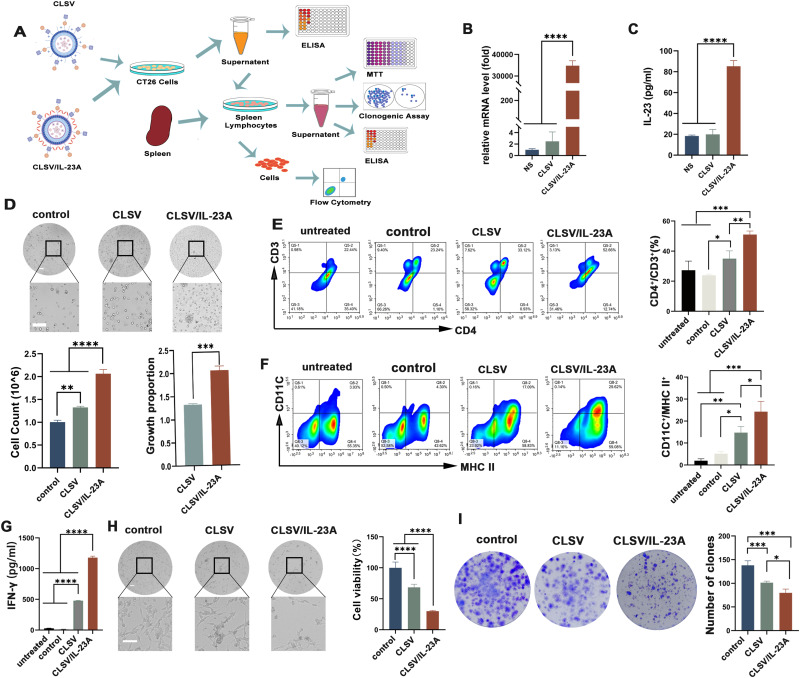
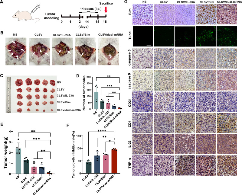
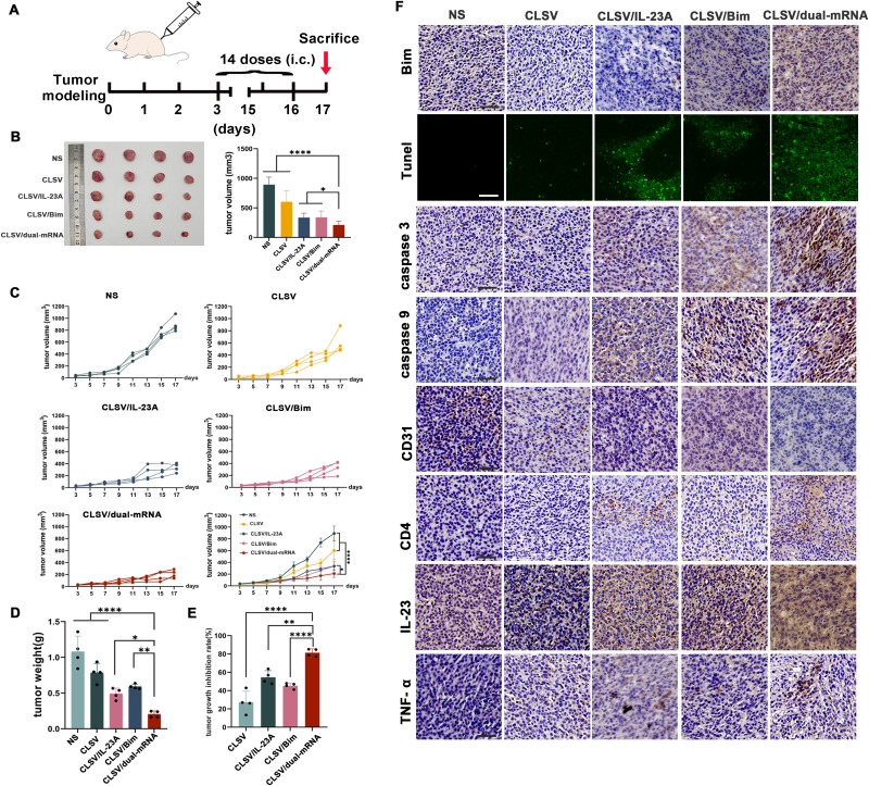
Results: The synthesized CLSV system had an average size of 241.17 nm and a potential of 39.53 mV. The CLSV system could not only encapsulate tumor cell lysates, but also deliver two mRNAs to tumor cells simultaneously, with a transfection efficiency of up to 60%. The CLSV system effectively activated the immune system such as dendritic cells to mature and activate, leading to an anti-tumor immune response. By loading Bim-encoded mRNA and IL-23A-encoded mRNA, CLSV/Bim and CLSV/IL-23A complexes were formed, respectively, to further induce apoptosis and anti-tumor immunity. The prepared CLSV/dual-mRNA complex showed significant anti-cancer effects in multiple CT26 mouse models.

Conclusion: Our results suggest that the prepared CLSV system is an ideal delivery system for dual-mRNA immunogene therapy.

Keywords: Bim and IL-23A mRNAs; dual-mRNA delivery; immune response; mRNA gene therapy; tumor cell lysates.


Conflict of interest statement

The authors declare that they have no conflicts of interest in this work.

Figures

Scheme 1
Scheme 1
A schematic view of the formulation and therapeutic processes of the CLSV/dual-mRNA complex.
Figure 1
Figure 1
Characterization of the CLSV system. (A) Size distribution of the DOTAP-mPEG-PCL (DMP) and CLSV systems. (B) Zeta potential of the DMP and CLSV systems. (C) TEM photomicrographs and the TEM histogram of the DMP (scale bar: 50 nm). (D) TEM photomicrographs and the TEM histogram of CLSV systems (scale bar: 200 nm). (E) Thermogravimetric analysis of CLSV before and after purification with control DMP. (F) Fourier-transform infrared spectroscopy (FTIR) spectra of CLSV (G) The size of CLSV nanoparticles in different time in normal saline. (H) The protein amounts of CT26 cell lysates released by CLSV nanoparticles in phosphate-buffered saline (PBS) at different time points. (I) Cell viability assay of PEI25K, DMP-CPPs, and CLSV on 293T cells (n = 3; ****P < 0.0001). (J) Gel electrophoresis to verify the murine IL-23A-encoded mRNA bands and Bim-encoded mRNA bands. (K) Gel retarding assay of the CLSV/mRNA complex. (L) RNase protection assay of the CLSV/mRNA complex. (M) the content of CLSV nanoparticles in different time (0 h, 0.25 h, 0.5 h, 1 h, 2 h, 6 h, 24 h) in mice blood.
Figure 2
Figure 2
In vitro gene transfection by the CLSV/mRNA complex. (A) Fluorescence microscopy images of CT26 and 293T cells (scale bar: 100 μm). (B) EGFP mRNA efficiency of CT26 cell transfection (n = 3; ****P < 0.0001) and mCherry mRNA efficiency (n = 3; ***P < 0.001 and ****P < 0.0001) analyzed by flow cytometry. (C) EGFP mRNA efficiency of 293T cell transfection (n = 3; ***P < 0.001 and ****P < 0.0001) and mCherry mRNA efficiency (n = 3; **P < 0.01, ***P < 0.001, and ****P < 0.0001) analyzed by flow cytometry. (D) Fluorescent images of internalization of the CLSV/mRNA complex after treatment with various inhibitors (scale bar: 100 μm). (E) The uptake rates were analyzed by flow cytometry (n = 3; **P < 0.01, ***P < 0.001, and ****P < 0.0001). (F) Transfection ability of CLSV nanoparticles in serum with different concentrations (scale bar: 100 μm). (G) EGFP mRNA transfection efficiency of CT26 cell in serum with different concentrations analyzed by flow cytometry.
Figure 3
Figure 3
CLSV system activates immunity in vitro and in vivo. (A) Colocalization study of BMDC uptake of CLSV (scale bar: 20 μm). (B and C) Expression of CD11C+ CD80+ CD86+ (B) and CD11C+ MHC II+ in BMDCs in vitro (C). (D and E) Ratio of the expression of CD11C+ CD80+ CD86+ (D) and CD11C+ MHC II+ by flow cytometry in BMDCs in vitro (E) (n = 3; *P < 0.05, **P < 0.01, ***P < 0.001, and ****P < 0.0001). (F and G) Expression of CD11C+, CD80+, CD86+, and MHC-II+ mouse lymph node lymphocytes (F) and mouse splenic lymphocytes (G) after treatment with DMP-CPPs, tumor cell lysate, and CLSV (n = 3; *P < 0.05, ***P < 0.001, and ****P < 0.0001).
Figure 4
Figure 4
CLSV/Bim complex inhibited CT26 cell proliferation in vitro. (A) Bim mRNA levels in CT26 cells after transfection (n = 3; ****P < 0.0001). (B) The level of the Bim protein. (C and D) CT26 cells after treatment with the CLSV/Bim complex (scale bar: 100 μm) and the cell viability in each group was measured using the MTT assay (n = 3; ****P < 0.0001). (E and F) Detection of the anti-proliferative effect of the CLSV/Bim complex using the clonogenic assay and inhibition rates were calculated on the basis of clone numbers (n = 3; ****P < 0.0001). (G) The CLSV/Bim complex efficiently induced apoptosis in CT26 cells as determined by flow cytometry (n = 3; ***P < 0.001).
Figure 5
Figure 5
Anti-tumor effects of the CLSV/IL-23A complex by lymphocyte-based stimulation in vitro. (A) Experimental design of the splenic lymphocyte supernatant anti-tumor assay. (B) Level of IL-23A mRNA in CT26 cells after transfection (n = 3; ****P < 0.0001). (C) Concentration of IL-23A in the supernatant after CLSV/IL-23A complex transfection of CT26 cells (n = 3; ****P < 0.0001). (D) Microscopic images of lymphocyte proliferation after stimulation with the supernatant of CT26 cells (scale bar: 100 μm; n = 3, **P < 0.01, ***P < 0.001, and ****P < 0.0001). (E and F) Activation of DCs and T cells detected by flow cytometry (n = 3; *P < 0.05, **P < 0.01, and ***P < 0.001). (G) IFN-γ level in the supernatant of lymphocytes as detected by ELISA (n = 3; ****P < 0.0001). (H) CT26 cells were treated with lymphocyte supernatant for viability assay (n = 3; scale bar: 100 μm; ****P < 0.0001) (I) A clonogenic assay was used to detect the anti-tumor effect of lymphocyte-mediated cytotoxicity (n = 3; *P < 0.05 and ***P < 0.001).
Figure 6
Figure 6
CLSV/dual-mRNA complex inhibited abdominal cavity metastatic tumor growth in vivo. (A) Schematic representation of the experimental design. (B) Images of representative mice from each treatment group. (C) Tumor nodules harvested from each group. (D) The average number of tumor nodules in each group (**P < 0.01 and ***P < 0.001). (E) The average tumor weight in each group (**P < 0.01 and ***P < 0.001). (F) The tumor growth inhibition rate in each group (*P < 0.05, **P < 0.01, and ****P < 0.0001). (G) Immunohistochemical evaluation of tumor tissues (scale bars: 50 μm).
Figure 7
Figure 7
CLSV/dual-mRNA complex suppressing the CT26 subcutaneous xenograft model in vivo. (A) Schematic representation of the experimental design. (B) Images of tumors collected from each group. (C) Tumor growth curves from each treatment group (*P < 0.05 and ****P < 0.0001). (D) Average tumor weights (*P < 0.05, **P < 0.01, and ****P < 0.0001). (E) The tumor growth inhibition rate in each group (**P < 0.01 and ****P < 0.0001). (F) Immunohistochemical evaluation of tumor tissue (scale bars: 50 μm).
Figure 8
Figure 8
Hematoxylin and eosin (H&E) analysis in vivo. (A) H&E analysis of the main organs in the CT26 abdominal cavity metastatic model (scale bars: 100 μm). (B) H&E analysis of the main organs in the CT26 subcutaneous xenograft model (scale bars: 100 μm).
Figure 9
Figure 9
Blood routine analysis of mice after intravenous injection of CLSV nanoparticles.

Similar articles

Cited by

References

    1. Siegel RL, Miller KD, Fuchs HE, Jemal A. Cancer Statistics, 2021. Ca a Cancer J Clinicians. 2021;71(1):7–33. doi:10.3322/caac.21654 - DOI - PubMed
    1. Herrmann J. Adverse cardiac effects of cancer therapies: cardiotoxicity and arrhythmia. Nat Rev Cardiol. 2020;17(8):474–502. doi:10.1038/s41569-020-0348-1 - DOI - PMC - PubMed
    1. Liu C, Shi Q, Huang X, Koo S, Kong N, Tao W. mRNA-based cancer therapeutics. Nat Rev Cancer. 2023;23(8):526–543. doi:10.1038/s41568-023-00586-2 - DOI - PubMed
    1. Qiu M, Li Y, Bloomer H, Xu Q. Developing biodegradable lipid nanoparticles for intracellular mRNA delivery and genome editing. Acc Chem Res. 2021;54(21):4001–4011. doi:10.1021/acs.accounts.1c00500 - DOI - PubMed
    1. Beck JD, Reidenbach D, Salomon N, et al. mRNA therapeutics in cancer immunotherapy. Mol Cancer. 2021;20(1):69. doi:10.1186/s12943-021-01348-0 - DOI - PMC - PubMed