. 2024 May 17:14:1408451.
doi: 10.3389/fcimb.2024.1408451. eCollection 2024.

Plasmodium vivax spleen-dependent protein 1 and its role in extracellular vesicles-mediated intrasplenic infections

Affiliations

Plasmodium vivax spleen-dependent protein 1 and its role in extracellular vesicles-mediated intrasplenic infections

Alberto Ayllon-Hermida et al. Front Cell Infect Microbiol. .

Abstract

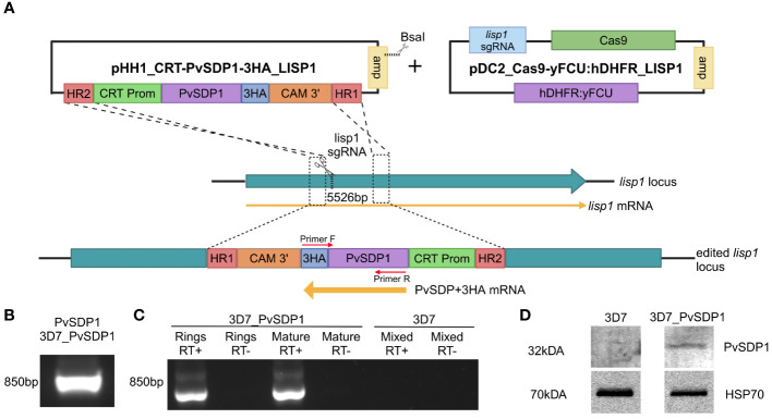
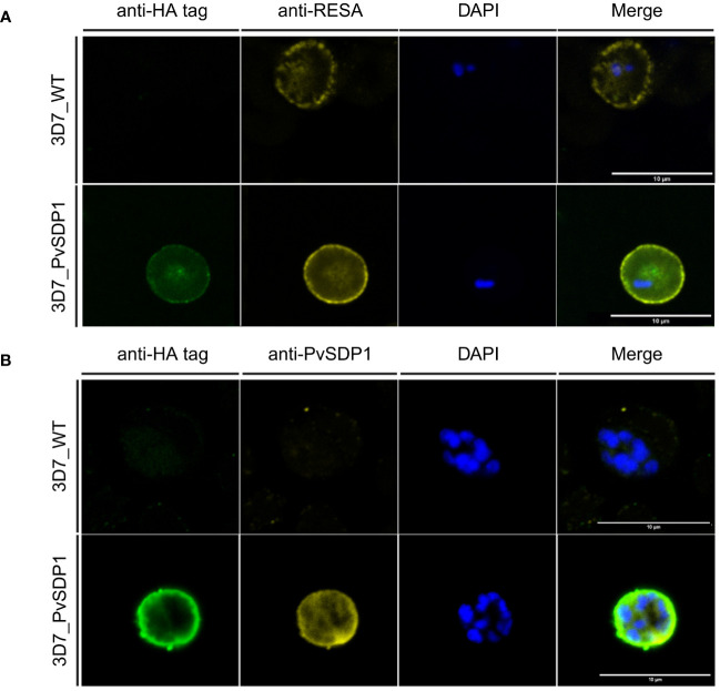
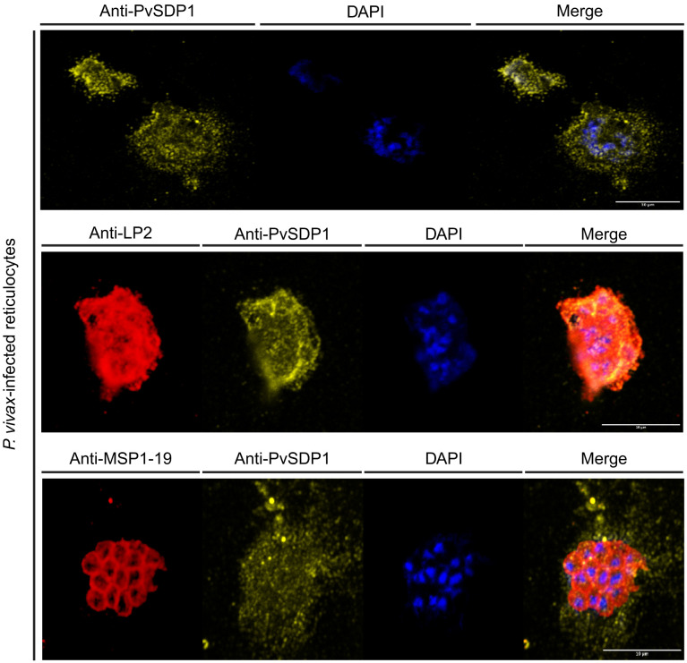
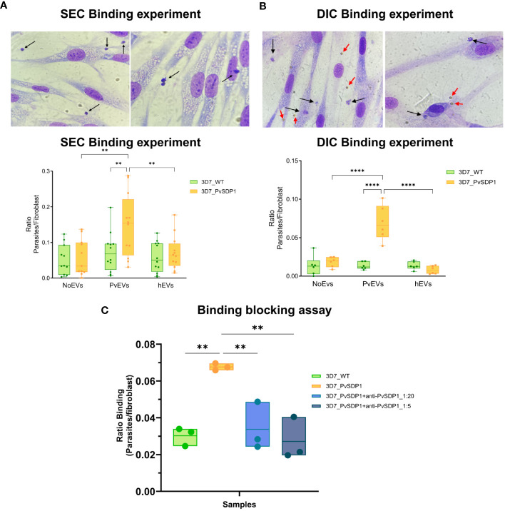
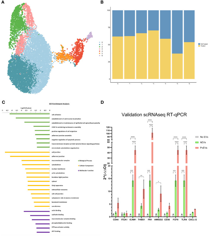
Recent studies indicate that human spleen contains over 95% of the total parasite biomass during chronic asymptomatic infections caused by Plasmodium vivax. Previous studies have demonstrated that extracellular vesicles (EVs) secreted from infected reticulocytes facilitate binding to human spleen fibroblasts (hSFs) and identified parasite genes whose expression was dependent on an intact spleen. Here, we characterize the P. vivax spleen-dependent hypothetical gene (PVX_114580). Using CRISPR/Cas9, PVX_114580 was integrated into P. falciparum 3D7 genome and expressed during asexual stages. Immunofluorescence analysis demonstrated that the protein, which we named P. vivax Spleen-Dependent Protein 1 (PvSDP1), was located at the surface of infected red blood cells in the transgenic line and this localization was later confirmed in natural infections. Plasma-derived EVs from P. vivax-infected individuals (PvEVs) significantly increased cytoadherence of 3D7_PvSDP1 transgenic line to hSFs and this binding was inhibited by anti-PvSDP1 antibodies. Single-cell RNAseq of PvEVs-treated hSFs revealed increased expression of adhesion-related genes. These findings demonstrate the importance of parasite spleen-dependent genes and EVs from natural infections in the formation of intrasplenic niches in P. vivax, a major challenge for malaria elimination.

Keywords: CRISPR/Ca9; Plasmodium vivax; extracellular vesicles (EVs); intrasplenic infections; single-cell RNASeq (scRNASeq); spleen fibroblasts.


Conflict of interest statement

The authors declare that the research was conducted in the absence of any commercial or financial relationships that could be construed as a potential conflict of interest.

Figures

Figure 1
Figure 1
Generation and characterization of the transgenic 3D7_PvSDP1 line. (A) Overview of the strategy followed for the lisp1 locus editing using pDC_Cas9-yFCU:hDHFR LISP1 plasmid through CRISPR/Cas9. Scissors indicate the position targeted by the guide RNA, where Cas9 cleavage is produced. HR1 and HR2 refers to homology regions. CRT promoter is used for active transcription throughout asexual cycle. 3HA tag is used as fusion protein to enable detection and tracking of PvSDP1. Created with BioRender.com. (B) PCR of genomic DNA from transgenic 3D7_PvSDP1 parasites to amplify both PVX_114580 and the 3HA tag within the fusion cassette. Primer localization marked with red arrows. (C) RT-PCR of RNA extracted from 3D7_PvSDP1 parasite line and 3D7_WT. (D) Western Blot of the 3D7_PvSDP1 transgenic line. Anti-HA antibody was used for PvSDP1 protein detection, and the anti P. falciparum HSP70 served as the loading control.
Figure 2
Figure 2
Immunofluorescence assay of 3D7_PvSDP1 transgenic line. (A) Rabbit anti-HA tag and mouse anti-RESA antibodies were used in combination with 3D7_PvSDP1 and 3D7 wild type (3D7_WT). In the immunofluorescence assay (IFA) images, the RESA antibody and HA-tag antibody co-localize exclusively at the membrane of the infected red blood cells (iRBC) in the transgenic 3D7_PvSDP1 line. The RESA antibody targets the membrane of 3D7_WT. (B) Anti-HA and anti-PvSDP1 antibodies were used in combination with 3D7_PvSDP1 and 3D7_WT. The HA-tag antibody and anti-PvSDP1 antibody co-localize at the membrane of the iRBC in 3D7_PvSDP1, with no signal detected in 3D7_WT. All scale bars represent 10 µm.
Figure 3
Figure 3
Immunofluorescence assay (IFA) on P. vivax field isolates. Anti-PvSDP1 antibody recognizes the SDP1 protein at the membrane of the infected reticulocyte in P. vivax field isolates. Anti-LP2 (targeting conserved motifs of the VIR superfamily) and anti-PvSDP1 co-localize at the membrane of the infected reticulocyte. Anti-MSP1-19 distinctly stains merozoites within the infected reticulocyte, while anti-PvSDP1 specifically targets the membrane of the infected reticulocyte. All scale bars represent 10 µm.
Figure 4
Figure 4
Binding of 3D7_PvSDP1 Transgenic Line to Human Spleen Fibroblasts (hSFs). (A) In the upper panel, Representative pictures illustrating binding of 3D7_PvSDP1 to hSFs after stimulation with SEC-purified PvEVs. Black arrows indicate parasites attached to hSFs. In Lower panel, the quantification of binding reveals a significantly increased binding capacity of the 3D7_PvSDP1 transgenic line to hSFs when cells have been stimulated with PvEVs (Two-Way ANOVA Sidak’s multiple comparisons test, **p<0.0021). (B) Upper panel are representative pictures depicting the binding of 3D7_PvSDP1 to hSFs after stimulation with DIC-purified PvEVs. Black arrows point to the parasite, and red arrows indicate the presence of magnetic Dynabeads. Lower panel shows the quantification of DIC binding, demonstrating an increased binding capacity of 3D7_PvSDP1 to hSFs when treated with PvEVs compared to hEVs and control PBS (Two-Way ANOVA Sidak’s Multiple Comparison test, ****p<0.0001). (C) Binding blocking assay. After a 1-hour incubation of parasites with two different dilutions of the anti-PvSDP1 antibody (1:5 and 1:20), the binding capacity of transgenic parasites to stimulated PvEVs in hSFs is significantly reduced by the action of the anti-PvSDP1 antibody (One-way ANOVA Tukey’s multiple comparison test, **p<0.002).
Figure 5
Figure 5
Single-Cell RNAseq Analysis. (A) UMAP Clusterization of the sequenced hSFs. (B) Bar plot illustrating the distribution of cells between not treated and PvEVs treated groups among the obtained clusters. (C) Gene Ontology (GO) analysis of Cluster 1. The DAVID Bioinformatic tool was employed for GO enrichment analysis. (D) RT-qPCR validation of selected adhesins. hSFs were stimulated with either PvEVs or hEVs. The results indicate upregulation of certain genes after stimulation with PvEVs and hEVs. The basal expression level in the absence of EVs was calculated individually for each gene (Two-way ANOVA Bonferroni’s multiple comparisons test, *p<0.0332, **p<0.0021, ***p<0.0002, ****p<0.0001).

Similar articles

Cited by

References

    1. Adjalley S., Lee M. C. S. (2022). CRISPR/cas9 editing of the plasmodium falciparum genome. Methods Mol. Biol. 2470, 221–239. doi: 10.1007/978-1-0716-2189-9_17/COVER - DOI - PubMed
    1. Angrisano F., Robinson L. J. (2022). Plasmodium vivax – How hidden reservoirs hinder global malaria elimination. Parasitol. Int. 87, 102526. doi: 10.1016/j.parint.2021.102526 - DOI - PubMed
    1. Aparici-Herraiz I., Gualdrón-López M., Castro-Cavadía C. J., Carmona-Fonseca J., Yasnot M. F., Fernandez-Becerra C., et al. . (2022). Antigen discovery in circulating extracellular vesicles from plasmodium vivax patients. Front. Cell Infect. Microbiol. 11. doi: 10.3389/fcimb.2021.811390 - DOI - PMC - PubMed
    1. Bernabeu M., Lopez F. J., Ferrer M., Martin-Jaular L., Razaname A., Corradin G., et al. . (2012). Functional analysis of Plasmodium vivax VIR proteins reveals different subcellular localizations and cytoadherence to the ICAM-1 endothelial receptor. Cell Microbiol. 14, 386–400. doi: 10.1111/j.1462-5822.2011.01726.x - DOI - PubMed
    1. Cabrera A., Neculai D., Kain K. C. (2014). CD36 and malaria: friends or foes? A decade of data provides some answers. Trends Parasitol. 30, 436–444. doi: 10.1016/j.pt.2014.07.006 - DOI - PubMed

MeSH terms