. 2024 May 17:15:1386238.
doi: 10.3389/fphar.2024.1386238. eCollection 2024.

LP340, a novel histone deacetylase inhibitor, decreases liver injury and fibrosis in mice: role of oxidative stress and microRNA-23a

Affiliations

LP340, a novel histone deacetylase inhibitor, decreases liver injury and fibrosis in mice: role of oxidative stress and microRNA-23a

Devadoss J Samuvel et al. Front Pharmacol. .

Abstract

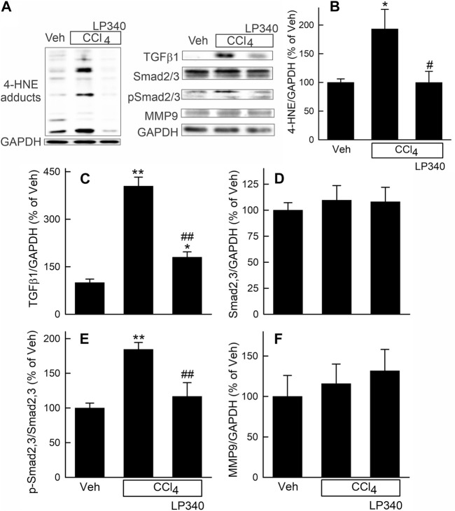
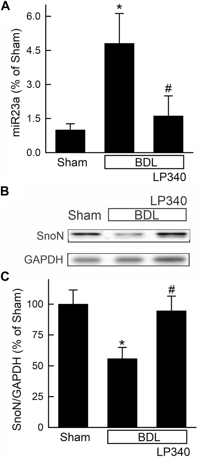
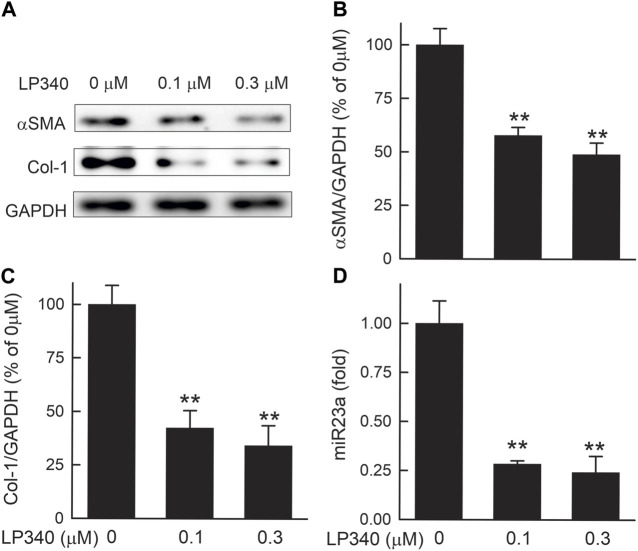
Effective therapy for liver fibrosis is lacking. Here, we examined whether LP340, the lead candidate of a new-generation of hydrazide-based HDAC1,2,3 inhibitors (HDACi), decreases liver fibrosis. Liver fibrosis was induced by CCl4 treatment and bile duct ligation (BDL) in mice. At 6 weeks after CCl4, serum alanine aminotransferase increased, and necrotic cell death and leukocyte infiltration occurred in the liver. Tumor necrosis factor-α and myeloperoxidase markedly increased, indicating inflammation. After 6 weeks, α-smooth muscle actin (αSMA) and collagen-1 expression increased by 80% and 575%, respectively, indicating hepatic stellate cell (HSC) activation and fibrogenesis. Fibrosis detected by trichrome and Sirius-red staining occurred primarily in pericentral regions with some bridging fibrosis in liver sections. 4-Hydroxynonenal adducts (indicator of oxidative stress), profibrotic cytokine transforming growth factor-β (TGFβ), and TGFβ downstream signaling molecules phospho-Smad2/3 also markedly increased. LP340 attenuated indices of liver injury, inflammation, and fibrosis markedly. Moreover, Ski-related novel protein-N (SnoN), an endogenous inhibitor of TGFβ signaling, decreased, whereas SnoN expression suppressor microRNA-23a (miR23a) increased markedly. LP340 (0.05 mg/kg, ig., daily during the last 2 weeks of CCl4 treatment) decreased 4-hydroxynonenal adducts and miR23a production, blunted SnoN decreases, and inhibited the TGFβ/Smad signaling. By contrast, LP340 had no effect on matrix metalloproteinase-9 expression. LP340 increased histone-3 acetylation but not tubulin acetylation, indicating that LP340 inhibited Class-I but not Class-II HDAC in vivo. After BDL, focal necrosis, inflammation, ductular reactions, and portal and bridging fibrosis occurred at 2 weeks, and αSMA and collagen-1 expression increased by 256% and 560%, respectively. LP340 attenuated liver injury, ductular reactions, inflammation, and liver fibrosis. LP340 also decreased 4-hydroxynonenal adducts and miR23a production, prevented SnoN decreases, and inhibited the TGFβ/Smad signaling after BDL. In vitro, LP340 inhibited immortal human hepatic stellate cells (hTERT-HSC) activation in culture (αSMA and collagen-1 expression) as well as miR23a production, demonstrating its direct inhibitory effects on HSC. In conclusions, LP340 is a promising therapy for both portal and pericentral liver fibrosis, and it works by inhibiting oxidative stress and decreasing miR23a.

Keywords: SnoN; TGFβ; histone deacetylase; liver fibrosis; microRNA-23a; oxidative stress.


Conflict of interest statement

CC is the co-founder of Lydex Pharmaceuticals where LP340 was synthesized. The remaining authors declare that the research was conducted in the absence of any commercial or financial relationships that could be construed as a potential conflict of interest. The author(s) declared that they were an editorial board member of Frontiers, at the time of submission. This had no impact on the peer review process and the final decision.

Figures

FIGURE 1
FIGURE 1
LP340 increases histone-3 acetylation and decreases alanine aminotransferase release and necrosis after CCl4 treatment. Male mice were injected with CCl4 (1:3 dilution in corn oil; 1.0 µL of dilution/g mouse, i.p.) or equal volume of corn oil once every 3 days for 6 weeks. LP340 (0.05 mg/kg, ig daily) or equal volume of vehicle (Veh) was administered during the last 2 weeks of CCl4 treatment. Blood and liver were collected after 6 weeks of CCl4 or corn oil treatment. (A) regimen of CCl4 and LP340 treatment. (B) representative immunoblots for acetylated histone-3 (acH3), acetylated tubulin (acTub), and housekeeping protein GAPDH; (C, D), quantification of acH3 and acTub immunoblots by densitometry. (E–G) representative images of liver histology. Bar is 50 µm. (H) serum ALT. *, p < 0.05 vs. vehicle; **, p < 0.01 vs. vehicle; ##, p < 0.01 vs. CCl4 without LP340. Data are means ± SEM (n = 3–4/group).
FIGURE 2
FIGURE 2
LP340 decreases inflammatory responses after CCl4 treatment. Male mice were treated, and livers were collected as described in Figure 1. (A) representative immunoblots for tumor necrosis factor-α (TNFα), myeloperoxidase (MPO), and housekeeping protein GAPDH. (B,C) quantification of TNFα and MPO immunoblots by densitometry. Veh, vehicle; **, p < 0.01 vs. vehicle; #, p < 0.05 vs. CCl4 without LP340; ##, p < 0.01 vs. CCl4 without LP340. Data are means ± SEM (n = 4/group).
FIGURE 3
FIGURE 3
LP340 decreases liver fibrosis after CCl4 treatment. Male mice were treated, and livers were collected as described in Figure 1. (A–C) representative images of tichrome-stained liver sections. Bar is 100 μm; (D–F) representative images of Sirius red/Fast green-stained liver sections. Bar is 100 µm. (G) representative immunoblots for α-smooth muscle actin (αSMA), collagen-1 (Col-1), and housekeeping protein GAPDH. (H,I) quantification of αSMA and Col-1 immunoblots by densitometry. Veh, vehicle; **, p < 0.01 vs. vehicle; #, p < 0.05 vs. CCl4 without LP340; ##, p < 0.01 vs. CCl4 without LP340. Data are means ± SEM (n = 4/group).
FIGURE 4
FIGURE 4
LP340 inhibits oxidative stress and TGFβ/Smad signaling but does not alter matrix metalloproteinase-9 expression after CCl4 treatment. Male mice were treated, and livers were collected as described in Figure 1. (A) representative immunoblots for 4-hydroxynonenal (4-HNE) adducts, transforming growth factor-β1 (TGFβ1), mothers against decapentaplegic homolog2,3 (Smad2,3), phospho-Smad2,3 (pSmad2,3), matrix metalloproteinase-9 (MMP9), and housekeeping protein GAPDH. (B–F) quantification of 4-HNE adducts, TGFβ1, Smad2,3, pSmad2,3 and MMP9 immunoblots by densitometry. Veh, vehicle; *, p < 0.05 vs. vehicle; **, p < 0.01 vs. vehicle; #, p < 0.05 vs. CCl4 without LP340; ##, p < 0.01 vs. CCl4 without LP340. Data are means ± SEM (n = 4/group).
FIGURE 5
FIGURE 5
LP340 decreases microRNA-23a and increases SnoN expression and SnoN/Smad4 complex formation after CCl4 treatment. Male mice were treated, and livers were collected as described in Figure 1. (A) hepatic microRNA-23a (miR23a) detected by qPCR. (B) representative immunoblots of Ski-novel protein (SnoN) and housekeeping protein GAPDH. (C) quantification of SnoN. (D) representative immunoblots of Smad4-bound SnoN detected after immunoprecipitation (IP). (E) quantification of Smad4-bound SnoN. *, p < 0.05 vs. vehicle; **, p < 0.01 vs. vehicle; #, p < 0.05 vs. CCl4 without LP340; ##, p < 0.01 vs. CCl4 without LP340. Data are means ± SEM (n = 4/group).
FIGURE 6
FIGURE 6
LP340 decreases liver injury and inflammation after bile duct ligation. Blood and livers were collected 2 weeks after BDL or sham operation (Sham). (A–C) representative images of H&E-stained liver sections. Arrows identify necro-inflammatory foci. Bar is 50 µm. (D) representative immunoblot images of TNFα and GAPDH; (E) quantification of TNFα immunoblots; (F) serum ALT; $, p = 0.052 vs. sham; *, p < 0.05 vs. sham; **, p < 0.01 vs. sham; ##, p < 0.01 vs. BDL without LP340. Data are means ± SEM (n = 3–4/group).
FIGURE 7
FIGURE 7
LP340 decreases ductular reactions after bile duct ligation. Livers were collected 2 weeks after BDL or sham operation (Sham). (A–C) representative images of liver histology after H&E staining. (D–F) representative images of immunohistological staining for CK19. Bars are 25 μm. n = 3–4/group.
FIGURE 8
FIGURE 8
LP340 decreases liver fibrosis after bile duct ligation. Livers were collected 2 weeks after BDL or sham operation (Sham). (A–C) representative images of trichrome-stained liver sections. Bar is 100 µm. (D–F) representative images of Sirius red/Fast green-stained liver sections. Bar 100 µm. (G) representative immunoblots for α-smooth muscle actin (αSMA), collagen-1 (Col-1), and housekeeping protein GAPDH. (H,I) quantification of αSMA and Col-1 immunoblots by densitometry. **, p < 0.01 vs. sham; ##, p < 0.01 vs. BDL without LP340. Data are means ± SEM (n = 4/group).
FIGURE 9
FIGURE 9
LP340 decreases oxidative stress and TGFβ/Smad signaling after bile duct ligation. Livers were collected 2 weeks after BDL or sham operation (Sham). (A,B) representative immunoblot for 4-HNE adducts, TGFβ1, Smad2,3, pSmad2,3 and housekeeping protein GAPDH. (C–F) quantification of 4-HNE adducts, TGFβ1, Smad2,3, pSmad2,3 immunoblots. *, p < 0.05 vs. sham; **, p < 0.01 vs. sham; &, p = 0.088 vs. sham; ##, p < 0.01 vs. BDL without LP340. Data are means ± SEM (n = 4/group).
FIGURE 10
FIGURE 10
LP340 decreases microRNA-23a and increases SnoN after bile duct ligation. Livers were collected 2 weeks after BDL or sham operation (Sham). (A) hepatic miR23a detected by qPCR. (B) representative immunoblots for SnoN and housekeeping protein GAPDH. (C) quantification of SnoN immunoblots. *, p < 0.05 vs. sham; #, p < 0.05 vs. BDL without LP340. Data are means ± SEM (n = 4/group).
FIGURE 11
FIGURE 11
LP340 suppresses hTERT-HSC activation and miR340 formation in vitro. hTERT-HSC were cultured in DMEM medium with 0.5% FBS with or without LP340 (0.1 and 0.3 µM) for 48 h. (A) representative immunoblots for α-smooth muscle actin (αSMA), collagen-1 (Col-1), and housekeeping protein GAPDH. (B,C) quantification of αSMA and Col-1 immunoblots by densitometry, (D) miR23a detected by qPCR. **, p < 0.01 vs. 0 µM LP340. Data are means ± SEM (n = 3/group).
FIGURE 12
FIGURE 12
Mechanisms by which LP340 ameliorates liver fibrosis. Chronic liver diseases increase ROS formation, which causes cell injury and death, stimulate inflammatory responses, and increase TGFβ. Inflammatory responses also stimulate TGFβ formation. TGFβ binds to its receptors, causing phosphorylation of Smad2 and Smad3. Phosphorylated Smad2 and Smad3 form a heterocomplex with Smad4, and this complex translocates to the nucleus to upregulate transcription of TGFβ target genes, which causes HSC activation and fibrosis. SnoN and Ski, endogenous inhibitors of TGFβ/Smad signaling, compete for Smad4 with pSmad2/3 thus inhibiting pSmad2/3 nuclear translocation and TGFβ target gene transcription. Alternatively or additionally, SnoN and Ski form an inhibitory complex with Smad4 that binds to the promoter of TGFβ target genes, thus inhibiting TGFβ target gene transcription. miR23a inhibits SnoN expression. LP340 decreases oxidative stress, most likely by increasing antioxidant protein formation. Inhibition of oxidative stress would decrease liver injury and subsequent proinflammatory and profibrotic responses. Moreover, LP340 suppresses miR23a formation, which increases SnoN and its binding to Smad4, thus inhibiting TGFβ/Smad signaling-induced HSC activation and liver fibrosis.

Similar articles

Cited by

References

    1. Allameh A., Niayesh-Mehr R., Aliarab A., Sebastiani G., Pantopoulos K. (2023). Oxidative stress in liver pathophysiology and disease. Antioxidants (Basel) 12 (9), 1653. 10.3390/antiox12091653 - DOI - PMC - PubMed
    1. Arthur M. J. (2002). Reversibility of liver fibrosis and cirrhosis following treatment for hepatitis C. Gastroenterology 122 (5), 1525–1528. 10.1053/gast.2002.33367 - DOI - PubMed
    1. Asrani S. K., Devarbhavi H., Eaton J., Kamath P. S. (2019). Burden of liver diseases in the world. J. Hepatol. 70 (1), 151–171. 10.1016/j.jhep.2018.09.014 - DOI - PubMed
    1. Bao L., Zhao J., Dai X., Wang Y., Ma R., Su Y., et al. (2014). Correlation between miR-23a and onset of hepatocellular carcinoma. Clin. Res. Hepatol. Gastroenterol. 38 (3), 318–330. 10.1016/j.clinre.2013.12.002 - DOI - PubMed
    1. Bataller R., Brenner D. A. (2005). Liver fibrosis. J. Clin. Invest. 115 (2), 209–218. 10.1172/JCI24282 - DOI - PMC - PubMed