. 2024 Jun;30(6):e14764.
doi: 10.1111/cns.14764.

SIRT1 mediates the excitability of spinal CaMKIIα-positive neurons and participates in neuropathic pain by controlling Nav1.3

Affiliations

SIRT1 mediates the excitability of spinal CaMKIIα-positive neurons and participates in neuropathic pain by controlling Nav1.3

Yuanzeng Wang et al. CNS Neurosci Ther. 2024 Jun.

Abstract

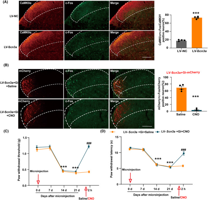
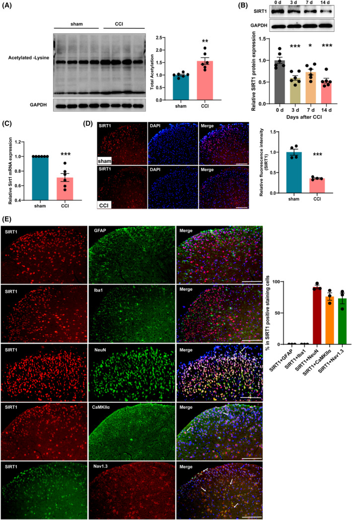
Aims: Neuropathic pain is a common chronic pain disorder, which is largely attributed to spinal central sensitization. Calcium/calmodulin-dependent protein kinase II alpha (CaMKIIα) activation in the spinal dorsal horn (SDH) is a major contributor to spinal sensitization. However, the exact way that CaMKIIα-positive (CaMKIIα+) neurons in the SDH induce neuropathic pain is still unclear. This study aimed to explore the role of spinal CaMKIIα+ neurons in neuropathic pain caused by chronic constriction injury (CCI) and investigate the potential epigenetic mechanisms involved in CaMKIIα+ neuron activation.

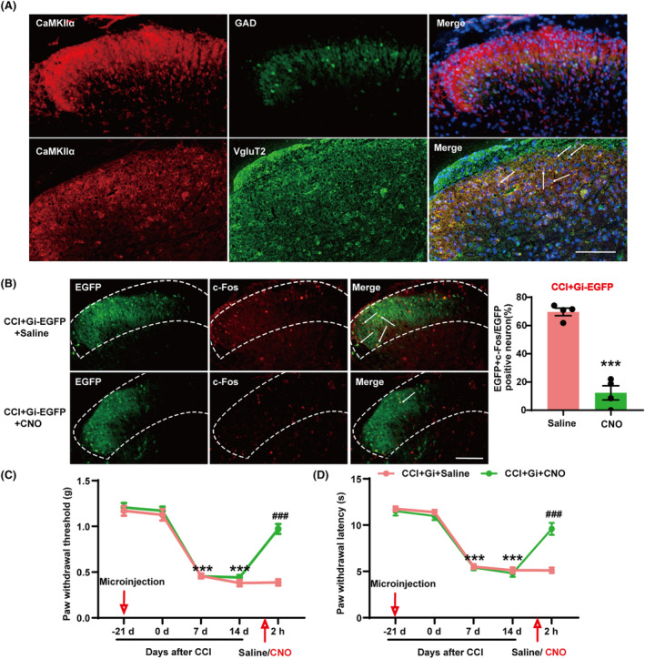
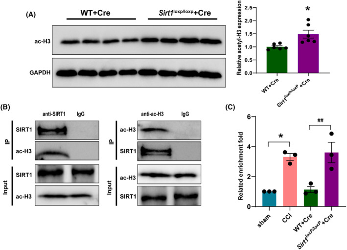
Methods: CCI-induced neuropathic pain mice model, Sirt1loxP/loxP mice, and chemogenetic virus were used to investigate whether the activation of spinal CaMKIIα+ neurons is involved in neuropathic pain and its involved mechanism. Transcriptome sequence, western blotting, qRT-PCR, and immunofluorescence analysis were performed to assay the expression of related molecules and activation of neurons. Co-immunoprecipitation was used to observe the binding relationship of protein. Chromatin immunoprecipitation (ChIP)-PCR was applied to analyze the acetylation of histone H3 in the Scn3a promoter region.

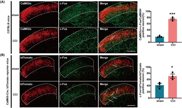
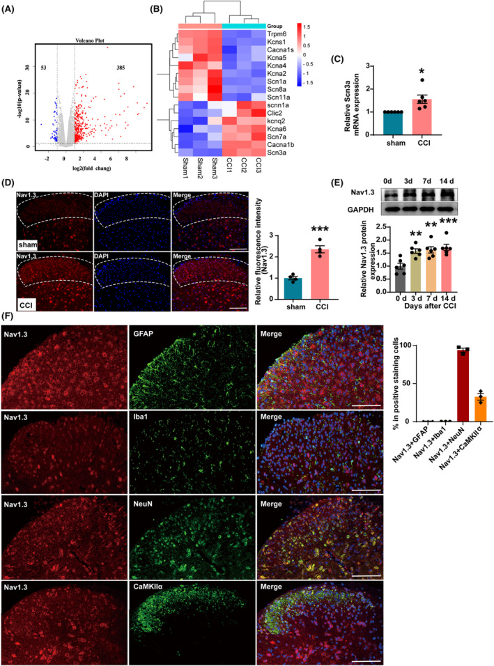
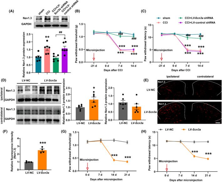
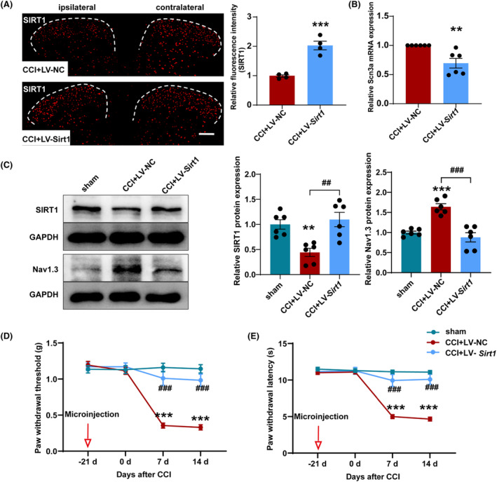
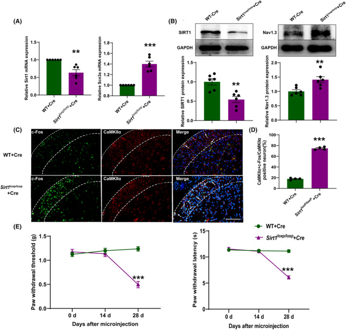
Results: The expression of sodium channel Nav1.3 was increased and the expression of SIRT1 was decreased in the spinal CaMKIIα+ neurons of CCI mice. CaMKIIα neurons became overactive after CCI, and inhibiting their activation relieved CCI-induced pain. Overexpression of SIRT1 reversed the increase of Nav1.3 and alleviated pain, while knockdown of SIRT1 or overexpression of Nav1.3 promoted CaMKIIα+ neuron activation and induced pain. By knocking down spinal SIRT1, the acetylation of histone H3 in the Scn3a (encoding Nav1.3) promoter region was increased, leading to an increased expression of Nav1.3.

Conclusion: The findings suggest that an aberrant reduction of spinal SIRT1 after nerve injury epigenetically increases Nav1.3, subsequently activating CaMKIIα+ neurons and causing neuropathic pain.

Keywords: CaMKIIα+ neurons; Nav1.3; SIRT1; neuropathic pain.


Conflict of interest statement

The authors declare no conflict of interest.

Figures

FIGURE 1
FIGURE 1
CCI surgery induced CaMKIIα+ neuron activation. (A) The percentage of CaMKIIα protein co‐labeled with c‐Fos increased in the SDH of CCI mice. ***p < 0.001 compared with sham. Scale bars = 200 μm. (B) The percentage of tdTomato‐labeled CaMKIIα+ neurons co‐labeled with c‐Fos increased in the SDH of CCI mice. *p < 0.05 compared with sham. Scale bars = 200 μm.
FIGURE 2
FIGURE 2
Inhibition of SDH CaMKIIα+ neurons alleviated CCI‐induced pain. (A) Co‐detection of CaMKIIα and VgluT2 and/or GAD in the SDH of mice. (B) The percentage of CaMKIIα+ neurons co‐labeled with c‐Fos in the SDH of CCI mice was reduced after activation of pAAV‐CaMKIIα‐hM4D(Gi)‐EGFP by CNO. ***p < 0.001 compared with CCI + Gi‐EGFP + Saline. (C) The paw withdrawal threshold was significantly increased after inhibition of CaMKIIα+ neurons in CCI mice. (D) The paw withdrawal latency was significantly increased after inhibition of CaMKIIα+ neuron activation in CCI mice. ***p < 0.001 compared with CCI + Gi‐EGFP + Saline 0 day ### p < 0.001 vs. CCI + Gi‐EGFP + Saline. SDH, spinal dorsal horn.
FIGURE 3
FIGURE 3
Nav1.3 was upregulated in the SDH of CCI mice. (A) Volcano plot showing the overall distribution of upregulated and downregulated mRNAs between the sham group and the CCI group mice. (B) Heatmap showing the differential expression of ion channel genes in the SDH of the sham and CCI mice. (C) The mRNA level of Scn3a was increased in the SDH of CCI mice. *p < 0.05 compared with sham. (D) Mean fluorescence intensity of the Nav1.3 immunostaining cells increased significantly in the ipsilateral SDH of CCI mice. ***p < 0.001 compared with sham. (E) Representative western blot and corresponding graphs showing that Nav1.3 protein level was increased in the SDH of CCI mice. **p < 0.01 and ***p < 0.001 compared with 0 day. (F) The photographs showed the double staining between NeuN or GFAP or Iba1 or CaMKIIα with Nav1.3 in SDH of mice. SDH: spinal dorsal horn. Scale bars = 200 μm.
FIGURE 4
FIGURE 4
Knockdown of Nav1.3 in SDH reversed mechanical and thermal hyperalgesia of CCI mice while overexpression of Nav1.3 decreased nociceptive thresholds. (A) Representative western blot and corresponding graphs showing that Nav1.3 protein level decreased in the SDH of CCI mice after microinjection of LV‐Scn3a shRNA. **p < 0.01 compared with sham; ## p < 0.01 compared with CCI + LV‐NC. (B, C) Microinjection of LV‐Scn3a shRNA into the ipsilateral SDH increased paw withdrawal threshold and paw withdrawal latency of CCI mice. ***p < 0.001 compared with sham; ### p < 0.001 compared with CCI + LV‐NC. (D) Representative western blot and corresponding graphs show that Nav1.3 protein level increased in the ipsilateral SDH of naive mice after microinjection of LV‐Scn3a. *p < 0.05 compared with LV‐NC. (E, F) Mean fluorescence intensity of the Nav1.3 immunostaining cells increased significantly in the ipsilateral SDH of naive mice after microinjection of LV‐Scn3a. ***p < 0.001 compared with LV‐NC. Scale bars = 200 μm. (G, H) Microinjection of LV‐Scn3a into the SDH decreased paw withdrawal threshold and paw withdrawal latency of naive mice. ***p < 0.001 compared with LV‐NC. SDH: spinal dorsal horn.
FIGURE 5
FIGURE 5
Inhibition of CaMKIIα+ neuron activation reversed the hyperalgesia induced by Nav1.3 overexpression in the SDH. (A) The percentage of CaMKIIα+ neurons co‐labeled with c‐Fos increased in the SDH of mice after microinjection of LV‐Scn3a. ***p < 0.001 compared with LV‐NC. Scale bars = 200 μm. (B) Activation of chemogenetic viruses by CNO reduced the percentage of CaMKIIα+ neurons co‐labeled with c‐Fos in the SHD of mice microinjected with LV‐Scn3a. ***p < 0.001 compared with LV‐Scn3a + Gi + Saline. Scale bars = 200 μm. (C, D) Activation of chemogenetic viruses by CNO increased paw withdrawal threshold and paw withdrawal latency of mice microinjected with LV‐Scn3a. ***p < 0.001 compared with LV‐Scn3a + Gi + CNO 21 d; ### p < 0.001 compared with LV‐Scn3a + Gi + Saline. SDH: spinal dorsal horn.
FIGURE 6
FIGURE 6
SIRT1 was decreased in the ipsilateral SDH of CCI mice. (A) Representative western blot and corresponding graphs showing that acetylated protein level increased in the SDH of CCI mice. **p < 0.01 compared with sham. (B) Representative western blot and corresponding graphs showing that SIRT1 protein level was decreased in the SDH of CCI mice. *p < 0.05 and ***p < 0.001 compared with 0 day. (C) The mRNA level of Sirt1 was decreased in the SDH of CCI mice. ***p < 0.001 compared with sham. (D) Mean fluorescence intensity of the SIRT1 immunostaining cells decreased significantly in the SDH of CCI mice. ***p < 0.001 compared with sham. Scale bars = 200 μm. (E) Co‐detection of SIRT1 and GFAP, Iba1, NeuN, CaMKIIα, or Nav1.3 in SDH. Scale bars = 200 μm. SDH, spinal dorsal horn.
FIGURE 7
FIGURE 7
Overexpression of SIRT1 in SDH increased nociceptive thresholds in CCI mice. (A) Mean fluorescence intensity of the SIRT1 immunostaining cells increased significantly in the SDH of CCI mice after microinjection of LV‐Sirt1. ***p < 0.001 compared with CCI + LV‐NC. Scale bars = 200 μm. (B) mRNA level of Scn3a decreased in the SDH of CCI mice after microinjection of LV‐Sirt1. **p < 0.01 compared with CCI + LV‐NC. (C) Representative western blot and corresponding graphs show that SIRT1 protein level increased and Nav1.3 protein level decreased in the SDH of CCI mice after microinjection of LV‐Sirt1. **p < 0.01 and ***p < 0.001 compared with sham; ## p < 0.01 and ### p < 0.001 compared with CCI + LV‐NC. (D) Microinjection of LV‐Sirt1 into the ipsilateral SDH increased paw withdrawal threshold of CCI mice. (E) Microinjection of LV‐Sirt1 into the ipsilateral SDH increased paw withdrawal latency of CCI mice. ***p < 0.001 compared with sham; ### p < 0.001 compared with CCI + LV‐NC. SDH: spinal dorsal horn.
FIGURE 8
FIGURE 8
Knockdown of SIRT1 in SDH‐induced hyperalgesia. (A) mRNA level of Sirt1 decreased and mRNA level of Scn3a increased in the SDH of Sirt1 loxP/loxP mice after microinjection of AAV‐Cre. **p < 0.01 and ***p < 0.001 compared with WT + Cre. (B) Representative western blot and corresponding graphs show that SIRT1 protein level decreased and Nav1.3 protein level increased in the SDH of Sirt1 loxP/loxP mice after microinjection of AAV‐Cre. **p < 0.01 compared with WT + Cre. (C‐D) The percentage of CaMKIIα co‐labeled with c‐Fos increased in the SDH of Sirt1 loxP/loxP mice after microinjection of AAV‐Cre. ***p < 0.001 compared with WT + Cre. Scale bars = 200 μm. Blue: DAPI. (E) Microinjection of AAV‐Cre into the superficial SDH decreased paw withdrawal threshold and paw withdrawal latency of Sirt1 loxP/loxP mice. ***p < 0.001 compared with WT + Cre. SDH: spinal dorsal horn.
FIGURE 9
FIGURE 9
The acetylation level of histone H3 in the Scn3a promoter region was increased in the SDH of CCI mice and after knockdown of Sirt1 in SDH. (A) Representative western blot and corresponding graphs showing that acetylated H3 protein level increased in the SDH of Sirt1 loxP/loxP mice after microinjection of AAV‐Cre. *p < 0.05 compared with WT + Cre. (B) SIRT1 was immunoprecipitated with acetylated H3 in SDH of mice. (C) The level of acetylated H3 in the Scn3a promoter region (−529 to −139 bp) upstream of the transcription start site was increased in the SDH of CCI mice and after knockdown of SIRT1 in SDH (n = 3, 12 mice in total); *p < 0.05 compared with sham; ## p < 0.01 compared with WT + Cre.

Similar articles

Cited by

References

    1. Bannister K, Sachau J, Baron R, Dickenson AH. Neuropathic pain: mechanism‐based therapeutics. In: Insel PA, ed. Annual Review of Pharmacology and Toxicology, Vol 60. Annual Reviews Inc.; 2020:257‐274. - PubMed
    1. Cavalli E, Mammana S, Nicoletti F, Bramanti P, Mazzon E. The neuropathic pain: an overview of the current treatment and future therapeutic approaches. Int J Immunopathol Pharmacol. 2019;33:1‐10. doi:10.1177/2058738419838383 - DOI - PMC - PubMed
    1. Cohen SP, Mao J. Neuropathic pain: mechanisms and their clinical implications. Br Med J. 2014;348:f7656. doi:10.1136/bmj.f7656 - DOI - PubMed
    1. Tsuda M, Koga K, Chen T, Zhuo M. Neuronal and microglial mechanisms for neuropathic pain in the spinal dorsal horn and anterior cingulate cortex. J Neurochem. 2017;141(4):486‐498. doi:10.1111/jnc.14001 - DOI - PubMed
    1. Todd AJ. Identifying functional populations among the interneurons in laminae I‐III of the spinal dorsal horn. Mol Pain. 2017;13:1‐19. doi:10.1177/1744806917693003 - DOI - PMC - PubMed

Publication types

LinkOut - more resources