

PD-L1 regulates tumor proliferation and T-cell function in NF2-associated meningiomas
- PMID: 38828669
- PMCID: PMC11145367
- DOI: 10.1111/cns.14784
PD-L1 regulates tumor proliferation and T-cell function in NF2-associated meningiomas
Abstract
Introduction: Programmed death-ligand 1 (PD-L1) expression is an immune evasion mechanism that has been demonstrated in many tumors and is commonly associated with a poor prognosis. Over the years, anti-PD-L1 agents have gained attention as novel anticancer therapeutics that induce durable tumor regression in numerous malignancies. They may be a new treatment choice for neurofibromatosis type 2 (NF2) patients.
Aims: The aims of this study were to detect the expression of PD-L1 in NF2-associated meningiomas, explore the effect of PD-L1 downregulation on tumor cell characteristics and T-cell functions, and investigate the possible pathways that regulate PD-L1 expression to further dissect the possible mechanism of immune suppression in NF2 tumors and to provide new treatment options for NF2 patients.
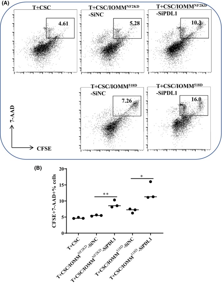
Results: PD-L1 is heterogeneously expressed in NF2-associated meningiomas. After PD-L1 knockdown in NF2-associated meningioma cells, tumor cell proliferation was significantly inhibited, and the apoptosis rate was elevated. When T cells were cocultured with siPD-L1-transfected NF2-associated meningioma cells, the expression of CD69 on both CD4+ and CD8+ T cells was partly reversed, and the capacity of CD8+ T cells to kill siPD-L1-transfected tumor cells was partly restored. Results also showed that the PI3K-AKT-mTOR pathway regulates PD-L1 expression, and the mTOR inhibitor rapamycin rapidly and persistently suppresses PD-L1 expression. In vivo experimental results suggested that anti-PD-L1 antibody may have a synergetic effect with the mTOR inhibitor in reducing tumor cell proliferation and that reduced PD-L1 expression could contribute to antitumor efficacy.
Conclusions: Targeting PD-L1 could be helpful for restoring the function of tumor-infiltrating lymphocytes and inducing apoptosis to inhibit tumor proliferation in NF2-associated meningiomas. Dissecting the mechanisms of the PD-L1-driven tumorigenesis of NF2-associated meningioma will help to improve our understanding of the mechanisms underlying tumor progression and could facilitate further refinement of current therapies to improve the treatment of NF2 patients.
Keywords: NF2; PDL1; PI3K/AKT/mTOR; immunosuppression; meningioma.
© 2024 The Author(s). CNS Neuroscience & Therapeutics published by John Wiley & Sons Ltd.
Conflict of interest statement
The authors declare that they have no competing interests.
Figures


Similar articles
-
Oncolytic reovirus enhances the effect of CEA immunotherapy when combined with PD1-PDL1 inhibitor in a colorectal cancer model.Immunotherapy. 2025 Apr;17(6):425-435. doi: 10.1080/1750743X.2025.2501926. Epub 2025 May 12. Immunotherapy. 2025. PMID: 40353308
-
Merlin immunoreactivity fails to predict neurofibromatosis type 2 mutations in human meningiomas.J Neuropathol Exp Neurol. 2025 Sep 1;84(9):825-830. doi: 10.1093/jnen/nlaf058. J Neuropathol Exp Neurol. 2025. PMID: 40447281 Free PMC article.
-
High matrix metalloproteinase-2 expression predicts poor prognosis of colon adenocarcinoma and is associated with PD-L1 expression and lymphocyte infiltration.PeerJ. 2025 Jun 30;13:e19550. doi: 10.7717/peerj.19550. eCollection 2025. PeerJ. 2025. PMID: 40611940 Free PMC article.
-
Comparison of efficacy and safety of PD-1/PD-L1 combination therapy in first-line treatment of advanced NSCLC: an updated systematic review and network meta-analysis.Clin Transl Oncol. 2024 Oct;26(10):2488-2502. doi: 10.1007/s12094-024-03442-3. Epub 2024 Apr 16. Clin Transl Oncol. 2024. PMID: 38625495
-
PD-L1 diagnostic tests: a systematic literature review of scoring algorithms and test-validation metrics.Diagn Pathol. 2018 Feb 9;13(1):12. doi: 10.1186/s13000-018-0689-9. Diagn Pathol. 2018. PMID: 29426340 Free PMC article.
Cited by
-
Schisanhenol Inhibits the Proliferation of Hepatocellular Carcinoma Cells by Targeting Programmed Cell Death-ligand 1 via the STAT3 Pathways.Anticancer Agents Med Chem. 2025;25(10):697-710. doi: 10.2174/0118715206349131241121091834. Anticancer Agents Med Chem. 2025. PMID: 39810520
-
lncRNAs as prognostic markers and therapeutic targets in cuproptosis-mediated cancer.Clin Exp Med. 2024 Sep 26;24(1):226. doi: 10.1007/s10238-024-01491-0. Clin Exp Med. 2024. PMID: 39325172 Free PMC article. Review.
-
Cas9 Mouse Model of Skull Base Meningioma Driven by Combinational Gene Inactivation in Meningeal Cells.CNS Neurosci Ther. 2025 Feb;31(2):e70287. doi: 10.1111/cns.70287. CNS Neurosci Ther. 2025. PMID: 39996452 Free PMC article.
References
-
- Perry A, Giannini C, Raghavan R, et al. Aggressive phenotypic and genotypic features in pediatric and NF2‐associated meningiomas: a clinicopathologic study of 53 cases. J Neuropathol Exp Neurol. 2001;60(10):994‐1003. - PubMed
-
- Aboukais R, Zairi F, Baroncini M, et al. Intracranial meningiomas and neurofibromatosis type 2. Acta Neurochir. 2013;155(6):997‐1001; discussion 1001. - PubMed
Publication types
MeSH terms
Substances
Grants and funding
- 82003023/National Natural Science Foundation of China
- 82373451/National Natural Science Foundation of China
- 81974387/National Natural Science Foundation of China
- JYY 2023-2/The Public Welfare Development and the Public Welfare Development and Reform Pilot Project of Beijing Medical Research Institute
LinkOut - more resources
Full Text Sources
Research Materials
Miscellaneous
