. 2024 Jun 3;134(11):e167811.
doi: 10.1172/JCI167811.

Increased endothelial sclerostin caused by elevated DSCAM mediates multiple trisomy 21 phenotypes

Affiliations

Increased endothelial sclerostin caused by elevated DSCAM mediates multiple trisomy 21 phenotypes

David M McKean et al. J Clin Invest. .

Abstract

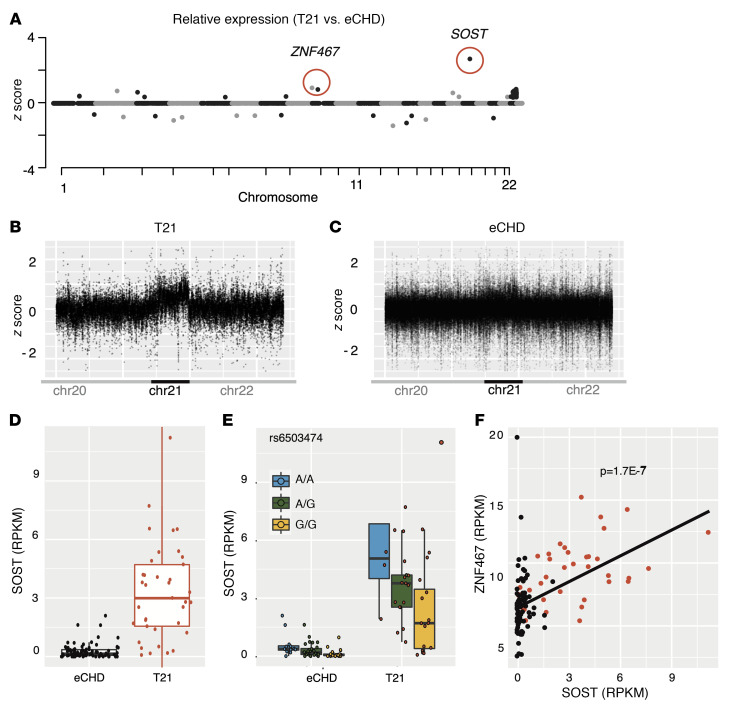
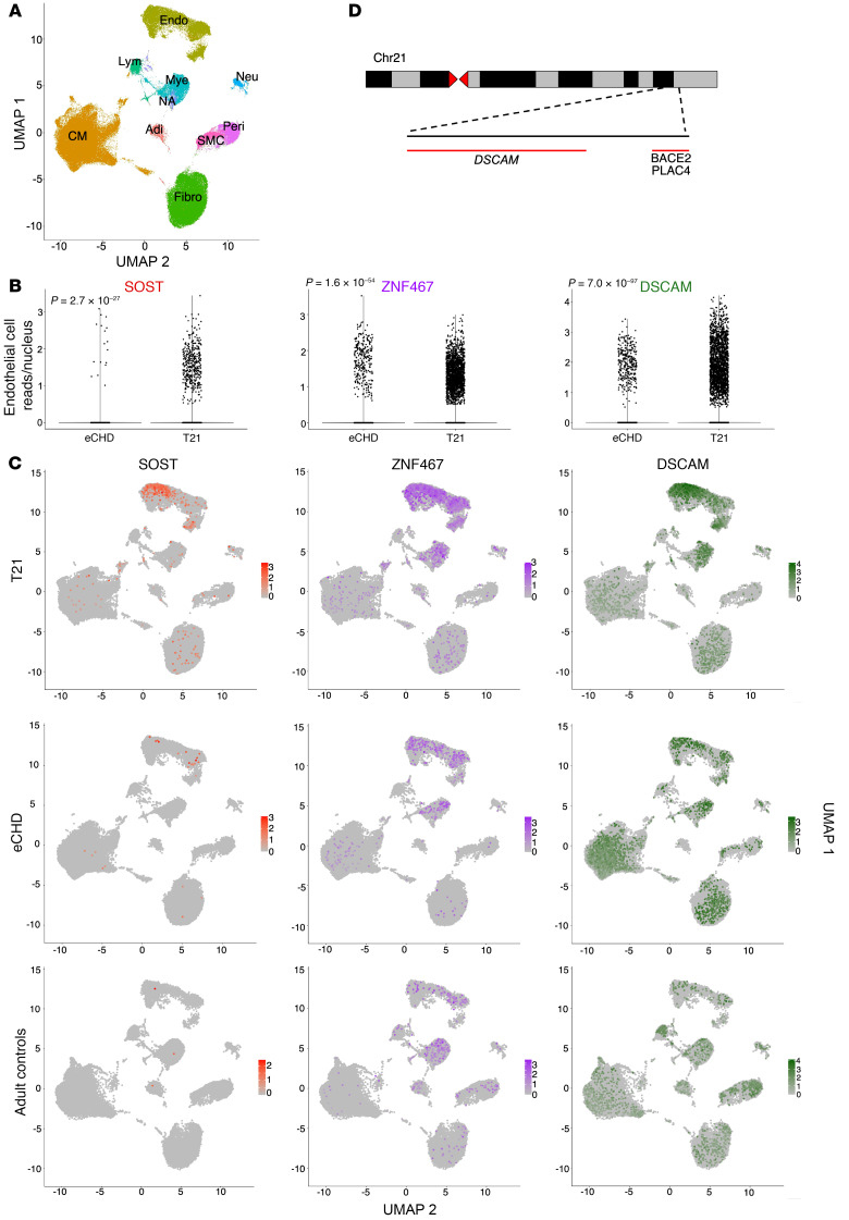
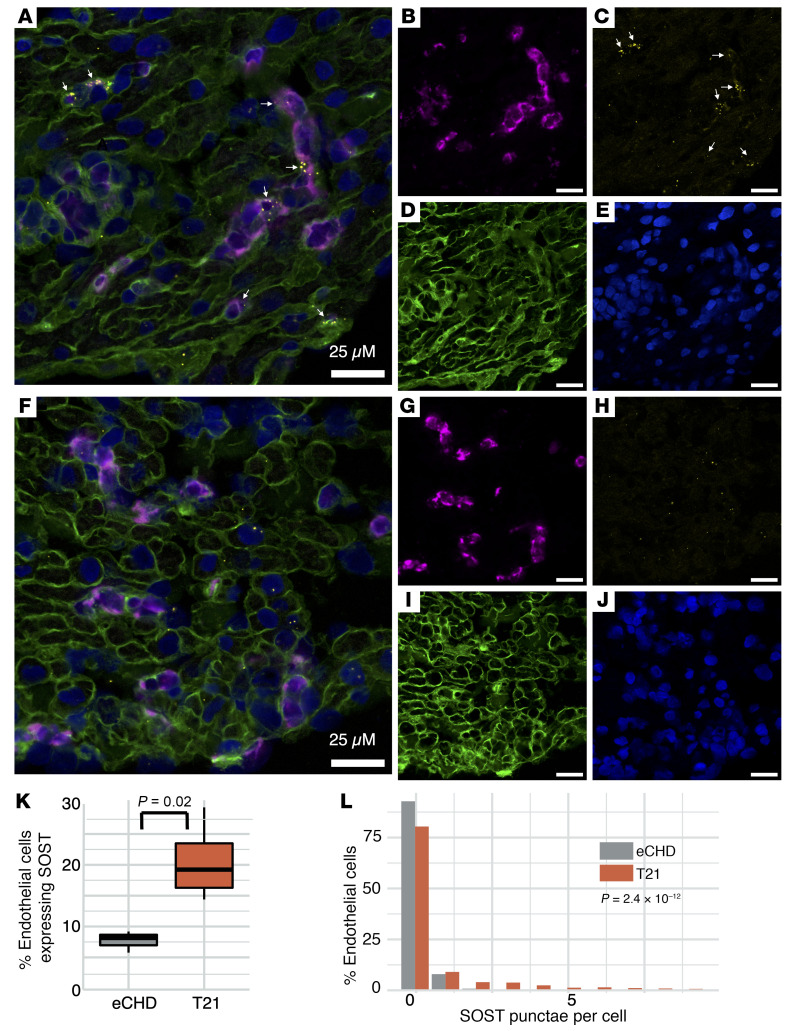
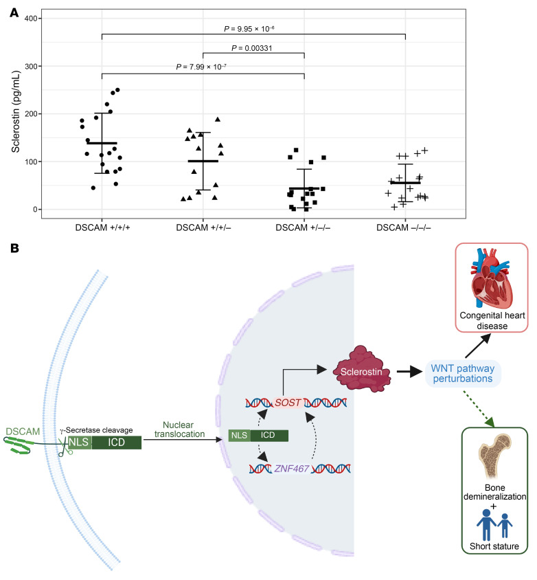
Trisomy 21 (T21), a recurrent aneuploidy occurring in 1:800 births, predisposes to congenital heart disease (CHD) and multiple extracardiac phenotypes. Despite a definitive genetic etiology, the mechanisms by which T21 perturbs development and homeostasis remain poorly understood. We compared the transcriptome of CHD tissues from 49 patients with T21 and 226 with euploid CHD (eCHD). We resolved cell lineages that misexpressed T21 transcripts by cardiac single-nucleus RNA sequencing and RNA in situ hybridization. Compared with eCHD samples, T21 samples had increased chr21 gene expression; 11-fold-greater levels (P = 1.2 × 10⁻⁸) of SOST (chr17), encoding the Wnt inhibitor sclerostin; and 1.4-fold-higher levels (P = 8.7 × 10⁻⁸) of the SOST transcriptional activator ZNF467 (chr7). Euploid and T21 cardiac endothelial cells coexpressed SOST and ZNF467; however, T21 endothelial cells expressed 6.9-fold more SOST than euploid endothelial cells (P = 2.7 × 10⁻²⁷). Wnt pathway genes were downregulated in T21 endothelial cells. Expression of DSCAM, residing within the chr21 CHD critical region, correlated with SOST (P = 1.9 × 10⁻⁵) and ZNF467 (P = 2.9 × 10⁻⁴). Deletion of DSCAM from T21 endothelial cells derived from human induced pluripotent stem cells diminished sclerostin secretion. As Wnt signaling is critical for atrioventricular canal formation, bone health, and pulmonary vascular homeostasis, we concluded that T21-mediated increased sclerostin levels would inappropriately inhibit Wnt activities and promote Down syndrome phenotypes. These findings imply therapeutic potential for anti-sclerostin antibodies in T21.

Keywords: Bone development; Cardiology; Cardiovascular disease; Genetic diseases; Genetics.


Figures

Figure 1
Figure 1. Genome-wide transcript analyses identifies increased expression of SOST and ZNF467 in T21 cardiac tissues.
(A) Graphical representation of the log2 fold change in RNA expression z score for all autosomal genes in 51 tissues from 49 individuals with T21 compared with and 236 tissues from 226 individuals with eCHD. ZNF467 is represented by the black dot within the red circle. (B and C) Standard (z) scores for differentially expressed chr21 genes in T21 (B) and eCHD (C) tissues, excluding genes with 1.35 < z score < 0.65 or 2-tailed t test P > 0.002. (D) Quantification of SOST expression (RPKM) demonstrates 11.03-fold-higher expression in T21 than eCHD (2-tailed t test P = 1.2 × 10⁻⁸) RA tissues. (E) Quantification of SOST expression (RPKM) is based on rs6503474 genotypes of RA tissues from eCHD (A/A n = 12; A/G n = 29; G/G n = 21) and T21 (A/A n = 7; A/G n = 20; G/G n = 10) patients and indicates a similar effect size in T21 (42%) and euploid (35%) tissues. (F) Expression of ZNF467 and SOST transcripts (RPKM) is correlated in eCHD (black) and T21 (red) tissues. In D and E, the box represents the first quartile (bottom) and third quartile (top) values, while the line represents 1.5 times the interquartile range beyond those values.
Figure 2
Figure 2. Single-nuclear RNA sequencing defines cell-type expression of SOST, ZNF467, and DSCAM in CHD tissues.
(A) A uniform manifold and projection graph of single-nuclear RNA sequencing right atria from participants with T21 (n = 10) and eCHD (n = 13) and healthy adults (n = 12) identifies 8 major cell types (Adi, adipocytes; CM, atrial cardiomyocytes; Endo, endothelial cells; Fibro, fibroblasts; Lym, lymphocytes; Mye, myeloid cells; NA, not assigned; Neu, neural cells; Peri, pericytes). (B) Comparison of endothelial cell transcript levels for SOST, ZNF467, and DSCAM in T21 and eCHD samples. More endothelial cells demonstrate expression (≥0.01 RPKM) of SOST, ZNF467, and DSCAM in T21 compared with eCHD tissues: SOST 2.8% vs. 0.2%; ZNF467 12.4% vs. 2.8%; DSCAM 14.2% vs. 3.2%. Boxes represent the first quartile (bottom) and third quartile (top) values, while the line represents 1.5 times the interquartile range beyond those values. Each dot represents a cell with the indicated number of reads; cells with no reads are clumped at the origin. (C) Feature plots demonstrating variable expression of SOST, ZNF467, and DSCAM in different cell lineages within T21, eCHD, and healthy adult RA. Note the different scales in the heat maps; scale bars: 25 μM. (D) Schematic representation of the 0.96 Mb CHD critical region on chr21 with protein-coding genes DSCAM, BACE2, PLAC4, and C21orf62.
Figure 3
Figure 3. Single-molecule RNA in situ hybridization identifies prominent SOST expression in endothelial cells.
(AJ) Fluorescently labeled RNA probes detect colocalization (white arrows) of SOST (red, C and H) and the endothelial cell–specific maker von Willebrand factor transcripts (VWF; cyan, B and G) in eCHD (n = 3, AE) and T21 (n = 4, FJ) RA tissues. Nuclei are DAPI-stained (blue, E and J), and cell membranes are defined by wheat germ agglutinin (WGA: green, D and I). Scale bars: 25 μm; original magnification, 63×. (K) The proportion of endothelial cells with coexpression of SOST and VWF expression is significantly higher in T21 (20.2%) compared with eCHD (7.7%) tissues. Box represents the first (bottom) and third quartile (top) values, while the line represents 1.5-times the interquartile range beyond those values. (L) SOST expression per endothelial cell is significantly higher in T21 (n = 514, mean number of red puncta 0.52) compared with eCHD (n = 337, mean number of red puncta 0.08; 2-tailed t test P = 2.4 × 10⁻¹² across all cells).
Figure 4
Figure 4. A model for SOST activation and Wnt inhibition in T21.
(A) DSCAM is necessary for increased sclerostin secretion in iPSC-derived endothelial cells as measured by an ELISA assay, grouped by DSCAM genotypes (+/+/+, n = 19; +/+/–, n = 14; +/–/–, n = 17; –/–/–, n = 17 replicates). Error bars represent mean ± SD in each group. P values were obtained by 2-tailed Student’s t test, with P < 0.0083 considered statistically significant following Bonferroni’s correction. Bold bars represent the mean, while the outer bars represent 1 standard deviation. (B) Cleavage of DSCAM, followed by nuclear translocation of the intercellular domain (ICD), which contains a nuclear localization signal (NLS), activates transcriptional responses (32). The expression of DSCAM, which resides within the chr21 CHD critical region, is correlated with both ZNF467 (chr1) and SOST (chr17) mRNA levels in cardiac endothelial cells from T21 heart tissues. The downstream effects of these molecular signals increase the binding of sclerostin to low-density lipoprotein receptor–related proteins 5 and 6 (LRP5, LRP6) and inhibit Wnt signaling through the frizzled receptor, resulting in attenuated expression of Wnt target genes. Reduced Wnt activity during development could promote CHD, limit skeletal bone maturation resulting in short stature, and increase risks for bone demineralization. Figure created with BioRender.com.

References

    1. Antonarakis SE, et al. Down syndrome. Nat Rev Dis Primers. 2020;6(1):9. doi: 10.1038/s41572-019-0143-7. - DOI - PMC - PubMed
    1. Bergström S, et al. Trends in congenital heart defects in infants with down syndrome. Pediatrics. 2016;138(1):e20160123. doi: 10.1542/peds.2016-0123. - DOI - PubMed
    1. Bull MJ. Down syndrome. N Engl J Med. 2020;382(24):2344–2352. doi: 10.1056/NEJMra1706537. - DOI - PubMed
    1. Pestel J, et al. Real-time 3D visualization of cellular rearrangements during cardiac valve formation. Development. 2016;143(12):2217–2227. doi: 10.1242/dev.133272. - DOI - PMC - PubMed
    1. Tan M, et al. Types and distribution of congenital heart defects associated with trisomy 21 in Singapore. J Paediatr Child Health. 2013;49(3):223–227. doi: 10.1111/jpc.12129. - DOI - PubMed

MeSH terms