. 2024 Jul 8;52(12):6748-6762.
doi: 10.1093/nar/gkae471.

Unlocking the potential of protein-derived peptides to target G-quadruplex DNA: from recognition to anticancer activity

Affiliations

Unlocking the potential of protein-derived peptides to target G-quadruplex DNA: from recognition to anticancer activity

Francesco Merlino et al. Nucleic Acids Res. .

Abstract

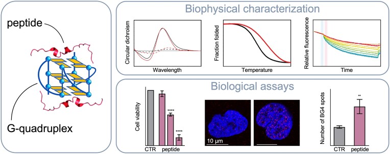
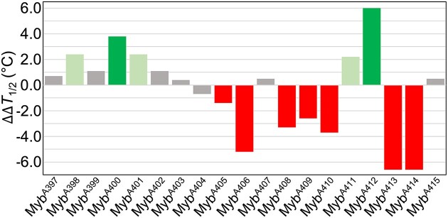
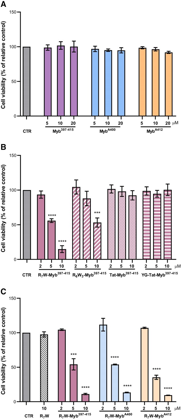
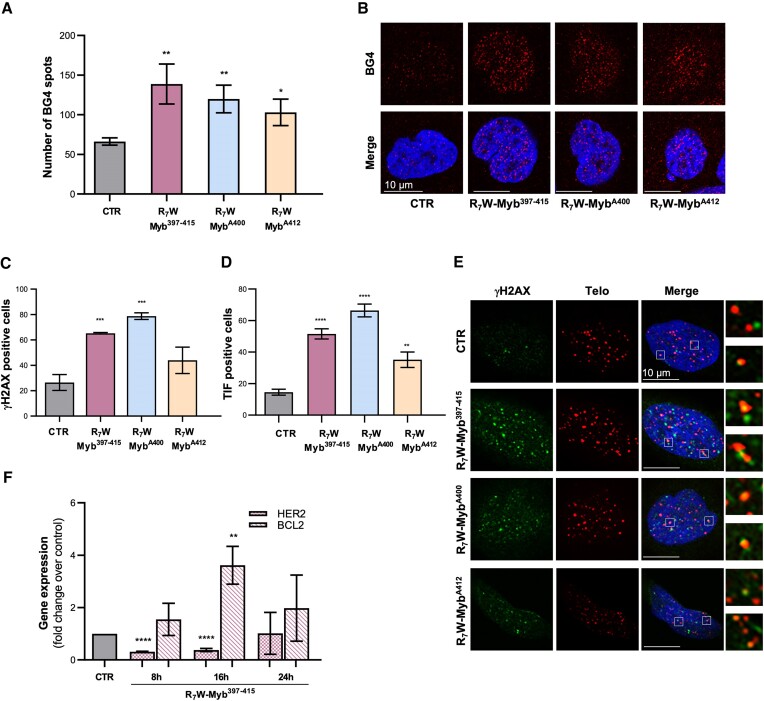
Noncanonical nucleic acid structures, particularly G-quadruplexes, have garnered significant attention as potential therapeutic targets in cancer treatment. Here, the recognition of G-quadruplex DNA by peptides derived from the Rap1 protein is explored, with the aim of developing novel peptide-based G-quadruplex ligands with enhanced selectivity and anticancer activity. Biophysical techniques were employed to assess the interaction of a peptide derived from the G-quadruplex-binding domain of the protein with various biologically relevant G-quadruplex structures. Through alanine scanning mutagenesis, key amino acids crucial for G-quadruplex recognition were identified, leading to the discovery of two peptides with improved G-quadruplex-binding properties. However, despite their in vitro efficacy, these peptides showed limited cell penetration and anticancer activity. To overcome this challenge, cell-penetrating peptide (CPP)-conjugated derivatives were designed, some of which exhibited significant cytotoxic effects on cancer cells. Interestingly, selected CPP-conjugated peptides exerted potent anticancer activity across various tumour types via a G-quadruplex-dependent mechanism. These findings underscore the potential of peptide-based G-quadruplex ligands in cancer therapy and pave the way for the development of novel therapeutic strategies targeting these DNA structures.


Figures

Graphical Abstract
Graphical Abstract
Figure 1.
Figure 1.
(A) Chemical structure of a G-tetrad showing the Hoogsteen hydrogen bonding between guanine bases, and schematic representation of the different G4 structural conformations investigated in this work: (B) parallel, (C) hybrid, and antiparallel (D) ‘chair’ and (E) ‘basket’ types. The anti and syn guanines are coloured in orange and magenta, respectively. The arrows indicate the direction of the DNA strands from the 5′ to 3′ end.
Figure 2.
Figure 2.
UVRR spectra to gain insights into the binding mode of Myb397–415 to c-KIT2 G4. From bottom to top: thymine (blue), adenine (red), cytosine (orange) and guanine (green) (as constituents of a nucleotide) normalized and weighted according to the respective number of nucleotides present in the c-KIT2 sequence; Myb397–415 (magenta); c-KIT2 (blue); c-KIT2/Myb397–415 complex (red); arithmetic sum of c-KIT2 and Myb397–415 spectra (red-dashed); normalized difference between the spectra of the complex and the arithmetic sum (dark grey). Spectra were normalized to the intensity of the c-KIT2 spectrum.
Figure 3.
Figure 3.
Raw ITC data (insets) and binding isotherms for titration of (A) c-KIT2 and (B) HER2 G4s with Myb397–415 peptide, and of c-KIT2 with (C) MybA400 and (D) MybA412 obtained at 25°C. The black dots represent the experimental data obtained by integrating the raw ITC data and subtracting the heat of peptide dilution into the buffer. The red lines represent the best-fit curve for the binding.
Figure 4.
Figure 4.
Bar graph depicting changes in c-KIT2 G4-stabilizing properties (ΔΔT1/2) of the indicated Ala-modified peptides with respect to Myb397–415, as determined by CD melting assay. The error in ΔΔT1/2 values does not exceed 0.8°C.
Figure 5.
Figure 5.
Selected peptides impair tumour cell viability. ALT-positive U2OS osteosarcoma cells were treated for 72 h with the indicated concentrations of the selected peptides, and cell viability was determined by crystal violet assay. (A) The Myb397–415 peptide and its derivatives (MybA400 and MybA412) were tested in their native form; (B) Myb397–415 was conjugated to four different CPPs (R6W3, R7W, Tat and YG-Tat) and the resulting molecules were evaluated; (C) Peptides were conjugated with R7W and the resulting derivatives (R7W-Myb397–415, R7W-MybA400 and R7W-MybA412) were tested. Results are expressed as the percentage of viable cells in treated samples over their untreated counterpart (CTR). Histograms show mean values ± SD of three independent experiments performed in triplicate; ***P< 0.001, ****P< 0.0001.
Figure 6.
Figure 6.
Mechanism of action of the selected peptides. (A, B) U2OS cells were subjected to 24 h of treatment with 10 μM of R7W-Myb397–415, R7W-MybA400 and R7W-MybA412 and G4-stabilizing activity was evaluated by IF confocal microscopy through the use of an antibody (BG4) able to specifically recognize G4 structures in cells. (A) Histograms showing the number of BG4 spots in treated over untreated (CTR) samples. Mean ± SD of three independent experiments performed in triplicate is shown; *P< 0.05, **P< 0.01. (B) Representative images of confocal sections (63×) from (A). For each condition, G4 structures (red) and merged images of DAPI counterstained nuclei (blue) with G4s are shown. Scale bar (10 μm) is reported in the images. (C–E) U2OS cells, subjected or not to treatment with the indicated peptides (10 μM for 24 h), were processed for telomeric FISH combined with immunofluorescence analysis of γH2AX, a common marker of DNA damage. (C) Histograms showing the percentage of γH2AX-positive cells. (D) Quantitative analysis of the percentage of Telomere Induced Foci (TIF)-positive cells. Cells with at least four γH2AX/Telo colocalizations were scored as TIF positive. (E) Representative confocal microscopy images (63×) used for the quantitative analyses reported in (C) and (D). γH2AX spots (green), telomere probe spots (red), DAPI stained nuclei (blue) and merged images are shown. For each experimental condition, 4× enlargements of merged fields are reported. Scale bars are shown in the images. (F) U2OS cells were treated with 10 μM of R7W-Myb397–415 and the expression levels of HER2 and BCL-2 were evaluated by real-time RT-PCR, at the indicated time-points. Results are expressed as fold change of gene expression in treated cells over their control counterpart (CTR). All the histograms show the mean ± SD of at least three independent experiments performed in triplicate; *P< 0.05, **P< 0.01, ***P< 0.001, ****P< 0.0001.

References

    1. Spiegel J., Adhikari S., Balasubramanian S.. The structure and function of DNA G-quadruplexes. Trends Chem. 2020; 2:123–136. - PMC - PubMed
    1. Biffi G., Tannahill D., McCafferty J., Balasubramanian S.. Quantitative visualization of DNA G-quadruplex structures in human cells. Nat. Chem. 2013; 5:182–186. - PMC - PubMed
    1. Rhodes D., Lipps H.J.. G-quadruplexes and their regulatory roles in biology. Nucleic Acids Res. 2015; 43:8627–8637. - PMC - PubMed
    1. Romano F., Di Porzio A., Iaccarino N., Riccardi G., Di Lorenzo R., Laneri S., Pagano B., Amato J., Randazzo A.. G-quadruplexes in cancer-related gene promoters: from identification to therapeutic targeting. Expert Opin. Ther. Pat. 2023; 33:745–773. - PubMed
    1. Cheung-Ong K., Giaever G., Nislow C.. DNA-damaging agents in cancer chemotherapy: serendipity and chemical biology. Chem. Biol. 2013; 20:648–659. - PubMed

LinkOut - more resources