. 2024 Jul 8;52(12):7031-7048.
doi: 10.1093/nar/gkae474.

The translocation activity of Rad54 reduces crossover outcomes during homologous recombination

Affiliations

The translocation activity of Rad54 reduces crossover outcomes during homologous recombination

Krishay Sridalla et al. Nucleic Acids Res. .

Abstract

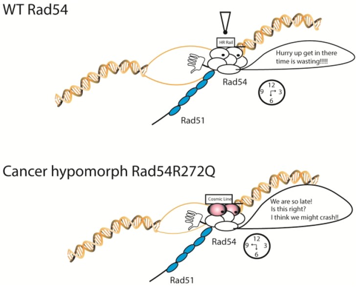
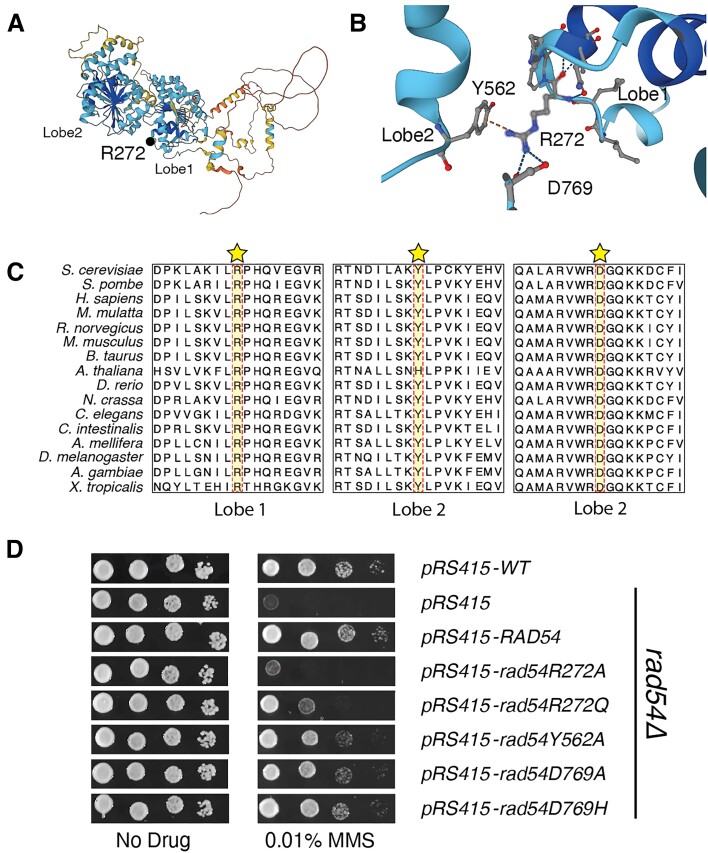
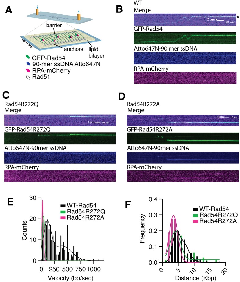
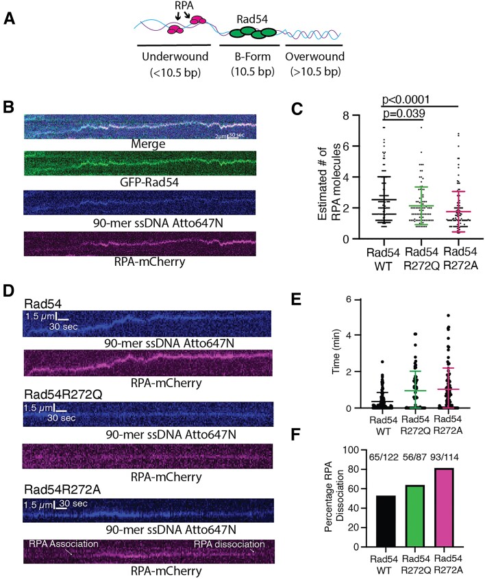
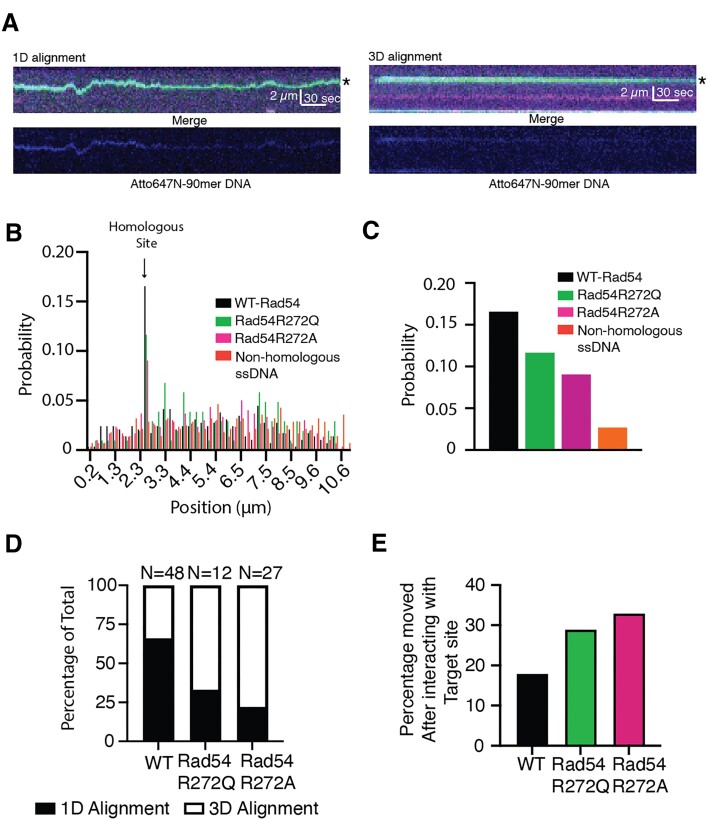
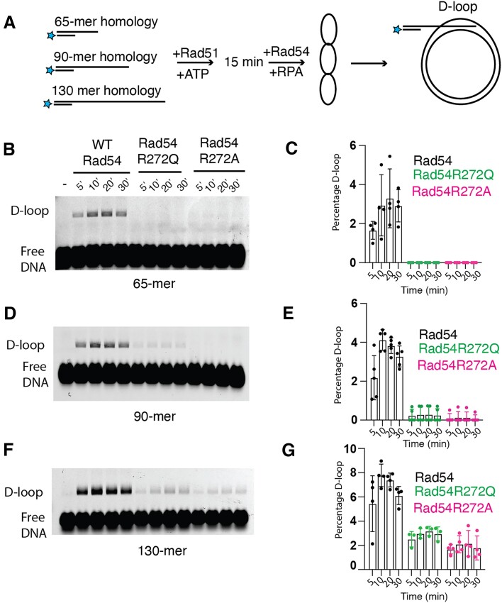
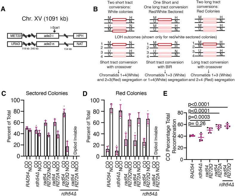
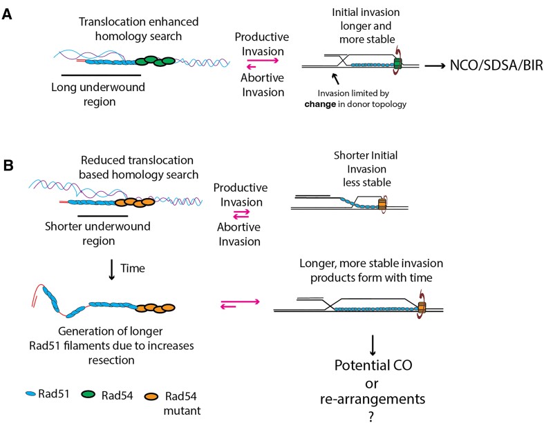
Homologous recombination (HR) is a template-based DNA double-strand break repair pathway that requires the selection of an appropriate DNA sequence to facilitate repair. Selection occurs during a homology search that must be executed rapidly and with high fidelity. Failure to efficiently perform the homology search can result in complex intermediates that generate genomic rearrangements, a hallmark of human cancers. Rad54 is an ATP dependent DNA motor protein that functions during the homology search by regulating the recombinase Rad51. How this regulation reduces genomic exchanges is currently unknown. To better understand how Rad54 can reduce these outcomes, we evaluated several amino acid mutations in Rad54 that were identified in the COSMIC database. COSMIC is a collection of amino acid mutations identified in human cancers. These substitutions led to reduced Rad54 function and the discovery of a conserved motif in Rad54. Through genetic, biochemical and single-molecule approaches, we show that disruption of this motif leads to failure in stabilizing early strand invasion intermediates, causing increased crossovers between homologous chromosomes. Our study also suggests that the translocation rate of Rad54 is a determinant in balancing genetic exchange. The latch domain's conservation implies an interaction likely fundamental to eukaryotic biology.


Figures

Graphical Abstract
Graphical Abstract
Figure 1.
Figure 1.
Disruption in the connection between Rad54 lobes leads to increased MMS sensitivity. (A) Alphafold2 structure of yeast Rad54. The protein is colored corresponding to the pLDDT format. A black dot illustrates the site of mutation. (B) Expanded view within Rad54 that corresponds to a motif that bridges the two RecA lobes. The R272 and D769 residues were mutated in the COSMIC database. (C) Three regions of an amino acid sequence alignment of several eukaryotic Rad54 proteins showing the conservation of the R272, Y562 and D769 residues. Protein sequences were obtained from Uniprot and aligned in Jalview (2.11.3.2) using MUSCLE with default settings. (D) Yeast complementation assay to evaluate Rad54 mutants for their ability to restore resistance to MMS. Alleles tested includes RAD54, rad54R272A, rad54R272Q, rad54Y562A, rad54D769A and rad54D769H.
Figure 2.
Figure 2.
Disruption of the Rad54 latch reduces translocation velocity of the Rad51/54 PSCs. (A) Cartoon diagram illustrating DNA curtains experiment to monitor Rad51/54 PSCs during the homology search step of HR. (B) Representative Kymograph illustrates GFP-Rad54 (Green) as part of the Rad51/54-PSC translocation along DNA in search of homology. The recipient 90-mer ssDNA Atto647N (middle) is labeled in blue, and RPA-mCherry (bottom) is included in the reaction (magenta). The Asterix denotes the sites of homology. (C) Kymographs for Rad54R272Q-PSCs with Merge, GFP-Rad54R272Q, Atto647N-90mer ssDNA, RPA-mCherry. (D) Kymographs for Rad54R272A-PSCs with Merge, GFP-Rad54R272A, Atto647N-90mer ssDNA, RPA-mCherry. (E) Distributions of measured velocities for WT-Rad54 (N = 248), Rad54R272Q (N = 65) and Rad54R272A (N = 58). A sum of two Gaussian distributions fits the data of WT-Rad54. (F) Distributions of measured translocation distances for WT-Rad54 (N = 248), Rad54R272Q (N = 65) and Rad54R272A (N = 58). A sum of two Gaussian distributions fits the data.
Figure 3.
Figure 3.
Mutations in Rad54 influence RPA dynamics on the PSC. (A) Cartoon schematic illustrating the proposed model for Rad54 movement on dsDNA. Movement creates underwound DNA behind and overwound DNA ahead. (B) Representative kymograph illustrating co-translocation of GFP-Rad54 (green), 90-mer ssDNA-Atto647N (blue), and RPA-mCherry (magenta). (C) Dot plot representing the estimated number of RPA molecules based on photobleaching measurements for an individual step size for WT-Rad54 (N = 119), Rad54R272Q (N = 87) (green) and Rad54R272A (N = 114) (magenta). The cross and error bars represent the mean and standard deviations of the data. (D) Representative kymographs for WT (top), Rad54R272Q (middle) and Rad54R272A (bottom). The 90-mer ssDNA-Atto647N from the PSC (blue) and RPA-mCherry (magenta) are shown. (E) Dot plot representing the time it takes for RPA to associate with Rad51/54-PSCs that are bound to dsDNA for WT-Rad54 (N = 108) (black), Rad54R272Q (N = 60) (green), Rad54R272A (N = 95) (magenta). The cross and error bars represent the mean and standard deviation of the data. (F) Bar graph representing the fraction of RPA molecules that bound to the PSC but then dissociated before the end of the experiment for WT-Rad54 (N = 122) (black), Rad54R272Q (N = 87) (green) and Rad54R272A (N = 114) (magenta).
Figure 4.
Figure 4.
Reduced Rad54 activity alters the efficiency of DNA sequence alignment. (A) Representative kymographs illustrating the alignment of the homologous sequence by 1D movement (left) and 3D movement (right). The kymographs represent a merged three-color image (top) of GFP-Rad54 (green), RPA-mCherry (magenta), and 90-mer ssDNA Atto647N (Blue) and an individual image of the Atto647N-90-mer (bottom). A star denotes the site of homology. (B) Graph representing the binding probability distribution of PSCs across lambda DNA after 15 min. An arrow marks the site of homology. The data are for Rad51/54-PSCs consisting of WT-Rad54 (N = 290) (black), Rad54R272Q (N = 103) (Green), and Rad54R272A (N = 298) (magenta). Non-homologous sequence (N = 280) (orange). (C) The expanded view of the graph in (B) represents only the homologous site. (D) Graph representing the percentage of sequence alignment events that occur via 1D movement versus 3D movement for WT-Rad54, Rad54R272Q and Rad54R272A. (E) Bar graph illustrating the number of PSCs that bind to the target site but then move after dwelling for at least 10 s.
Figure 5.
Figure 5.
Rad54 mutants have a diminished ability to form D-loops in vitro. (A) Cartoon diagram illustrating experiment to measure WT-Rad54, Rad54R272Q and Rad54R272A activity for D-loop formation with different lengths of homology. (B) Representative gel illustrating D-loop formation for WT-Rad54, Rad54R272Q and Rad54R272A with a 65-mer of homology to the pUC19 plasmid. (C) The bar graph quantifies the D-loop formation percentage for WT-Rad54, Rad54R272Q, and Rad54R272A. The error bars represent the standard deviation of 4 independent experiments. (D) Representative gel illustrating D-loop formation for WT-Rad54, Rad54R272Q and Rad54R272A with a 90-mer of homology to the pUC19 plasmid. (E) The bar graph quantifies the percentage of D-loop formation for WT-Rad54, Rad54R272Q and Rad54R272A. The error bars represent the standard deviation of 5 independent experiments. (F) Representative gel illustrating D-loop formation for WT-Rad54, Rad54R272Q and Rad54R272A with a 130-mer of homology to the pUC19 plasmid. (G) The bar graph quantifies the D-loop formation percentage for WT-Rad54, Rad54R272Q and Rad54R272A. The error bars represent the standard deviation of 3–4 independent experiments.
Figure 6.
Figure 6.
Mutations in Rad54 lead to increased crossover outcomes. (A) Schematic diagram illustrating the double strand break reporter construct used in these experiments. (B) Schematic diagram illustrating the possible recombination outcomes using this assay. The general outcome from recombination can be inferred from the color of the colony and the antibiotic sensitivity. (C) Graph illustrating the CO and NCO outcomes for sectored colonies of WT, rdh54Δ, rad54R272Q, rad54R272A, rad54R272Q rdh54Δ and rad54R272A rdh54Δ. The error bars represent the standard deviation of six independent experiments. (D) Graph illustrating the CO and NCO outcomes ratio for solid red colonies of WT, rad54R272Q, rad54R272A, rdh54Δ, rad54R272Q rdh54Δand rad54R272A rdh54Δ. The error bars represent the standard deviation of six independent experiments. (E) Graph illustrating crossover outcomes as a percentage of total recombination events for WT, rad54R272Q, rad54R272A, rdh54Δ, rad54R272Q rdh54Δ and rad54R272A rdh54Δ. The bars represent the mean, and the error bars represent the standard deviation from six independent experiments.
Figure 7.
Figure 7.
Model for reduction in abortive invasion. (A) Schematic diagram illustrating that longer tracts of underwound DNA generated by Rad54 lead to faster invasions and NCO repair or SDSA. (B) Mutant versions of Rad54 lack general translocation activity and produce shorter regions of underwound DNA, leading to less stable invasion products. As an indirect consequence, ssDNA gets longer and generates longer Rad51 filaments, which leads to higher stability after invasion. This increases the probability of crossover formation.

Similar articles

Cited by

References

    1. Jasin M., Rothstein R.. Repair of strand breaks by homologous recombination. Cold Spring Harb. Perspect. Biol. 2013; 5:a012740. - PMC - PubMed
    1. Kowalczykowski S.C. An overview of the molecular mechanisms of recombinational DNA repair. Cold Spring Harb. Perspect. Biol. 2015; 7:a016410. - PMC - PubMed
    1. Symington L.S., Gautier J.. Double-strand break end resection and repair pathway choice. Annu. Rev. Genet. 2011; 45:247–271. - PubMed
    1. Symington L.S., Rothstein R., Lisby M.. Mechanisms and regulation of mitotic recombination in Saccharomyces cerevisiae. Genetics. 2014; 198:795–835. - PMC - PubMed
    1. Cejka P., Symington L.S.. DNA end resection: mechanism and control. Annu. Rev. Genet. 2021; 55:285–307. - PubMed

MeSH terms

LinkOut - more resources