. 2024 Jul 8;52(12):6733-6747.
doi: 10.1093/nar/gkae461.

Site-specific regulation of RNA editing with ribose-modified nucleoside analogs in ADAR guide strands

Affiliations

Site-specific regulation of RNA editing with ribose-modified nucleoside analogs in ADAR guide strands

Victorio Jauregui-Matos et al. Nucleic Acids Res. .

Abstract

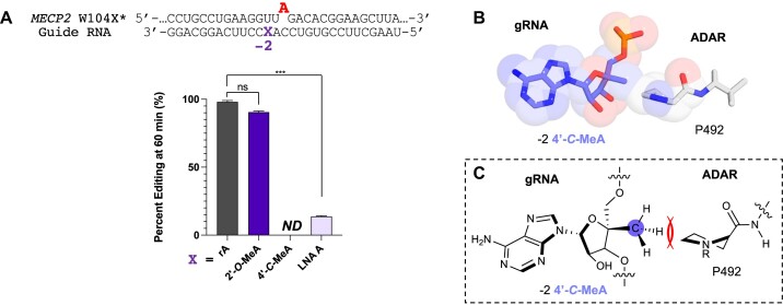
Adenosine Deaminases Acting on RNA (ADARs) are enzymes that catalyze the conversion of adenosine to inosine in RNA duplexes. These enzymes can be harnessed to correct disease-causing G-to-A mutations in the transcriptome because inosine is translated as guanosine. Guide RNAs (gRNAs) can be used to direct the ADAR reaction to specific sites. Chemical modification of ADAR guide strands is required to facilitate delivery, increase metabolic stability, and increase the efficiency and selectivity of the editing reaction. Here, we show the ADAR reaction is highly sensitive to ribose modifications (e.g. 4'-C-methylation and Locked Nucleic Acid (LNA) substitution) at specific positions within the guide strand. Our studies were enabled by the synthesis of RNA containing a new, ribose-modified nucleoside analog (4'-C-methyladenosine). Importantly, the ADAR reaction is potently inhibited by LNA or 4'-C-methylation at different positions in the ADAR guide. While LNA at guide strand positions -1 and -2 block the ADAR reaction, 4'-C-methylation only inhibits at the -2 position. These effects are rationalized using high-resolution structures of ADAR-RNA complexes. This work sheds additional light on the mechanism of ADAR deamination and aids in the design of highly selective ADAR guide strands for therapeutic editing using chemically modified RNA.


Figures

Graphical Abstract
Graphical Abstract
Figure 1.
Figure 1.
Site-specific LNA modification can reduce or block editing at various off-target sites by ADAR2 on the SRC transcript. (A) Left top: schematic of target:guide strand duplex indicating nomenclature and numbering scheme used in this work; bottom left: Target RNA and guide RNA hybrid construct; red A: adenosine target; grey: bystander sites; bold: fragment studied for the LNA modifications. Right: structure of LNA in blue. (B) Editing yield at 60 min with no LNA modification. Red bars indicate editing for target A; off-target sites are displayed in light grey bars. (C–I) Editing yield at 60 min for different guide oligonucleotides bearing LNA (blue and italics L) at different sites on the guide RNA (−3, −2, −1, −4, +3, +2 and +1, respectively). Reactions for each time point were carried out with a ratio of 5:50 nM of RNA hybrid to ADAR2 WT. Error bars represent the standard deviation of three technical replicates. N: No detected editing.
Figure 2.
Figure 2.
Structure of hADAR2-R2D E488Q bound to the GLI1 32 bp RNA at 2.8 Å resolution (19). (A) View of the structure perpendicular to the dsRNA helical axis, the pink and slate blue region shows the kink in the guide RNA and the widening of the major groove opposite the editing site induced by ADAR2. The warm pink shows the target RNA and the light grey the guide RNA. (B) Schematic of target and guide RNA hybrid for ADAR editing in a 5′-UA-3′ sequence context indicating nomenclature and numbering scheme used in this work. −1 position highlighted in pink and −2 in blue. (C) Space filling model of the −1 nucleotide 4′-C closely approaching the γ and δ carbons of I491 in ADAR’s flipping loop. (D) Space filling model of the −2 nucleotide 4′-C closely approaching P492’s side chain. (E) The −1 A (pink) adopts a C2’-endo sugar pucker with a high anti glycosidic bond angle (χ = −90.6°) compared to an ideal A-form nucleotide (black). (F) The −2 G (slate blue) adopts an RNA canonical C3’-endo sugar pucker (black).
Figure 3.
Figure 3.
Proposed structures of the possible methyl groups rearranged from the 2′-C-4′-C-oxymethylene linkage cleavage of LNA at the −1 position. LNA is fixed to an RNA C3’-endo sugar pucker conformation. Right: 2′-O-Me A; Left: 4′-C-MeA and proposed steric clash with I491 or P492. 4′-C-Me A expected to adopt a DNA-like C2’-endo sugar pucker conformation.
Scheme 1.
Scheme 1.
Synthesis of the 4′-C-MeA phosphoramidite. Reagents and conditions: (a) SnCl4, N6-benzoyladenine, CH2Cl2, 0–25°C, 63%; (b) K2CO3, MeOH, 25°C, 88%; (c) 1 M Tetrabutylammonium fluoride/THF, 25°C, 79%; (d) 1M BCl3/CH2Cl2, −78°C, 41%; (e) DMTr-Cl, pyridine, DMAP, 25°C, 70%; (f) t-Bu(Me)2Si-Cl, pyridine, AgNO3, THF, 0–25°C, 64%; (g) 2-cyanoethyl-N,N-diisopropylchlorophosphoramidite, (i-Pr)2NEt, 25°C, 77%.
Figure 4.
Figure 4.
(A) Top: Target sequence derived from the mRNA for human MECP2 proximal to the W104X mutation associated with Rett syndrome and guide RNA used here to direct corrective editing at the premature stop codon. The target adenosine is indicated in red. X indicates the −1 position of the guide strand relative to the target adenosine. Right: Comparison of different ribose modifications at the −1 nucleotide on selective editing at the target site plotted as editing percent at 60 min. Left: Deamination rate comparison of canonical A and 4′-C-MeA. Reactions for each timepoint were carried out with a ratio of 5:15 nM of RNA hybrid to ADAR2 WT. (B) Top: target sequence derived from the mRNA for mouse IDUA proximal to the W392X truncation mutation. Bottom: Effect of different ribose modifications at the −1 nucleotide on ADAR1p110 and ADAR2 WT editing at the target site plotted as editing percent at 120 and 60 min, respectively. Reactions for each timepoint were carried out with a ratio of 15:150 nM of RNA to ADAR1p110 and 5:15 nM for ADAR2 WT. Error bars represent the standard deviation of three technical replicates. A two-tailed Welch's t test was conducted, where *p < 0.05, **p < 0.01, ***p< 0.001 from rA -1 gRNA; ns: no significant difference. (C) Structures of rA, 2′-O-MeA, 4′-C-MeA, LNA A and UNA A. ND: no detected editing.
Figure 5.
Figure 5.
(A) Comparison of different ribose modifications at the −2 nucleotide on editing at the human modified MECP2* mRNA target site plotted as editing percent at 60 min. Left: reactions for each timepoint were carried out with a ratio of 5:15 nM of RNA hybrid to ADAR2 WT. Error bars represent the standard deviation of three technical replicates. A two-tailed Welch's t test was conducted, where *P < 0.05, **P < 0.01, ***P< 0.001 from rA -1 gRNA; ns: no significant difference. ND: no detected editing. (B) Stick and space-filling model overlay of the −2 4′-C-MeA closely approaching the methylene group in Pro 492 of ADAR’s flipping loop (19). (C) Chemical structure of model of Pro 492 and -2 4′-C-MeA in the guide RNA highlighting the location of the predicted steric clash.
Figure 6.
Figure 6.
Site-specific 4′-C-MeA localization in the guide RNA affects ADAR editing of target and proximal off-target sites on the SRC transcript. (A) Top: schematic of target:guide strand duplex indicating target (red), 5′ adjacent off-target site (blue) and other bystander sites (grey). Guide RNA with X indicating −2 position relative to the 5′ adjacent off-target site (blue), −3 relative to the target (red) and −4 and + 1 relative to other bystander sites. Bottom: ADAR editing yield at 120 min when X = no modification (left) and when X = 4′-C-methyladenosine. Red bars indicate editing for target A; −2 off-target site editing is displayed in blue bars and remaining bystander sites editing in grey bars. Reactions for each timepoint were carried out with a ratio of 5:50 nM of RNA hybrid to ADAR2 WT. Error bars represent the standard deviation of three technical replicates. N: No detected editing. (B) Structure of hADAR2-R2D E488Q bound to the GLI1 32 bp RNA with 8-azanebularine (N), an ADAR transition state adenosine analog at 2.8 Å resolution; stick model of the -3 nucleotide 4′-C closely approaching the side chains of R481, I456 and F457 in ADAR’s flipping loop (19).
Figure 7.
Figure 7.
Site-specific inhibition of 3′ adjacent bystander editing of the modified mouse IDUA W392X* site (5′-UAA-3′) employing 4′-C-methyladenosine (4′-C-MeA) modification at the −1 nucleotide with varying orphan bases and ADAR isoforms. (A) Top: 5′-UAA-3′ target RNA from modified IDUA with target adenosine (red) and 3′ flanking bystander edit site (blue). X: −1 sugar modification (rA or 4′-C-MeA), Y: orphan base (rC or rU). Bottom: ADAR1p110, ADAR2 WT and hyperactive mutant ADAR2 E488Q editing at RNA:enzyme ratios of 15:150, 5:15 and 5:15 nM, respectively. Red bars represent % edited of target A and blue bars % editing of bystander site. Error bars represent the standard deviation of three technical replicates. A two-tailed Welch's t test was conducted, where *P < 0.05, **P < 0.01, ***P< 0.001; ns: no significant difference. ND: No detected editing.
Figure 8.
Figure 8.
Site-specific inhibition of 3′ adjacent bystander editing in a therapeutic transcript employing 4′-C-methyladenosine (4′-C-MeA) and locked nucleic acid (LNA A). Top: 5′-UAA-3′ target RNA from the human protein phosphatase 2 regulatory subunit B’delta (PPP2R5D) E200K point mutation with the target adenosine (red) and 3′ flanking bystander edit site (blue). X: -1 sugar modification (rA, 4′-C-MeA or LNAA). Bottom: ADAR1p110, ADAR2 WT and hyperactive mutant ADAR2 E488Q editing at RNA:enzyme ratios of 5:50 nM at 120 min. Red bars represent % edited of target A and blue bars % editing of bystander site. Error bars represent the standard deviation of three technical replicates. A two-tailed Welch's t test was conducted, where *P < 0.05, **P < 0.01, ***P< 0.001; ns: no significant difference. ND: No detected editing.
Figure 9.
Figure 9.
Effect of −1 and −2 adenosine analogs in directed editing guide strands targeting an endogenous target in human cells. (A) Modification pattern for guide oligonucleotides used in cellular editing and sequence of the endogenous 3′-UTR β-actin transcript and editing site (red) where X (−1) and Y (−2) are modified. (B) Structure and denominations of chemical modifications used. (C) Percent editing of endogenous β-actin target site in HEK293T cells with overexpressed hADAR2 and varying sugar modifications at the −1 and −2 position of the guide RNA after 48h. Error bars, SD (n = 3 biological replicates). ND: no detected editing. A two-tailed Welch's t test was conducted, where *P < 0.05, **P < 0.01, ***P< 0.001 from rA -1 gRNA; ns: no significant difference.

Similar articles

Cited by

References

    1. Pullirsch D., Jantsch M.F.. Proteome diversification by adenosine to inosine RNA-editing. RNA Biol. 2010; 7:205–212. - PubMed
    1. Yang Y., Okada S., Sakurai M.. Adenosine-to-inosine RNA editing in neurological development and disease. RNA Biol. 2021; 18:999–1013. - PMC - PubMed
    1. Montiel-Gonzalez M.F., Diaz Quiroz J.F., Rosenthal J.J.C.. Current strategies for site-directed RNA editing using ADARs. Methods. 2019; 156:16–24. - PMC - PubMed
    1. Higuchi M., Maas S., Single F.N., Hartner J., Rozov A., Burnashev N., Feldmeyer D., Sprengel R., Seeburg P.H.. Point mutation in an AMPA receptor gene rescues lethality in mice deficient in the RNA-editing enzyme ADAR2. Nature. 2000; 406:78–81. - PubMed
    1. Khosravi H.M., Jantsch M.F.. Site-directed RNA editing: recent advances and open challenges. RNA Biol. 2021; 18:41–50. - PMC - PubMed

MeSH terms