. 2024 Jun 15;13(6):bio060412.
doi: 10.1242/bio.060412. Epub 2024 Jun 18.

The Endosomal Sorting Complex, ESCRT, has diverse roles in blood progenitor maintenance, lineage choice and immune response

Affiliations

The Endosomal Sorting Complex, ESCRT, has diverse roles in blood progenitor maintenance, lineage choice and immune response

Arindam Ray et al. Biol Open. .

Abstract

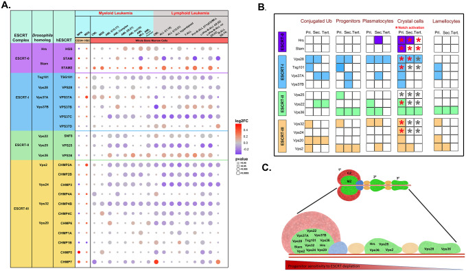
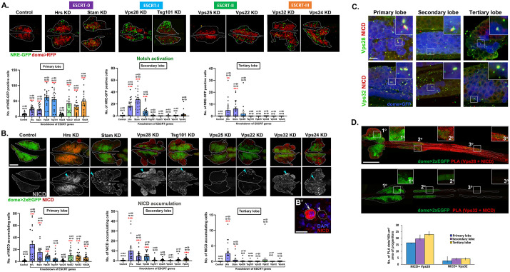
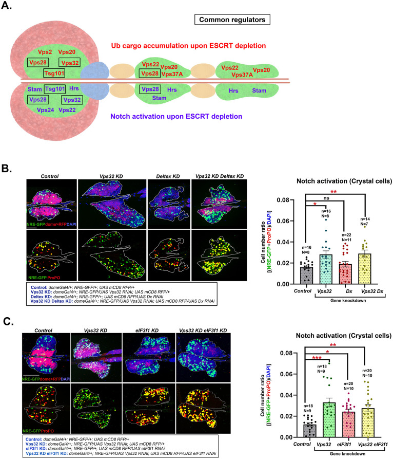
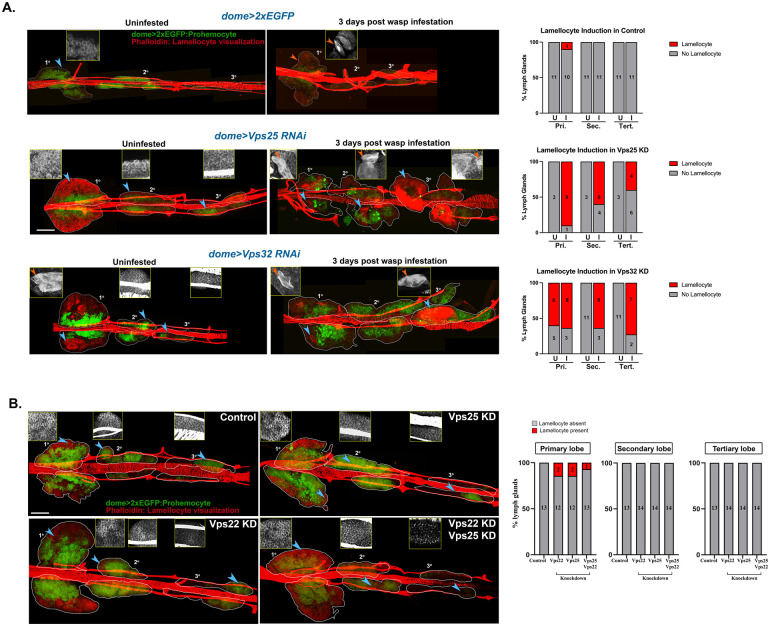
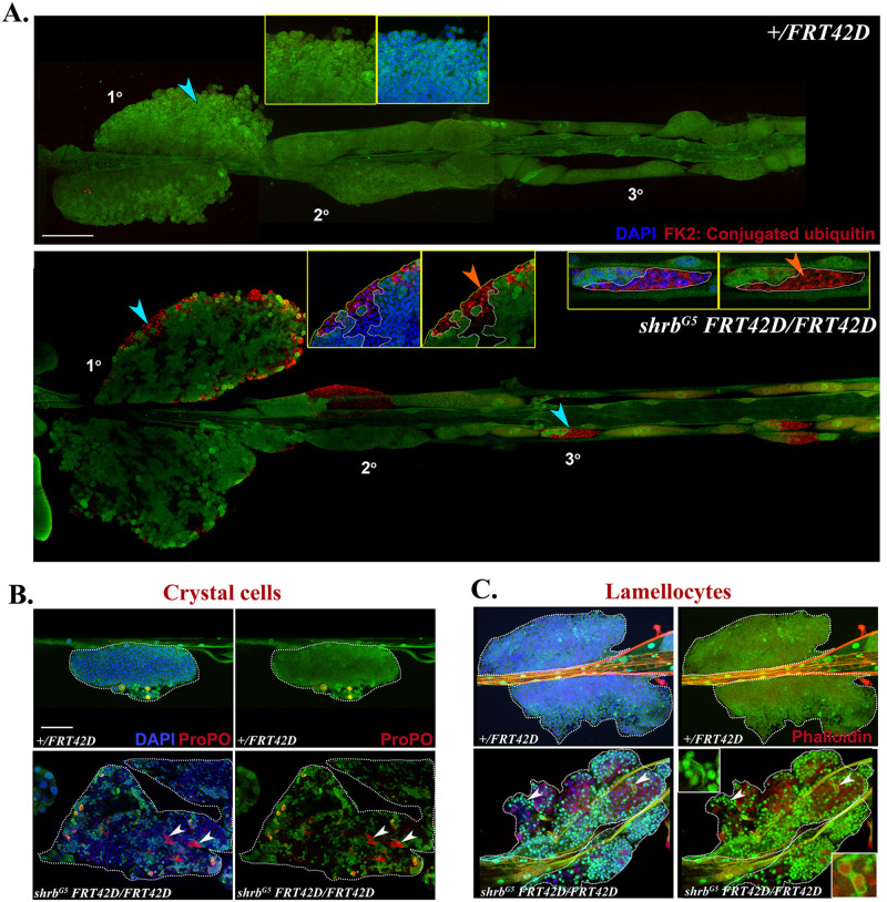
Most hematological malignancies are associated with reduced expression of one or more components of the Endosomal Sorting Complex Required for Transport (ESCRT). However, the roles of ESCRT in stem cell and progenitor maintenance are not resolved. Parsing signaling pathways in relation to the canonical role of ESCRT poses a challenge. The Drosophila hematopoietic organ, the larval lymph gland, provides a path to dissect the roles of cellular trafficking pathways such as ESCRT in blood development and maintenance. Drosophila has 13 core ESCRT components. Knockdown of individual ESCRTs showed that only Vps28 and Vp36 were required in all lymph gland progenitors. Using the well-conserved ESCRT-II complex as an example of the range of phenotypes seen upon ESCRT depletion, we show that ESCRTs have cell-autonomous as well as non-autonomous roles in progenitor maintenance and differentiation. ESCRT depletion also sensitized posterior lobe progenitors to respond to immunogenic wasp infestation. We also identify key heterotypic roles for ESCRT in position-dependent control of Notch activation to suppress crystal cell differentiation. Our study shows that the cargo sorting machinery determines the identity of progenitors and their adaptability to the dynamic microenvironment. These mechanisms for control of cell fate may tailor developmental diversity in multiple contexts.

Keywords: Cargo sorting; ESCRT; Hematopoiesis; Lamellocytes; Lineage choice; Non-canonical function of ESCRT; Notch signaling; Ubiquitin accumulation.


Conflict of interest statement

Competing interests The authors declare no competing or financial interests.

Figures

Fig. 1.
Fig. 1.
Dysregulation of ESCRT components perturbs hematopoietic homeostasis. (A) Change in expression levels of ESCRT components as found in CD34+ hematopoietic stem cells from MPN (Baumeister et al., 2021) and MDS patients (Pellagatti et al., 2010) as well as in whole marrow cells obtained from leukemia patients (BloodSpot, MILE study) is visualized using a bubble plot, across all classes of leukemia described in the study. Shades of red indicate overexpression while shades of blue indicate downregulation of individual components. The intensity of color is correlative to the log2 fold change while the size of the individual bubble indicates the P-value. The left-most column lists the Drosophila homologs of the human ESCRT proteins. (B) Comprehensive summary chart of the effects of individual ESCRT depletion on the various aspects of hematopoiesis as indicated. Presence or absence of a phenotype is depicted by colored or white boxes, respectively. Red asterisk indicates Notch pathway activation and empty asterisk indicates no change, for components that were tested. (C) Schematic representation of lymph gland lobes from anterior to posterior (left to right). Green region (medullary zone) in primary and posterior lobes indicate the progenitor pool depleted of ESCRT components (13 genes from ESCRT-0, I, II and III). Red region (cortical zone) in the anterior lobe represents the mature hemocytes. ESCRT components mentioned in each lobe are those whose depletion affected crystal cell differentiation. Red monochrome heatmap indicates position-dependent progenitor sensitivity to depletion of ESCRT.
Fig. 2.
Fig. 2.
ESCRT components regulate Notch activation and NICD trafficking in the lymph gland. (A) Whole-mount larval lymph gland showing NRE-GFP+ve (Notch reporter) cells (green) and dome+ve progenitors (red) in primary lobes upon progenitor-specific knockdown of eight representative ESCRT components indicated [Hrs, Stam (ESCRT-0); Vps28, Tsg101 (ESCRT-I); Vps25, Vps22 (ESCRT-II); Vps32, Vps24 (ESCRT-III)]. Scale bar: 100 µm. Bar diagrams show quantification of the number of NRE-GFP positive cells in primary, secondary and tertiary lobes upon knockdown of the same eight ESCRT components. (B) Whole-mount larval lymph gland showing NICD expression (shown in red in the upper panel and in gray scale in the lower panel) in primary lobes upon progenitor-specific knockdown of the same eight aforementioned ESCRT components. Progenitors are marked by dome>2xEGFP (green). Scale bar: 100 µm. (B′) Magnified view showing lymph gland hemocytes with (arrow) or without (arrowhead) NICD accumulation. Scale bar: 10 µm. One-way ANOVA was performed to determine statistical significance. *P<0.05, **P<0.01, ***P<0.001. (C) Immunostaining for NICD (red) and ESCRT components Vps28 and Vps32 (green) showing colocalization in dome+ve progenitors (blue) across primary, secondary and tertiary lobes of the lymph gland. Scale bar: 10 μm. (D) PLA dots (red) mark the interaction of NICD with Vps28 and Vps32 across three lobes of the lymph gland. Progenitors are marked by dome>2×EGFP. Insets show a magnified view of the progenitors from primary, secondary and tertiary lobes. Scale bar: 200 μm. Bar diagram shows quantification of the number of PLA dots per 100 μm2 area of the progenitors. N=5 larvae.
Fig. 3.
Fig. 3.
Non-canonical ESCRT functions may be at play for maintenance of hematopoietic homeostasis. (A) Schematic showing ESCRT components that affect ubiquitinated cargo sorting upon their depletion (in red) in the upper half and components that affect notch activation upon their depletion (in purple) in the lower half of the lymph gland schematic, in the respective lymph gland lobes. Black boxes indicate ESCRT proteins that affect both the processes in the respective lymph gland lobes. (B) Whole-mount larval lymph gland showing NRE-GFP and ProPO staining mark Notch activation and crystal cell differentiation, respectively in the primary lobe of control, Vps32 KD, eIF3f1 KD and Vps32 KD eIF3f1 KD lymph gland. Detailed genotypes are mentioned below the image panel of C and D. Bar diagrams show quantification of the fraction of NRE-GFP and ProPO positive cells in the primary lobe. n indicates the number of individual lobes analyzed and N indicates the number of larvae analyzed. Error bars represent s.e.m.; one-way ANOVA was performed to determine the statistical significance. *P<0.05, **P<0.01.
Fig. 4.
Fig. 4.
ESCRT components are non-compensatory in function and regulate prohemocyte sensitivity to immunological cues. (A) Whole-mount lymph glands of Vps25 KD (domeGal4 UAS 2xEGFP; UAS Vps25 RNAi; +/+) and Vps32 KD (domeGal4 UAS 2xEGFP; UAS Vps32 RNAi; +/+) larvae uninfested or 3 days after wasp infestation. Phalloidin staining shows presence of lamellocytes (marked by orange arrowhead in the inset). Bar diagram shows quantification of the percentage of lymph glands showing lamellocyte differentiation in primary, secondary and tertiary lobes upon knockdown of ESCRT components (Vps25 and Vps32), with and without immune challenge. Values in the columns indicate the number of larvae analyzed for presence or absence of lamellocytes. (B) Whole-mount larval lymph gland showing Phalloidin staining (red) to visualize elongated morphology of lamellocytes upon progenitor-specific knockdown of ESCRT-II components [Vps22 (domeGal4 UAS 2xEGFP; UAS Vps22 RNAi; +/+), Vps25 (domeGal4 UAS 2xEGFP; UAS Vps25 RNAi; +/+) and both Vps25 and Vps22 together (domeGal4 UAS 2xEGFP; UAS Vps25 RNAi/ UAS Vps22 RNAi; +/+)]. Blue arrowheads mark the region from primary, secondary or tertiary lobes, magnified in the insets. The inset panel shows enlarged view of Phalloidin staining with or without lamellocytes. Scale bar: 100 µm. Bar diagram shows quantification of the percentage of lymph glands showing lamellocyte differentiation in primary, secondary and tertiary lobes upon knockdown of ESCRT-II components, without any immune challenge. Values in the columns indicate the number of larvae analyzed for presence or absence of lamellocytes.
Fig. 5.
Fig. 5.
ESCRT cell-autonomously regulates ubiquitinated cargo sorting in the lymph gland progenitors but may regulate differentiation in non-autonomous manner as well. (A) Whole-mount lymph glands showing immunostaining for conjugated ubiquitin (red) in control (domeGal4/+; neoFRT42D/+; UAS mCD8 RFP/+) and progenitor-specific mutant clone of representative ESCRT component Vps32/shrub (domeGal4/+; shrbG5 neoFRT42D/neoFRT42D; UAS mCD8 RFP/UAS FLP). Area marked by blue arrowheads are magnified in the insets to show homozygous mitotic clones (GFP-ve patch, demarcated by dotted white line). Orange arrowheads indicate ubiquitin accumulation in the mutant cells. DAPI marks the nuclei. (B) Primary lobe of control and Vps32 mutant clone showing ProPO staining (red) to mark crystal cells. Arrowheads mark the crystal cells which are GFP-ve (homozygous mutant). (C) Phalloidin staining in the same genotypes shows GFP+ elongated and coalescing cells marked by arrowheads (insets) in the mutant clone. Scale bar in all image panels: 100 µm.

Similar articles

References

    1. Adoro, S., Park, K. H., Bettigole, S. E., Lis, R., Shin, H. R., Seo, H., Kim, J. H., Knobeloch, K. P., Shim, J. H. and Glimcher, L. H. (2017). Post-translational control of T cell development by the ESCRT protein CHMP5. Nat. Immunol. 18, 780-790. 10.1038/ni.3764 - DOI - PubMed
    1. Alfred, V. and Vaccari, T. (2016). When membranes need an ESCRT: endosomal sorting and membrane remodelling in health and disease. Swiss Med. Wkly. 146, w14347. 10.4414/smw.2016.14347 - DOI - PubMed
    1. Avet-Rochex, A., Boyer, K., Polesello, C., Gobert, V., Osman, D., Roch, F., Auge, B., Zanet, J., Haenlin, M. and Waltzer, L. (2010). An in vivo RNA interference screen identifies gene networks controlling Drosophila melanogaster blood cell homeostasis. BMC Dev. Biol. 10, 65. 10.1186/1471-213X-10-65 - DOI - PMC - PubMed
    1. Baumeister, J., Maie, T., Chatain, N., Gan, L., Weinbergerova, B., De Toledo, M. A. S., Eschweiler, J., Maurer, A., Mayer, J., Kubesova, B.et al. (2021). Early and late stage MPN patients show distinct gene expression profiles in CD34(+) cells. Ann. Hematol. 100, 2943-2956. 10.1007/s00277-021-04615-8 - DOI - PMC - PubMed
    1. Benmimoun, B., Polesello, C., Haenlin, M. and Waltzer, L. (2015). The EBF transcription factor Collier directly promotes Drosophila blood cell progenitor maintenance independently of the niche. Proc. Natl. Acad. Sci. USA 112, 9052-9057. 10.1073/pnas.1423967112 - DOI - PMC - PubMed

Publication types

MeSH terms

Substances

LinkOut - more resources