Review
. 2024 Jun 3;9(6):488-502.
doi: 10.1530/EOR-23-0178.

Practical management of renal cell carcinoma: integrating current approaches with advances in bone metastasis treatment

Affiliations
Review

Practical management of renal cell carcinoma: integrating current approaches with advances in bone metastasis treatment

Irina-Anca Eremia et al. EFORT Open Rev. .

Abstract

Renal cell carcinoma (RCC) is a common type of tumor that can develop in the kidney. It is responsible for around one-third of all cases of neoplasms. RCC manifests itself in a variety of distinct subtypes. The most frequent of which is clear cell RCC, followed by papillary and chromophobe RCC. RCC has the potential for metastasis to a variety of organs; nevertheless, bone metastases are one of the most common and potentially fatal complications. These bone metastases are characterized by osteolytic lesions that can result in pathological fractures, hypercalcemia, and other complications, which can ultimately lead to a deterioration in quality of life and an increase morbidity. While nephrectomy remains a foundational treatment for RCC, emerging evidence suggests that targeted therapies, including tyrosine kinase inhibitors and T cell checkpoint inhibitors, may offer effective alternatives, potentially obviating the need for adjuvant nephrectomy in certain cases of metastatic RCC Bone metastases continue to be a difficult complication of RCC, which is why more research is required to enhance patient outcome.

Keywords: bone metastases; fatal complications; renal cell carcinoma.


Conflict of interest statement

The authors declare that there is no conflict of interest that could be perceived as prejudicing the impartiality of the work reported here.

Figures

Figure 1
Figure 1
The case of a 56-year-old man with a vascularized metastasis from renal cell carcinoma in the proximal humerus. A–C showcase the tumor's rich blood supply, indicating potential surgical bleeding risks. (D) Post-treatment image revealing a significant decrease in vascularization following angiography and embolization, thus confirming the procedure's success and indicating reduced intraoperative bleeding risks.
Figure 2
Figure 2
The case of a 62-year-old woman. (A) An unexpected solitary metastasis in the proximal femur discovered due to a fracture after a minor fall. ( B) A transverse CT scan, showing the fracture paths and exact tumor location. (C and D) Sagittal and frontal views of the tumor, illustrating its three-dimensional nature and proximity to nearby structures. (E) Post-surgery image depicting the tumor’s removal and the femur’s reconstruction using a modular prosthesis, emphasizing the surgery’s success in restoring bone structure and function.
Figure 3
Figure 3
The case of a 59-year-old male patient with an unremarkable medical history and no previous hospitalizations. The patient arrived at the emergency department with a sudden onset of pain and limited ability to move the right hip. These symptoms occurred while trying to chase after public transportation. Upon initial radiographic examination of the pelvis, a pathological fracture was observed at the specific location of interest (A). Following further medical examinations, which included extensive laboratory tests and a thoracoabdominopelvic CT scan, no abnormalities were found except for a cancerous growth in the left kidney. Due to the metastasis being limited to the upper third of the right femur, it was decided to surgically remove the kidney lesion as a whole, and then reconstruct the upper part of the femur using a modular prosthesis (B).
Figure 4
Figure 4
A 69-year-old male with advanced metastases in the proximal femur and sacrum. The image shows significant osteolysis, which is a strong indication of an imminent pathological fracture. (A) Depicts the presence of an osteolytic tumor in the femur, whereas B and C exhibit CT scans that illustrate the magnitude of metastasis in the femur and sacrum. The bone scintigraphy (D) provides additional evidence of the extensive distribution of the disease. Due to the presence of an inoperable tumor in the femur and sacrum, as shown in Fig. 4, the patient received a comprehensive treatment plan aimed at alleviating symptoms and enhancing quality of life. Due to the unavailability of surgical alternatives, the patient underwent a treatment regimen consisting of a combination of radiation therapy and radiofrequency ablation. The objective of this approach was to alleviate pain, decrease the size of the tumor, and stabilize the bone structure in order to prevent fractures. The radiation therapy was carefully designed to optimize the dosage delivered to the tumor while minimizing the impact on nearby healthy tissues, thereby effectively reducing pain and preventing additional deterioration of the bone.
Figure 5
Figure 5
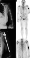
The case of a 66-year-old woman, with complex health issues, who has suffered a pathological fracture in the humerus due to metastasis. (A) Highlights the osteolytic metastasis in the humerus. (B) Scintigraphic image showing systemic spread. (C) Postoperative image showing an intramedullary rod used for osteosynthesis, allowing immediate arm movement.
Figure 6
Figure 6
The case of a 78-year-old patient who has a confirmed diagnosis of renal carcinoma and is also experiencing widespread metastatic disease, as confirmed by whole-body bone scintigraphy (A). The patient received treatment at our oncology clinic, where a team of specialists closely observed the progression of the disease. Due to the patient’s high likelihood of experiencing fractures caused by disease, especially in the right femur where metastatic involvement was found (C), and in multiple vertebral bodies, it was decided to perform palliative surgical procedures with the goal of enhancing the patient's quality of life and reducing discomfort. In order to mitigate the imminent danger of a femur fracture, a procedure called comfort osteosynthesis was conducted using gamma nails (D). This technique was selected due to its minimally invasive characteristics and its ability to effectively stabilize metastatic lesions. Furthermore, the stabilization of the vertebral column was accomplished by means of segmentation (as shown in E), which additionally aids in pain reduction and provides structural support. The patient's metastatic burden, as revealed in CT imaging (B), was too extensive to allow for curative resection and reconstruction. The extensive tumor infiltration in the femoral region emphasizes the need for a cautious surgical strategy. Due to the patient's advanced age and the existence of multiple comorbidities, which made her especially susceptible, it was determined that a forceful surgical procedure was not feasible. The therapeutic approach was customized to prioritize the management of symptoms and the preservation of functional independence for as long as feasible, recognizing the inherent complexities in treating metastatic renal carcinoma with substantial skeletal involvement.

Similar articles

Cited by

References

    1. Alzubaidi AN Sekoulopoulos S Pham J Walter V Fuletra JG & Raman JD. Incidence and distribution of new renal cell carcinoma cases: 27-year trends from a statewide cancer registry. Journal of Kidney Cancer and VHL 202297–12. (10.15586/jkcvhl.v9i2.219) - DOI - PMC - PubMed
    1. Bray F Ferlay J Soerjomataram I Siegel RL Torre LA & Jemal A. Global cancer statistics 2018: GLOBOCAN estimates of incidence and mortality worldwide for 36 cancers in 185 countries. CA: A Cancer Journal for Clinicians 201868394–424. (10.3322/caac.21492) - DOI - PubMed
    1. Petard JJ, Leray E, Rioux-Leclercq N, Cindolo L, Ficarra V, Zisman A, De La Taille A, Tostain J, Artibani W, Abbou CC, et al.Prognostic value of histologic subtypes in renal cell carcinoma: a multicenter experience. Journal of Clinical Oncology 2005232763–2771. (10.1200/JCO.2005.07.055) - DOI - PubMed
    1. Choueiri TK & Motzer RJ. Systemic therapy for metastatic renal-cell carcinoma. New England Journal of Medicine 2017376354–366. (10.1056/NEJMra1601333) - DOI - PubMed
    1. Cancer Genome Atlas Research Network, Linehan WM, Spellman PT, Ricketts CJ, Creighton CJ, Fei SS, Davis C, Wheeler DA, Murray BA, Schmidt L, et al.Comprehensive molecular characterization of papillary renal-cell carcinoma. New England Journal of Medicine 2016374135–145. (10.1056/NEJMoa1505917) - DOI - PMC - PubMed

LinkOut - more resources