. 2024 Nov 1;14(11):2224-2242.
doi: 10.1158/2159-8290.CD-24-0519.

Clinical Validation of a Cell-Free DNA Fragmentome Assay for Augmentation of Lung Cancer Early Detection

Affiliations

Clinical Validation of a Cell-Free DNA Fragmentome Assay for Augmentation of Lung Cancer Early Detection

Peter J Mazzone et al. Cancer Discov. .

Abstract

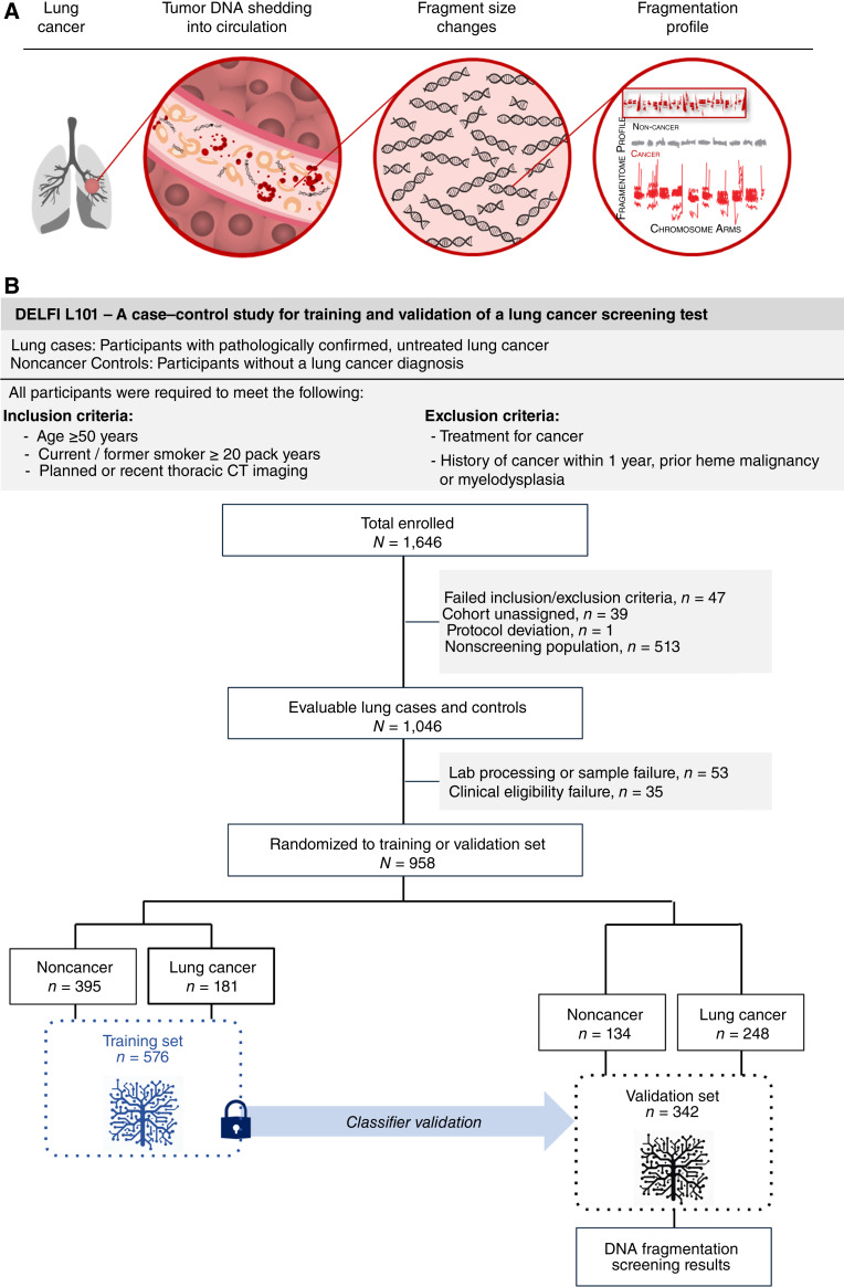
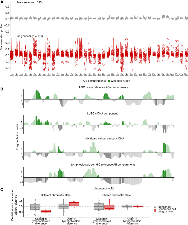
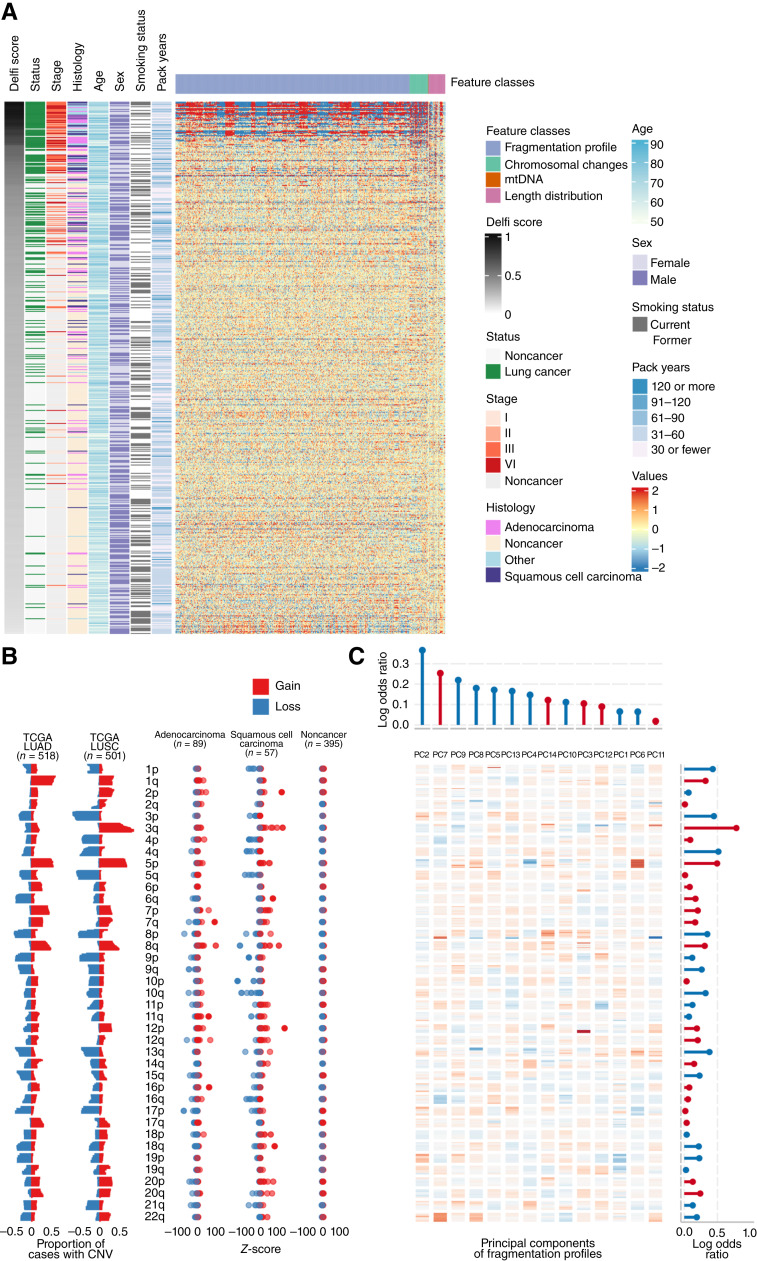
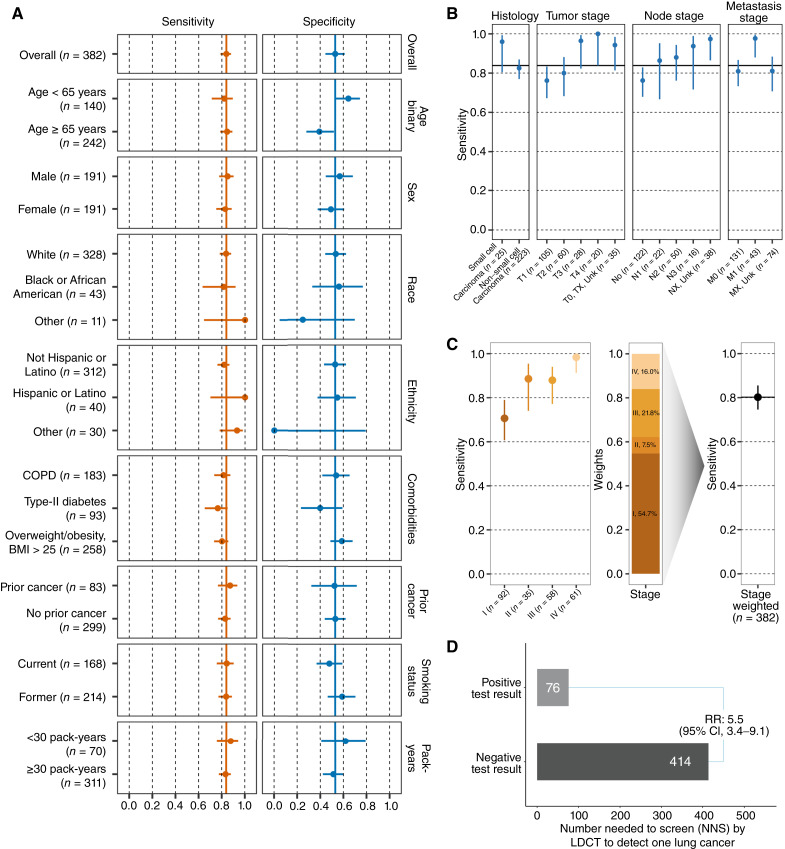
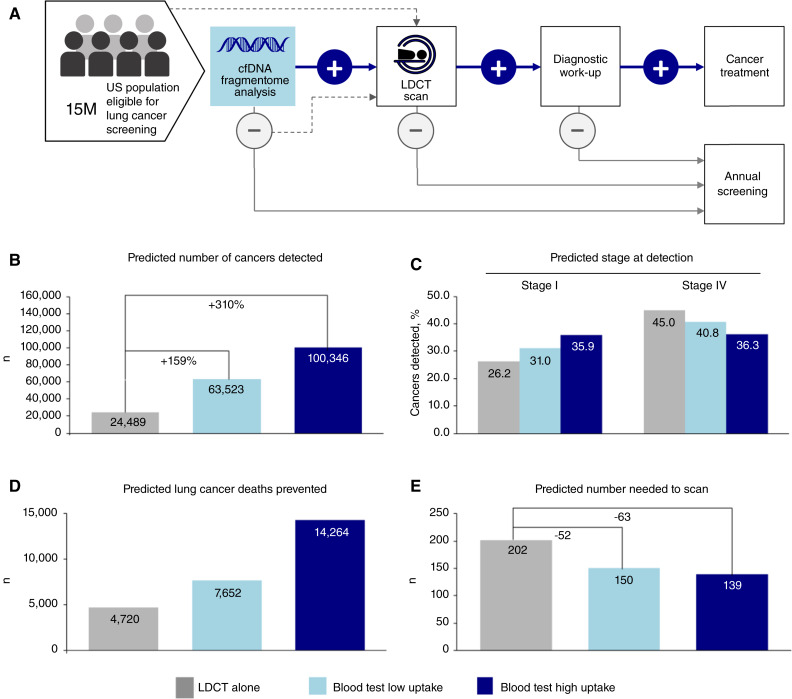
Lung cancer screening via annual low-dose computed tomography has poor adoption. We conducted a prospective case-control study among 958 individuals eligible for lung cancer screening to develop a blood-based lung cancer detection test that when positive is followed by a low-dose computed tomography. Changes in genome-wide cell-free DNA fragmentation profiles (fragmentomes) in peripheral blood reflected genomic and chromatin characteristics of lung cancer. We applied machine learning to fragmentome features to identify individuals who were more or less likely to have lung cancer. We trained the classifier using 576 cases and controls from study samples and validated it in a held-out group of 382 cases and controls. The validation demonstrated high sensitivity for lung cancer and consistency across demographic groups and comorbid conditions. Applying test performance to the screening eligible population in a 5-year model with modest utilization assumptions suggested the potential to prevent thousands of lung cancer deaths. Significance: Lung cancer screening has poor adoption. Our study describes the development and validation of a novel blood-based lung cancer screening test utilizing a highly affordable, low-coverage genome-wide sequencing platform to analyze cell-free DNA fragmentation patterns. The test could improve lung cancer screening rates leading to substantial public health benefits. See related commentary by Haber and Skates, p. 2025.


Conflict of interest statement

J. Carey, J. Catallini, C. Cisar, L. Cotton, N. Dracopoli, A. Gershman, S. Kotagiri, A. Leal, K. Lumbard, L.K. Millberg, J. Nawrocki, C. Portwood, A. Rangnekar, A. Ryan, C.A. Schonewolf, C.C. Sheridan, N. Trivedi, T. Wu, Y. Zong, and P. Bach report employment with DELFI Diagnostics and/or ownership of stock in DELFI Diagnostics. A. Leal reports being a co-founder of DELFI Diagnostics; serving as a consultant; and being an inventor on patent applications submitted by Johns Hopkins University. P. Mazzone reports research support at his institution from Adela, Biodesix, DELFI Diagnostics, Exact Sciences, Nucleix, PrognomiQ, Veracyte, Mandel Accelerator Award, Moore Foundation, and PCORI. J.D. Ortendahl was an employee of Partnership for Health Analytic Research, which was paid to develop the economic model in this article, while this research was underway. J.D. Ortendahl reports current employment with Stratevi, which is paid by pharmaceutical companies to conduct health economic research. M.S. Ahluwalia reports grant funding from Seagen; consulting for Bayer, Kiyatec, Insightec, GSK, Xoft, Nuvation, SDP Oncology, Apollomics, Prelude, Janssen, Voyager Therapeutics, ViewRay, Caris Life Sciences, Pyramid Biosciences, Varian Medical Systems, Cairn Therapeutics, AnHeart Therapeutics, TherAguix, Menarini Ricerche, Sumitomo Pharma Oncology, Autem Therapeutics, GT Medical Technologies, AlloVir, Equillium Bio, and VBI Vaccines; residing on the scientific advisory boards for Modifi Biosciences and Bugworks; and holding shares in MimiVax, CytoDyn, MedInnovate Advisors LLC, and TriSalus Life Sciences. L. Pike reports funding from DELFI Diagnostics to conduct this study at Memorial Sloan Kettering Cancer Center. R. Scharpf reports being a founder and consultant of DELFI Diagnostics and ownership of DELFI Diagnostics stock. Under a license agreement between DELFI Diagnostics and Johns Hopkins University, the university and R. Scharpf are entitled to royalty distributions related to technology described in this article. Additionally, the university owns equity in DELFI Diagnostics. He also serves as the head of Data Science. This arrangement has been reviewed and approved by Johns Hopkins University in accordance with its conflict of interest policies. J.C. Thompson reports a consulting role with AstraZeneca and funding from DELFI Diagnostics and Incyte. A. Vachani reports serving as an unpaid scientific advisor to DELFI Diagnostics; receiving fees as an advisor from Johnson & Johnson; and grants from PreCyte, Inc., Optellum Ltd., Median Technologies, and NCCN/AstraZeneca. L.V. Sequist reports funding from DELFI Diagnostics, AstraZeneca, and Novartis. K.-K. Wong reports a sponsored research agreement from DELFI Diagnostics. V. Velculescu reports being a founder of DELFI Diagnostics; serving on the board of directors; and ownership of DELFI Diagnostics stock, which is subject to certain restrictions under university policy. Additionally, Johns Hopkins University owns equity in DELFI Diagnostics. V. Velculescu divested his equity in Personal Genome Diagnostics (PGDx) to LabCorp in February 2022 and is an inventor on patent applications submitted by Johns Hopkins University related to cancer genomic analyses and cell-free DNA for cancer detection that have been licensed to one or more entities, including DELFI Diagnostics, LabCorp, Qiagen, Sysmex, Agios, Genzyme, Esoterix, Ventana, and ManaT Bio. Under the terms of these license agreements, the university and inventors are entitled to fees and royalty distributions. V. Velculescu is an advisor to Viron Therapeutics and Epitope. These arrangements have been reviewed and approved by Johns Hopkins University in accordance with its conflict-of-interest policies. No disclosures were reported by the other authors.

Figures

Figure 1.
Figure 1.
Overall approach to clinical validation of a cell-free DNA fragmentome assay for augmentation of lung cancer early detection. A, Illustration representing the DELFI approach for lung cancer through noninvasive assessment of cell-free DNA fragmentation profiles (ratio of short to long cfDNA fragments). Nucleosomal DNA with variable length of linker DNA is released by dying lung cancer cells into the circulation. Genome-wide mapping of the cfDNA fragments demonstrates more aberrant profiles with cancer cell cfDNA fragments compared with the cfDNA in noncancer individuals. B, The DNA Evaluation of Fragments for Early Interception–Lung Cancer Training Study, DELFI-L101 study was a prospective case–control study (NCT04825834, including two institutional supplementary protocols NCT00301119 and NCT01775072). The flow diagram illustrates the inclusion and exclusion of L101 participants based on clinical, sample, and assay eligibility criteria and the assignment of evaluable participants to the classifier training (n = 576) and clinical validation (n = 382) sets. Machine learning of genome-wide cfDNA fragmentation profiles from the training set was used to develop a locked classifier that was evaluated in the clinical validation set.
Figure 2.
Figure 2.
Genome-wide fragmentation profiles are altered in patients with cancer and reflect underlying chromatin structure. A, The fragmentation profile (ratio of short to long cfDNA fragments in 5 Mb bins) across the genome was evaluated in the classifier training plasma samples of lung cancer (n = 181) and noncancer individuals (n = 395). The noncancer individuals had similar fragmentation profiles, whereas patients with lung cancer exhibited significant variation. B, Comparison of cfDNA profiles with Hi-C A/B chromatin compartment reference data from lung cancer tissue or peripheral blood cells. Track 1 shows A/B compartments extracted from LUSC cancer tissue (48). Track 2 shows a median lung cancer component extracted from the LUSC plasma samples of 7 patients with lung cancer from the classifier training set with high tumor fraction by ichorCNA (49). The 7 LUSC cases with high ichorCNA have values of 0.051, 0.439, 0.230, 0.259 0.439, 0.167, and 0.057. Track 3 shows the median profile for 10 noncancer plasma samples from the training set. Track 4 shows A/B compartments for lymphoblast cells (48). These four tracks show chromosome 22 as an example, with darker shading indicating informative regions of the genome where the two reference tracks differ in domain (open/closed). C, 100-kb regions were selected using the reference LUSC and lymphoblast A/B tracks as having the same chromatin state or opposite chromatin state. Within these regions, the deviation of the fragmentation value from a noncancer cfDNA reference (n = 10) was plotted per region per individual (noncancer n = 20, LUSC n = 7). Values around 0 have little variation from the noncancer reference. Negative values indicate a region has a more open chromatin state than the reference and positive values indicate a region has a more closed chromatin state than the reference. These data suggest that although cfDNA profiles of healthy individuals reflect the chromatin structure of blood cells, those of patients with lung cancer represent a mixture of cfDNA patterns of chromatin compartments from lung cancer as well as blood cells.
Figure 3.
Figure 3.
High-dimensional fragmentation features reflect lung cancer biology and are incorporated in the machine learning classifier. A, Heatmap representation of the deviation of cfDNA fragmentation features across the genome for the classifier training set with lung cancer or noncancer individuals compared with the mean of classifier training noncancer individuals. Each row represents a sample, whereas columns show individual genomic features. The cross-validated DELFI score and clinical characteristics are indicated to the left of the fragmentation deviation heatmap. B, Left, TCGA-derived observations of chromosomal arm gains (red) and losses (blue) in lung adenocarcinoma (LUAD; n = 518) and squamous cell cancer tissues (LUSC; n = 501). Right, the observed chromosome arm gains (red) and losses (blue) in the classifier training individuals separated by histology. C, A heatmap representation of the principal component eigenvectors of the fragmentation profile features. Regression coefficients from the final classifier indicating how the principal components of the fragmentation profiles and z-scores of the chromosomal arms were combined are provided in the top and right margins of the heatmap, respectively. Positive values for the coefficients are represented in red, whereas negative values are represented in blue. Agreement across copy number chromosomal gains and losses in TCGA lung cancers, observed z-scores in the cfDNA of patients with lung cancer, and chromosome arm model coefficients reflect biologic consistency between chromosomal changes in lung cancer, cfDNA fragmentation profiles, and classifier features.
Figure 4.
Figure 4.
Performance of blood-based lung cancer screening test. A, Sensitivity and specificity of the test in the clinical validation set (N = 382) overall and by clinical subgroup. Point estimates are reported with 95% Wilson confidence intervals. Overall sensitivity and specificity denoted by solid vertical lines. B, Sensitivity of the test in the lung cancer cases in the clinical validation set (N = 248) evaluated across cancer histology, and T, N, and M categories. Point estimates are reported with 95% Wilson confidence intervals. Overall sensitivity of 84% denoted by the solid horizontal line. C, Left, sensitivity of the test in the lung cancer cases in the clinical validation set (N = 246) by cancer group stage. Middle, bar plot showing the stage distribution of lung cancer as observed in populations undergoing lung cancer screening with LDCT (based on NLST study) that are used to weigh observed stage-specific sensitivities. Right, lung cancer screening relevant stage-weighted sensitivity in clinical validation set. D, Comparison of the NNS with LDCT conditioned on test positive or negative result when applied in the lung cancer screening eligible population. Test performance showed consistency across clinical subgroups and expected increased performance with increasing burden of disease (tumor (T), node (N), metastasis (M) and group staging). After weighting, the stage distribution to reflect a screening population, test performance remained high and demonstrated the ability to reliably identify those individuals more likely to have lung cancer detected on LDCT.
Figure 5.
Figure 5.
Population health benefits of a blood-based test in lung cancer screening. A, Care pathway reflecting the recommended standard of care for lung cancer screening with LDCT that is received by 6%–10% of eligible individuals annually, as well as potential pathway employing initial blood-based test and follow-on events. B, The predicted number of cancers detected by screening scenario: LDCT alone (“base case”); LDCT + low test uptake; LDCT + high test uptake. C, Predicted cancers diagnosed at stage I versus Stage IV by screening scenario: LDCT alone (“base case”); LDCT + low test uptake; LDCT + high test uptake. D, Predicted decrease in lung cancer deaths represented by screening scenario: LDCT alone (“base case”); LDCT + low test uptake; LDCT + high test uptake. E, Simulated comparison of the predicted number needed to scan with LDCT to detect one lung cancer: LDCT alone (“base case”); LDCT + low test uptake; LDCT + high test uptake. Population-level modeling demonstrates significant health benefits when a blood-based test is available as an alternative for lung cancer screening.

References

    1. American lung association . [cited 2024 May 31]. Available from: https://www.lung.org/getmedia/647c433b-4cbc-4be6-9312-2fa9a449d489/solc-....
    1. Siegel RL, Miller KD, Wagle NS, Jemal A. Cancer statistics, 2023. CA Cancer J Clin 2023;73:17–48. - PubMed
    1. Sung H, Ferlay J, Siegel RL, Laversanne M, Soerjomataram I, Jemal A, et al. . Global cancer statistics 2020: GLOBOCAN estimates of incidence and mortality Worldwide for 36 cancers in 185 countries. CA Cancer J Clin 2021;71:209–49. - PubMed
    1. National Lung Screening Trial Research Team; Aberle DR, Adams AM, Berg CD, Black WC, Clapp JD, Fagerstrom RM, et al. . Reduced lung-cancer mortality with low-dose computed tomographic screening. N Engl J Med 2011;365:395–409. - PMC - PubMed
    1. de Koning HJ, van der Aalst CM, de Jong PA, Scholten ET, Nackaerts K, Heuvelmans MA, et al. . Reduced lung-cancer mortality with volume CT screening in a randomized trial. N Engl J Med 2020;382:503–13. - PubMed

Publication types