. 2024 Jul 23;98(7):e0076924.
doi: 10.1128/jvi.00769-24. Epub 2024 Jun 3.

A full-length glycoprotein mRNA vaccine confers complete protection against severe fever with thrombocytopenia syndrome virus, with broad-spectrum protective effects against bandaviruses

Affiliations

A full-length glycoprotein mRNA vaccine confers complete protection against severe fever with thrombocytopenia syndrome virus, with broad-spectrum protective effects against bandaviruses

Jia Lu et al. J Virol. .

Abstract

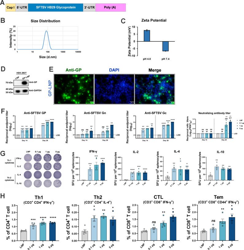
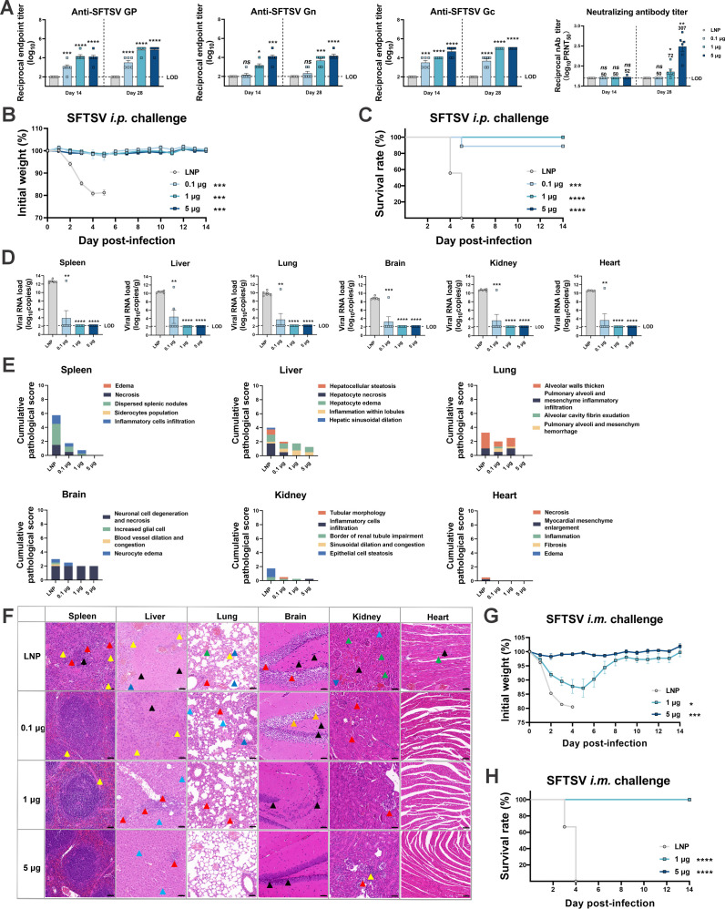
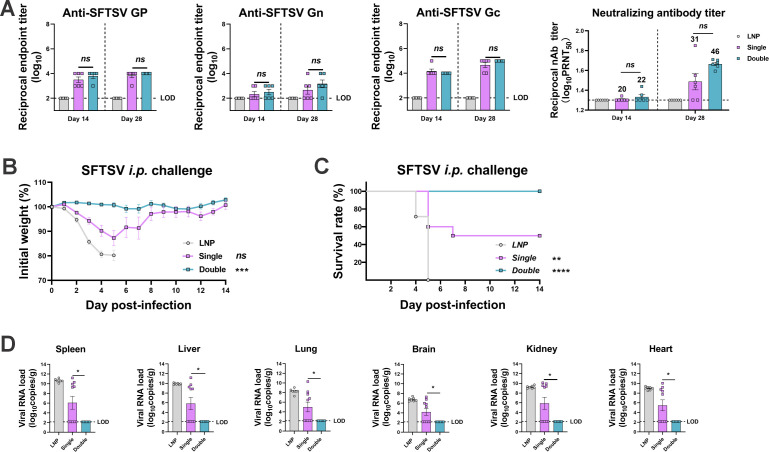
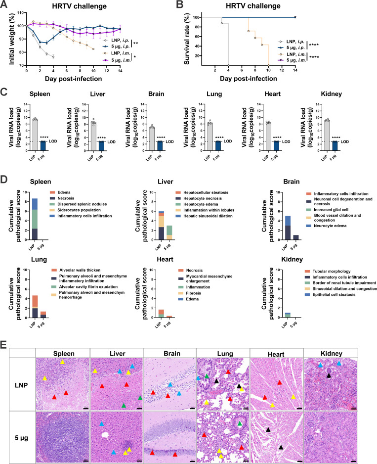
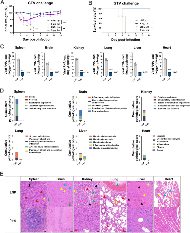
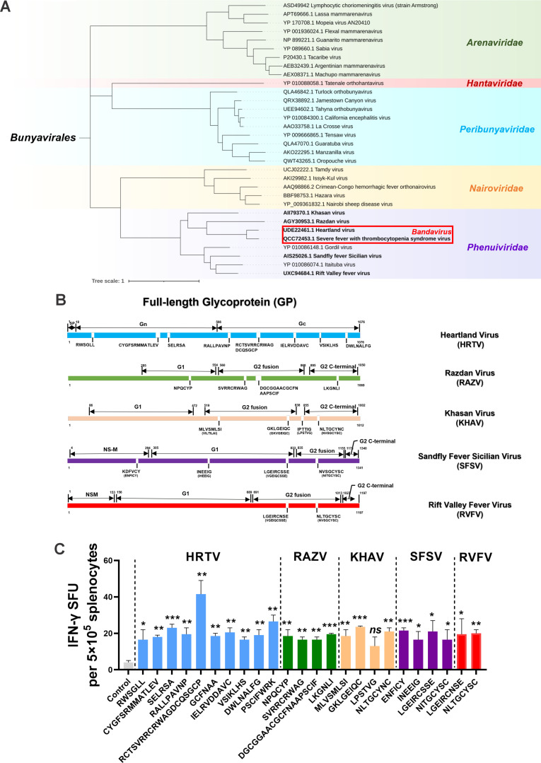
Highly pathogenic viruses from family Phenuiviridae, which are mainly transmitted by arthropods, have intermittently sparked epidemics worldwide. In particular, tick-borne bandaviruses, such as severe fever with thrombocytopenia syndrome virus (SFTSV), continue to spread in mountainous areas, resulting in an average mortality rate as high as 10.5%, highlighting the urgency and importance of vaccine development. Here, an mRNA vaccine developed based on the full-length SFTSV glycoprotein, containing both the receptor-binding domain and the fusion domain, was shown to confer complete protection against SFTSV at a very low dose by triggering a type 1 helper T cell-biased cellular immune response in rodents. Moreover, the vaccine candidate elicited long-term immunity and protection against SFTSV for at least 5 months. Notably, it provided complete cross-protection against other bandaviruses, such as the Heartland virus and Guertu virus, in lethal challenge models. Further research revealed that the conserved epitopes among bandaviruses within the full-length SFTSV glycoprotein may facilitate broad-spectrum protection mediated by the cellular immune response. Collectively, these findings demonstrate that the full-length SFTSV glycoprotein mRNA vaccine is a promising vaccine candidate for SFTSV and other bandaviruses, and provide guidance for the development of broad-spectrum vaccines from conserved antigens and epitopes.

Importance: Tick-borne bandaviruses, such as SFTSV and Heartland virus, sporadically trigger outbreaks in addition to influenza viruses and coronaviruses, yet there are no specific vaccines or therapeutics against them. mRNA vaccine technology has advantages in terms of enabling in situ expression and triggering cellular immunity, thus offering new solutions for vaccine development against intractable viruses, such as bandaviruses. In this study, we developed a novel vaccine candidate for SFTSV by employing mRNA vaccination technology and using a full-length glycoprotein as an antigen target. This candidate vaccine confers complete and durable protection against SFTSV at a notably low dose while also providing cross-protection against Heartland virus and Guertu virus. This study highlights the prospective value of full-length SFTSV-glycoprotein-based mRNA vaccines and suggests a potential strategy for broad-spectrum bandavirus vaccines.

Keywords: broad-spectrum protection; cellular immunology; conserved epitope; glycoprotein; severe fever with thrombocytopenia syndrome virus.


Conflict of interest statement

The authors declare no conflict of interest.

Figures

Fig 1
Fig 1
Immunogenicity evaluation of the GP mRNA vaccine in BALB/c mice. (A) Schematic diagram of the full-length SFTSV GP mRNA vaccine. (B and C) Size distribution and zeta potential of the mRNA-LNPs. (D and E) The expression of SFTSV GP mRNA in HEK-293T cells was detected by Western blotting with glyceraldehyde-phosphate dehydrogenase used as the control (also shown in Fig. S2), and visualized in the cytoplasm using immunofluorescence with nuclei stained with 4′,6-diamidino-2-phenylindole. (F–H) Female BALB/c mice were allocated into the following four groups with five per group: empty LNPs, 0.1, 1.0, or 5.0 μg of GP-LNPs. Vaccinations followed a standard prime-boost regimen with a 3-week interval. The levels of binding antibodies against SFTSV GP, Gn, and Gc were determined by enzyme-linked immunosorbent assay with a starting dilution of 1:100 (LOD). Neutralizing antibodies were ascertained using a plaque reduction neutralization test, with geometric mean titers indicated above the corresponding bars (LOD of 1:30) (F). The cellular immune response was detected by either ELISpot or flow cytometry. Representative images (left panel) and spots of IFN-γ, IL-2, IL-4, and IL-10 (right panel) are shown (G), and the proportions of Th1 cells, Th2 cells, CTLs, and Tem cells were analyzed via ICS assay (H). *P < 0.05, **P < 0.01, ***P < 0.001, ****P < 0.0001. CTL, cytotoxic T cell; DAPI, 4′,6-diamidino-2-phenylindole; ELISpot, enzyme-linked immunospot assay; ICS, intracellular cytokine staining assay; GP, glycoprotein; IFN-γ, interferon gamma; IL, interleukin; LNP, lipid nanoparticle; LOD, limit of detection; ns, not significant; SFTSV, severe fever with thrombocytopenia syndrome virus; Tem, effector memory T; Th1, type 1 helper T; Th2, type 2 helper T; UTR, untranslated region.
Fig 2
Fig 2
Dose-dependent efficacy of the GP mRNA vaccine in A129 mice. A129 mice received vaccinations with empty LNPs, 0.1, 1.0, or 5.0 µg of GP-LNPs with a regular two-shot regimen, followed by i.p. challenge with 100 TCID50 of SFTSV 10 days after the booster vaccination. (A) Binding and neutralizing antibodies were detected using ELISAs for GP, Gn, or Gc and PRNT, respectively. The LODs for the ELISA and PRNT were 1:100 and 1:50, respectively. (B and C) Body weight and survival were monitored over 14 days post-challenge. (D) The viral loads in the spleen, liver, lung, brain, kidney, and heart were detected by reverse transcription-quantitative polymerase chain reaction based on the standard curve method with a pair of primers targeting the NP gene. The LOD for qPCR was 138 copies per reaction, and converted copies are presented. (E) Cumulative pathological scores for different tissues from each mouse were calculated based on pathological indicators; the details of the scoring criteria are provided in the supplemental materials. (F) Representative images from each group were analyzed by hematoxylin and eosin staining. The colored arrows in the images marking obvious pathological features and their detailed meanings are listed as follows: (i) lungs: red indicates alveolar epithelial cells proliferated, alveolar atrophy, and alveolar walls thickened; green denotes congested and dilated capillaries within the alveolar wall; blue indicates protein mucus present in the bronchus lumen; yellow denotes inflammatory cell infiltration; (ii) liver: red indicates hepatocellular necrosis, nuclear fragmentation, and karyopyknosis; green denotes hepatocyte steatosis; black indicates hepatic sinusoidal dilatation; blue denotes hepatocyte edema; yellow indicates inflammatory cell infiltration; (iii) kidneys: red denotes glomerular atrophy and decreased number of mesangial cells; green indicates glomeruli were lobulated or had interstitial hemorrhages; black denotes degeneration of renal tubular epithelial cells; blue denotes protein mucus present in the kidney tubules; yellow denotes inflammatory cell infiltration; (iv) spleen: red denotes necrosis, nuclear fragmentation, and karyopyknosis of lymphocytes; black indicates scattered splenic nodules and blurred boundary between the red pulp; yellow denotes inflammatory cell infiltration; (v) brain: red indicates neuronal cell degeneration, karyopyknosis, and basophilia were enhanced; blue denotes neuronal cell edema; yellow denotes increased number of glial cells; green indicates congested and dilated blood vessels; (vi) heart: black denotes myocardial fibrosis and rupture; blue: indicates disordered arrangement of myocardial fibers and connective tissue hyperplasia; yellow denotes inflammatory cell infiltration. (G and H) A129 mice received vaccinations with empty LNPs, 1 or 5 µg of GP-LNPs with a regular two-shot regimen, followed by i.m. challenge with 1 × 10⁵ TCID50 of SFTSV 10 days after the booster vaccination. Body weight and survival were monitored for 14 days. ELISA, enzyme-linked immunosorbent assay; i.m., intramuscular; i.p., intraperitoneal; PRNT, plaque reduction neutralization test; qPCR, quantitative PCR; TCID50, 50% tissue culture infectious dose.
Fig 3
Fig 3
Efficacy of the GP mRNA vaccine in single-dose and double-dose regimens. A129 mice received vaccinations with empty LNPs or 1 µg of GP-LNPs in a single- or double-dose regimen, followed by i.p. challenge with 100 TCID50 of SFTSV. (A) Binding antibodies against SFTSV GP, Gn, and Gc were detected by ELISA (LOD, 1:100). Neutralizing antibodies were detected by PRNT, with the GMTs indicated above the corresponding bars (LOD, 1:20). (B and C) Curves of body weight and survival, which were monitored for 14 days post-challenge. (D) Viral loads in the spleen, liver, lung, brain, kidney, and heart were detected by reverse transcription-quantitative PCR (LOD, 138 copies). *P < 0.05, **P < 0.01, ***P < 0.001, ****P < 0.0001.
Fig 4
Fig 4
Long-term immune response and protection provided by the GP mRNA vaccine. A129 mice were vaccinated with empty LNPs or 5 µg of GP-LNPs following a two-shot regimen and were then were challenged with 100 TCID50 of SFTSV (i.p.) 21 weeks later. (A) The titers of binding antibodies specific to SFTSV GP, Gn, or Gc lasting for 20 weeks were detected by ELISA (LOD, 1:100). (B) The neutralizing antibody titers within 20 weeks were detected by PRNT (LOD, 1:30). (C and D) The body weight and survival rate were monitored for 14 days post-challenge. **P < 0.01, ***P < 0.001, ****P < 0.0001.
Fig 5
Fig 5
Cross-protection offered by the GP mRNA vaccine against Heartland virus. A129 or AG129 mice were vaccinated with empty LNPs or 5 µg of GP-LNPs in a two-shot regimen and were subsequently challenged with HRTV (A129, i.p., 1 × 10⁷ TCID50; AG129, i.m., 1 × 10⁵ TCID50). (A and B) Body weight and survival were monitored for 14 days post-challenge. (C) viral loads in the tissues after i.p. challenge were quantified using reverse transcription-quantitative PCR, as described above (LOD, 629 copies). (D and E) Cumulative pathological scores and representative images from each tissue are presented. A detailed description of the pathological analysis is listed in the supplemental materials, and annotations for the colored arrows can be referred to in the Fig. 2 legend. *P < 0.05, **P < 0.01, ****P < 0.0001. HRTV, Heartland virus.
Fig 6
Fig 6
Cross-protection of the GP mRNA vaccine against Guertu virus. A129 mice were vaccinated with empty LNPs or 5 µg of GP-LNPs using a two-shot regimen and were subsequently challenged with GTV (i.p., 100 TCID50; i.m., 1 × 10⁵ TCID50). (A and B) Body weight and survival were monitored for 14 days post-challenge. (C) Viral loads in tissues after i.p. challenge were quantified using reverse transcription-quantitative PCR, as described above (LOD, 232 copies). (D and E) Cumulative pathological scores and representative images from each tissue are presented. A detailed description of the pathological analysis is listed in the supplemental materials, and annotations for the colored arrows can be referred to in the Fig. 2 legend. **P < 0.01, ***P < 0.001, ****P < 0.0001. GTV, Guertu virus.
Fig 7
Fig 7
Identification of the potential protective epitopes from SFTSV GP. (A) Phylogenetic tree of the pathogens in the order Bunyavirales and pathogens from the genus Bandavirus, family Phenuiviridae, is highlighted. (B) Mapping of conserved and similar peptides from the SFTSV GP protein sequences among pathogenic Phenuiviridae pathogens. (C) IFN-γ secretion upon restimulation with each peptide (50 µg/mL) was detected by ELISpot, followed by statistical analyses comparing with the control. *P < 0.05, **P < 0.01, ***P < 0.001.

Similar articles

Cited by

References

    1. Shi J, Hu Z, Deng F, Shen S. 2018. Tick-borne viruses. Virol Sin 33:21–43. doi:10.1007/s12250-018-0019-0 - DOI - PMC - PubMed
    1. Huang Y-J, Higgs S, Vanlandingham DL. 2019. Emergence and re-emergence of mosquito-borne arboviruses. Curr Opin Virol 34:104–109. doi:10.1016/j.coviro.2019.01.001 - DOI - PubMed
    1. Ayhan N, Charrel RN. 2017. Of phlebotomines (sandflies) and viruses: a comprehensive perspective on a complex situation. Curr Opin Insect Sci 22:117–124. doi:10.1016/j.cois.2017.05.019 - DOI - PubMed
    1. Zhou H, Xu L, Shi W. 2023. The human-infection potential of emerging tick-borne viruses is a global public health concern. Nat Rev Microbiol 21:215–217. doi:10.1038/s41579-022-00845-3 - DOI - PubMed
    1. Mansfield KL, Jizhou L, Phipps LP, Johnson N. 2017. Emerging tick-borne viruses in the twenty-first century. Front Cell Infect Microbiol 7:298. doi:10.3389/fcimb.2017.00298 - DOI - PMC - PubMed

MeSH terms

LinkOut - more resources