Review
. 2024 Jun 13;67(11):8502-8518.
doi: 10.1021/acs.jmedchem.4c00231. Epub 2024 Jun 3.

VHH Nanobody Versatility against Pentameric Ligand-Gated Ion Channels

Affiliations
Review

VHH Nanobody Versatility against Pentameric Ligand-Gated Ion Channels

Dorota Nemecz et al. J Med Chem. .

Erratum in

Abstract

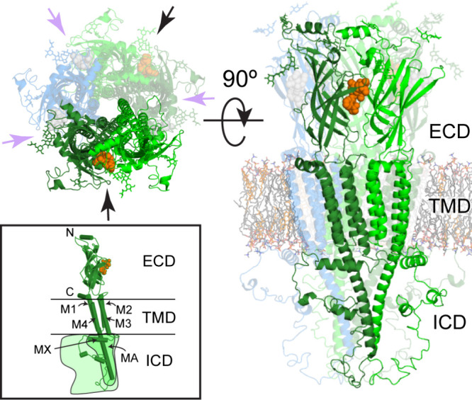
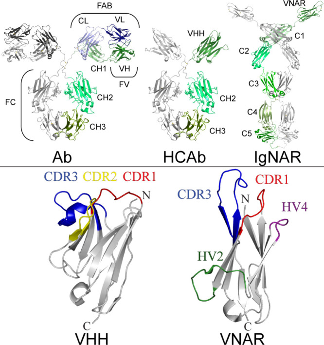
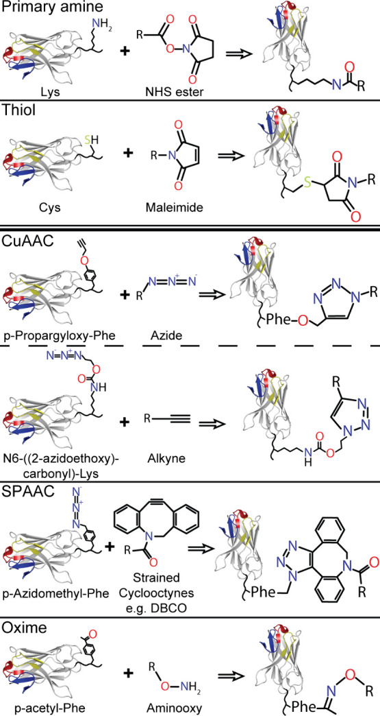
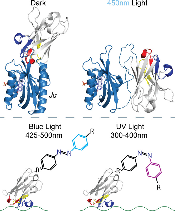
Pentameric ligand-gated ion channels provide rapid chemical-electrical signal transmission between cells in the central and peripheral nervous system. Their dysfunction is associated with many nervous system disorders. They are composed of five identical (homomeric receptors) or homologous (heteromeric receptors) subunits. VHH nanobodies, or single-chain antibodies, are the variable domain, VHH, of antibodies that are composed of the heavy chain only from camelids. Their unique structure results in many specific biochemical and biophysical properties that make them an excellent alternative to conventional antibodies. This Perspective explores the published VHH nanobodies which have been isolated against pentameric ligand-gated ion channel subfamilies. It outlines the genetic and chemical modifications available to alter nanobody function. An assessment of the available functional and structural data indicate that it is feasible to create therapeutic agents and impart, through their modification, a given desired modulatory effect of its target receptor for current stoichiometric-specific VHH nanobodies.


Conflict of interest statement

The authors declare no competing financial interest.

Figures

Figure 1
Figure 1
PLGIC structure. Top down (left) and side (right) view of a cartoon representation of a homomeric α7-nAChR (PDBs 7koo merged with 7rpm). Subunits are colored for a typical heteromeric receptor, where the dark-green (principal) and green (complementary) subunits compose an orthosteric binding site (black arrows and orange spheres [methyllycaconitine from PDB 3sh1]). The complementary (green) subunits in heteromeric systems need not be the same subunit. Purple arrows indicate other subunit interfaces that may act as an allosteric or different orthosteric site depending on what type of subunit occupies the blue-colored subunit. Helices are labeled in the cartoon inset depicting a single subunit, where those in the ICD are only found in cationic receptors. The two schematic outlines also depict how the size of the ICD may vary.
Figure 2
Figure 2
Structural comparison of antibody types and nanobodies. Top: Structures of an IgG2 class antibody (Ab, modified PDB 1igt), camelid heavy chain only antibody (HCAb, VHH from PDB 4pir and FC from PDB 1igt), and a shark immunoglobulin novel antigen receptor (IgNAR, using PDBs 4q97, 4q9b, 4q9c, 2mkl, and 2ywz). Each structure has a single heavy chain colored in shades of green with the respective domains labeled, whereas the Ab also has the respective light chain colored (blues). Bottom: Comparison between representative VHH-Nb (from PDB 6i53) and VNAR (PDB 2ywz) structures, with N- and C-terminals indicated, and the complementarity determining regions (CDRs), along with the hypervariable regions (HVs), of VNAR color coded and labeled.
Figure 3
Figure 3
Alignment of all available pLGIC Nb sequences. For clarity, the signal peptide and C-terminal tags have been removed from all sequences. The consensus sequence represents all residues with >50% identity between the sequences with X for those residues, which are more variable. Almost all of these fall within the CDRs 1, 2, and 3, which are indicated with the red, yellow, and blue boxes, respectively. Gaps in the alignment are highlighted in gray for clarity, and residues noted in the text with direct (yellow), backbone (cyan), and mixed (half-cyan/half-yellow) interactions are also highlighted. Sequences, listed in the receptor order found in the text (with 5-HT3A and AB divided), have the published subunit selectivity denoted between the receptor and VHH name. PAM Nbs for each receptor are listed first, then silent or NAM Nbs in order of potency. Nb38 sequence derived from PDB 6i53 and Nb25 from PDB 5ojm. All 5-HT3R VHH sequences are from ref (38). Both nAChR VHH sequences are from ref (39). ELIC PAM VHH is from PDB 6ssi, ELIC NAM VHH is from PDB 6ssp, and finally ELIC Nb72 is from PDB 6hjy.
Figure 4
Figure 4
VHH binding surface on GABAARs. Side view, cartoon representation, of two subunits in a given GABAAR subunit-interface zoomed in on the ECD, with the bound VHH name above the structure and subunits labeled to the side. Color-coded surface representation of residues within 4 Å of the bound VHH, color-coded as follows: white for non-CDR proximity; red, CDR1; yellow, CDR2; blue, CDR3; and overlapping residue interactions as orange, CDR1 and 2; purple, CDR1 and 3; green, CDR2 and 3; and brown, CDR1 and 2 and 3. PDBs: 6i53 and 7qnb.
Figure 5
Figure 5
GABAAR VHH pharmacophore interactions. Detailed interactions of each CDR from Mb38 with the GABAAR α1-β3 subunit interface (top) and from Mb25 with the GABAAR β3−β3 subunit interface (bottom) depicted in Figure 4. CDR2 is the principal component of binding for Mb38, and CDR3 for Mb25. Each panel is labeled with its respective CDR and color coded as in Figure 4, with the exception of oxygen, nitrogen, and sulfur atoms, which are colored red, blue, and yellow, respectively. Residues within 4 Å of a CDR have their surface shown and are labeled. Residue labels in bold indicate a direct interaction with the CDR. Polar interactions are shown with black dashed lines with distances indicated. CDR residues are labeled with a black background and colored and represented as sticks according to interaction type (yellow/side chain and cyan/backbone). Acceptor, red; donor, blue; and hydrophobic, yellow; van der Waalls interacting atoms from the subunit residues are depicted as color-coded dots.
Figure 6
Figure 6
VHH binding with the 5-HT3AR. (A) VHH binding surface on the 5-HT3AR. Side view, cartoon representation of two subunits involved in the subunit–interface binding, zoomed in on the ECD, with color-coded surface representation of residues within 4 Å of bound VHH15S (following the same color code as Figure 4). PDB 4pir. (B) VHH15s vs VHH7. Interacting residues of VHH15S from (A) are shown in stick representation (removed in inset). Line (VHH15S) and stick (VHH7) representations of mutated residues are shown and labeled. Inset: Slightly rotated, for clarity, zoomed representation of I203 and the end of the β9–10 loop from the principal A subunit, with residues’ side chains within 4 Å of I203 (R76, I51, I31, and Y24) and the A104I mutation (F29, V2, and Y113) also labeled and shown in line representation.
Figure 7
Figure 7
VHH15S pharmacophore interactions. Detailed interactions of each CDR from VHH15S with the 5-HT3AR interface depicted in Figure 6. CDR1 and 2 envelop the β9–10 loop as the principal component of binding. Each panel, labeled by its respective CDR, is depicted, color coded, and labeled as in Figure 5.
Figure 8
Figure 8
VHH binding surface on the α7-nAChR. Top down view of α7-nAChR-ECD structures with bound VHH name above the structures and color-coded surface representation of residues within 4 Å of this VHH following the same color code as Figure 4. Five VHHs are bound to the pentamer. For clarity a single VHH binding site is denoted for each below the fully bound representation. PDBs (left/right): 8ce4/8c9x.
Figure 9
Figure 9
VHHE3 and VHHC4 pharmacophore interactions. Detailed interactions of each CDR from VHHE3 (top) and VHHC4 (bottom) with the α7-nAChR interface depicted in Figure 8. CDR3 makes the principal component of binding to the apical main immunogenic region, whereas CDR2 contains the functionally distinguishing A56R variation. Each panel, labeled by its respective CDR, is depicted, color coded, and labeled as in Figure 5, with the addition that aromatic side chain interactions are shown with a green dashed line and the distance indicated, and aromatic interacting atoms from the subunit residues are depicted as green dots.
Figure 10
Figure 10
VHH binding surface on ELIC. Color-coded surface representation of residues within 4 Å of the bound VHH follows the same color code as Figure 4. Top row: Side view, cartoon representation, of two subunits involved in the subunit-interface binding, zoomed in on the ECD, with the bound VHH name listed above the structure. PDBs (left/right): 6ssi/6hjy. Bottom: Top-down view of ELIC. Only one VHH is bound per pentamer. PDB 6ssp.
Figure 11
Figure 11
Nb–Nb fusions. Examples of Nb–Nb fusion types using VHHs (top row) that bind to different epitopes (yellow symbols). Example of two types of bivalent molecules (middle row), where identical VHHs are fused together. Left: Binding to a unique, star, epitope on a receptor. Higher affinity is achieved through association of the linked VHH, which was already in close proximity, when the bound VHH dissociates (black arrows) and vice versa (gray arrows, faded VHH). Right: Top-down view. Binding to an, oval, epitope that occurs five times on a homopentameric receptor. An increased affinity is achieved from the rapid reassociation (gray arrow, faded VHH) of a single dissociated VHH (black arrow), during which the other linked VHH stays associated. Biparatopic VHH fusion (bottom row, left) is similar to this but the fusion consists of two unique VHHs that bind to different epitopes on the same receptor. Right: Bispecific VHH fusion consists of two unique VHHs binding to two unique proteins (pLGIC and albumin [white]).
Figure 12
Figure 12
MegaBody38 with c7HopQ. CDRs 1, 2, and 3 are color coded red, yellow, and blue, respectively on Nb38 (from PDB 6i53). β-sheets A and B (darker) on Nb38 are labeled, as well as linker-connected (and truncated) N- and C-termini of HopQ (brown, from PDB 6qd6).
Figure 13
Figure 13
Chemical conjugation methods of Nbs. Options for chemical conjugations include conjugation to naturally occurring functional groups (above double line), such as primary amine and thiol groups, or non-natural amino acid incorporation (below double line, with some of the common non-natural moieties represented). These incorporated amino acids are displayed on a C-terminal Nb linker, with the Nb CDRs color coded for orientation. The bio-orthogonal pair is listed and shown in the middle with the corresponding Nb conjugation product on the right after the arrow.
Figure 14
Figure 14
Opto-Nb constructions. Photoactivation may modify the Nb (model from PDB 8ce4 with CDRs 1, 2, and 3 colored red, yellow, and blue respectively) activity directly (top) or may modify a chemically attached photoreactive ligand (bottom). Top: Truncated A.s.-LOV2 (modified from PDB 2v0u, colored sky-blue with flavin mononucleotide, light-blue, displayed in stick representation) inserted between NbA74 and NbK75, shown in dark/inactivated (left) form or light/activated (right) form, where the Jα helix undocks and loses structure adding flexibility. Bottom: Azo-benzene moiety, chemically linked (R′) using any of the aforementioned chemical modifications in this Perspective with rigid linkers that may be used to allow for a ligand (R) to interact with a Nb target protein (represented by the green wavy line) after conformational change under UV light.

Similar articles

References

    1. Nemecz Á.; Prevost M. S.; Menny A.; Corringer P.-J. Emerging Molecular Mechanisms of Signal Transduction in Pentameric Ligand-Gated Ion Channels. Neuron 2016, 90 (3), 452–470. 10.1016/j.neuron.2016.03.032. - DOI - PubMed
    1. Hu H.; Ataka K.; Menny A.; Fourati Z.; Sauguet L.; Corringer P.-J.; Koehl P.; Heberle J.; Delarue M. Electrostatics, Proton Sensor, and Networks Governing the Gating Transition in GLIC, a Proton-Gated Pentameric Ion Channel. Proc. Natl. Acad. Sci. U. S. A. 2018, 115 (52), E12172–E12181. 10.1073/pnas.1813378116. - DOI - PMC - PubMed
    1. Changeux J.-P. The Nicotinic Acetylcholine Receptor: A Typical “Allosteric Machine. Philos. Trans. R. Soc. London B. Biol. Sci. 2018, 373 (1749), 20170174 10.1098/rstb.2017.0174. - DOI - PMC - PubMed
    1. Lara C. O.; Burgos C. F.; Moraga-Cid G.; Carrasco M. A.; Yévenes G. E. Pentameric Ligand-Gated Ion Channels as Pharmacological Targets Against Chronic Pain. Front. Pharmacol. 2020, 11, 167. 10.3389/fphar.2020.00167. - DOI - PMC - PubMed
    1. Rao R.; Shah S.; Bhattacharya D.; Toukam D. K.; Cáceres R.; Pomeranz Krummel D. A.; Sengupta S. Ligand-Gated Ion Channels as Targets for Treatment and Management of Cancers. Front. Physiol. 2022, 13, 839437 10.3389/fphys.2022.839437. - DOI - PMC - PubMed

Publication types

MeSH terms

Substances