

Accuracy and feasibility in building a personalized 3D printed femoral pseudoaneurysm model for endovascular training
- PMID: 38829913
- PMCID: PMC11146720
- DOI: 10.1371/journal.pone.0304506
Accuracy and feasibility in building a personalized 3D printed femoral pseudoaneurysm model for endovascular training
Abstract
Background: The use of three-dimensional(3D) printing is broadly across many medical specialties. It is an innovative, and rapidly growing technology to produce custom anatomical models and medical conditions models for medical teaching, surgical planning, and patient education. This study aimed to evaluate the accuracy and feasibility of 3D printing in creating a superficial femoral artery pseudoaneurysm model based on CT scans for endovascular training.
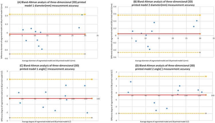
Methods: A case of a left superficial femoral artery pseudoaneurysm was selected, and the 3D model was created using DICOM files imported into Materialise Mimics 22.0 and Materialise 3-Matic software, then printed using vat polymerization technology. Two 3D-printed models were created, and a series of comparisons were conducted between the 3D segmented images from CT scans and these two 3D-printed models. Ten comparisons involving internal diameters and angles of the specific anatomical location were measured.
Results: The study found that the absolute mean difference in diameter between the 3D segmented images and the 3D printed models was 0.179±0.145 mm and 0.216±0.143mm, respectively, with no significant difference between the two sets of models. Additionally, the absolute mean difference in angle was 0.99±0.65° and 1.00±0.91°, respectively, and the absolute mean difference in angle between the two sets of data was not significant. Bland-Altman analysis confirmed a high correlation in dimension measurements between the 3D-printed models and segmented images. Furthermore, the accuracy of a 3D-printed femoral pseudoaneurysm model was further tested through the simulation of a superficial femoral artery pseudoaneurysm coiling procedure using the Philips Azurion7 in the angiography room.
Conclusions: 3D printing is a reliable technique for producing a high accuracy 3D anatomical model that closely resemble a patient's anatomy based on CT images. Additionally, 3D printing is a feasible and viable option for use in endovascular training and medical education. In general, 3D printing is an encouraging technology with diverse possibilities in medicine, including surgical planning, medical education, and medical device advancement.
Copyright: © 2024 Lee et al. This is an open access article distributed under the terms of the Creative Commons Attribution License, which permits unrestricted use, distribution, and reproduction in any medium, provided the original author and source are credited.
Conflict of interest statement
The authors have declared that no competing interests exist.
Figures


Similar articles
-
A systematic evaluation of medical 3D printing accuracy of multi-pathological anatomical models for surgical planning manufactured in elastic and rigid material using desktop inverted vat photopolymerization.Med Phys. 2021 Jun;48(6):3223-3233. doi: 10.1002/mp.14850. Epub 2021 Apr 16. Med Phys. 2021. PMID: 33733499
-
Accuracy evaluation of patient-specific 3D-printed aortic anatomy.Ann Anat. 2021 Mar;234:151629. doi: 10.1016/j.aanat.2020.151629. Epub 2020 Nov 1. Ann Anat. 2021. PMID: 33137459
-
Fabrication and assessment of 3D printed anatomical models of the lower limb for anatomical teaching and femoral vessel access training in medicine.Anat Sci Educ. 2016 Jan-Feb;9(1):71-9. doi: 10.1002/ase.1538. Epub 2015 Jun 24. Anat Sci Educ. 2016. PMID: 26109268
-
Three-Dimensional Modeling in Training, Simulation, and Surgical Planning in Open Vascular and Endovascular Neurosurgery: A Systematic Review of the Literature.World Neurosurg. 2021 Oct;154:53-63. doi: 10.1016/j.wneu.2021.07.057. Epub 2021 Jul 19. World Neurosurg. 2021. PMID: 34293525
-
A Systematic Review of Three-Dimensional Printing in Liver Disease.J Digit Imaging. 2018 Oct;31(5):692-701. doi: 10.1007/s10278-018-0067-x. J Digit Imaging. 2018. PMID: 29633052 Free PMC article.
References
-
- Moonen HPFX, Koning OHJ, van den Haak RF, et al.. Short-term outcome and mid-term access site complications of the percutaneous approach to endovascular abdominal aortic aneurysm repair (PEVAR) after introduction in a vascular teaching hospital. Cardiovasc Interv Ther 2019; 34:226. doi: 10.1007/s12928-018-0547-4 - DOI - PubMed
-
- Standard for diagnostic arteriography in adults. Standards of Practice Committee of the Society of Cardiovascular and Interventional Radiology.J Vasc Interv Radiol. 1993; 4: 385–395 - PubMed
