. 2024 Jun 11;121(24):e2400378121.
doi: 10.1073/pnas.2400378121. Epub 2024 Jun 3.

Epitranscriptomic cytidine methylation of the hepatitis B viral RNA is essential for viral reverse transcription and particle production

Affiliations

Epitranscriptomic cytidine methylation of the hepatitis B viral RNA is essential for viral reverse transcription and particle production

Pei-Yi Alma Su et al. Proc Natl Acad Sci U S A. .

Abstract

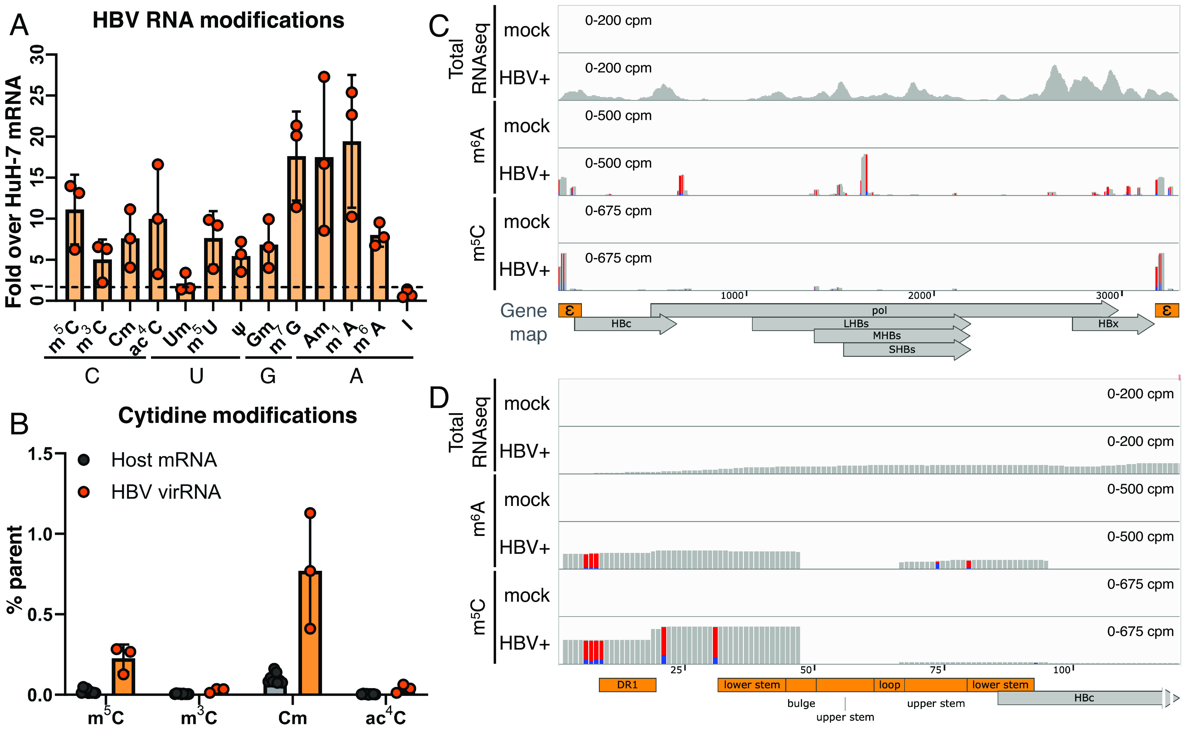
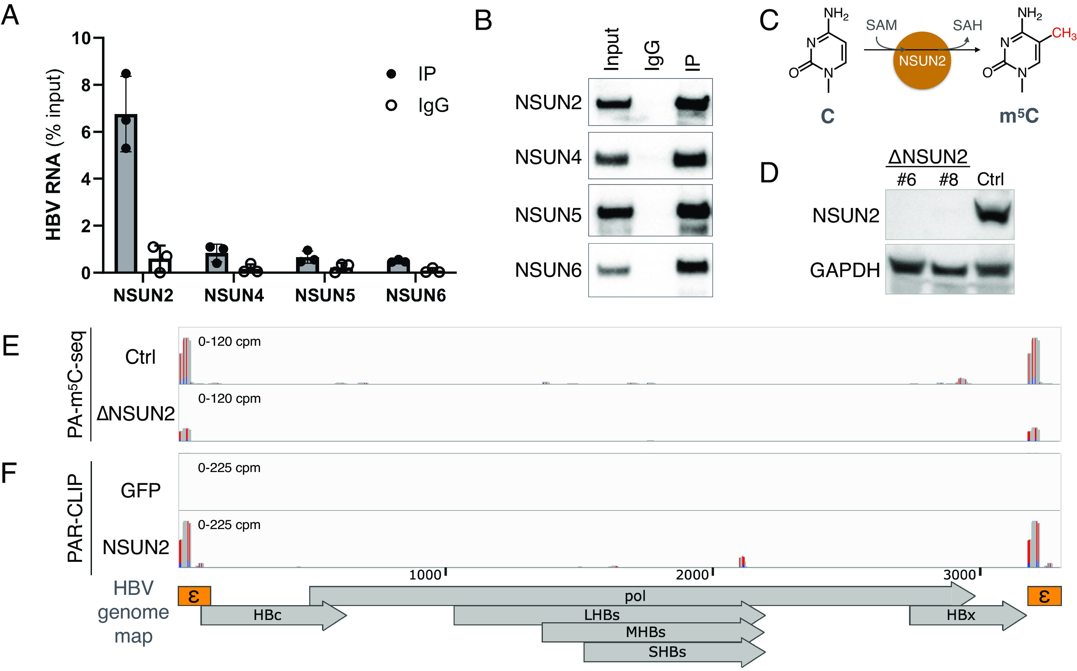
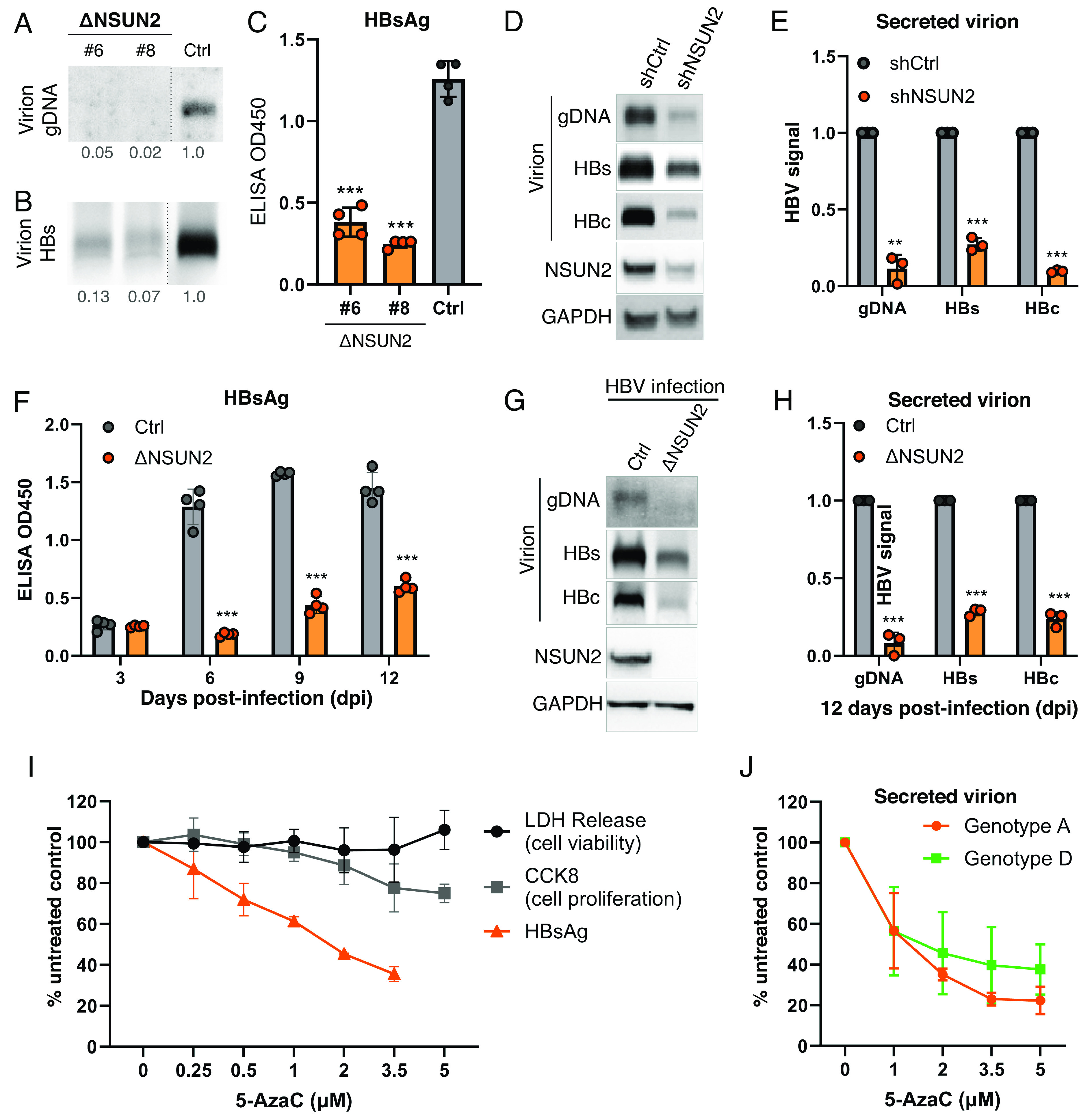
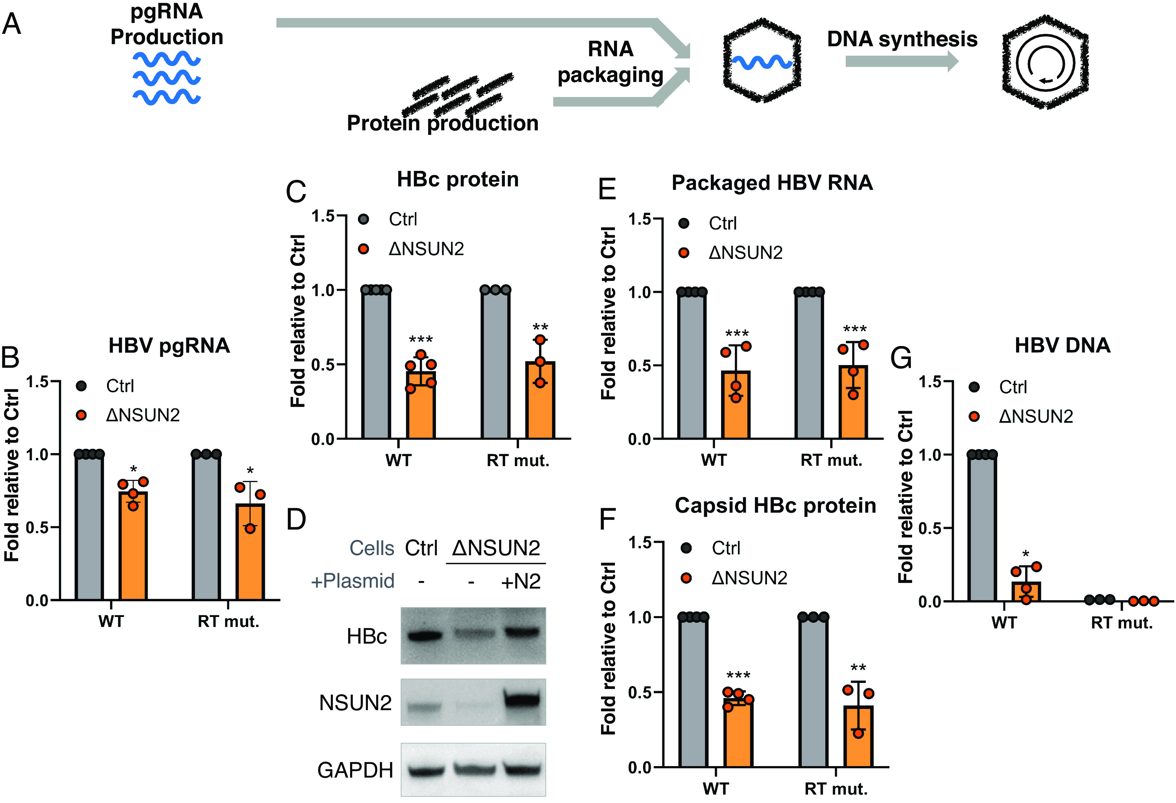
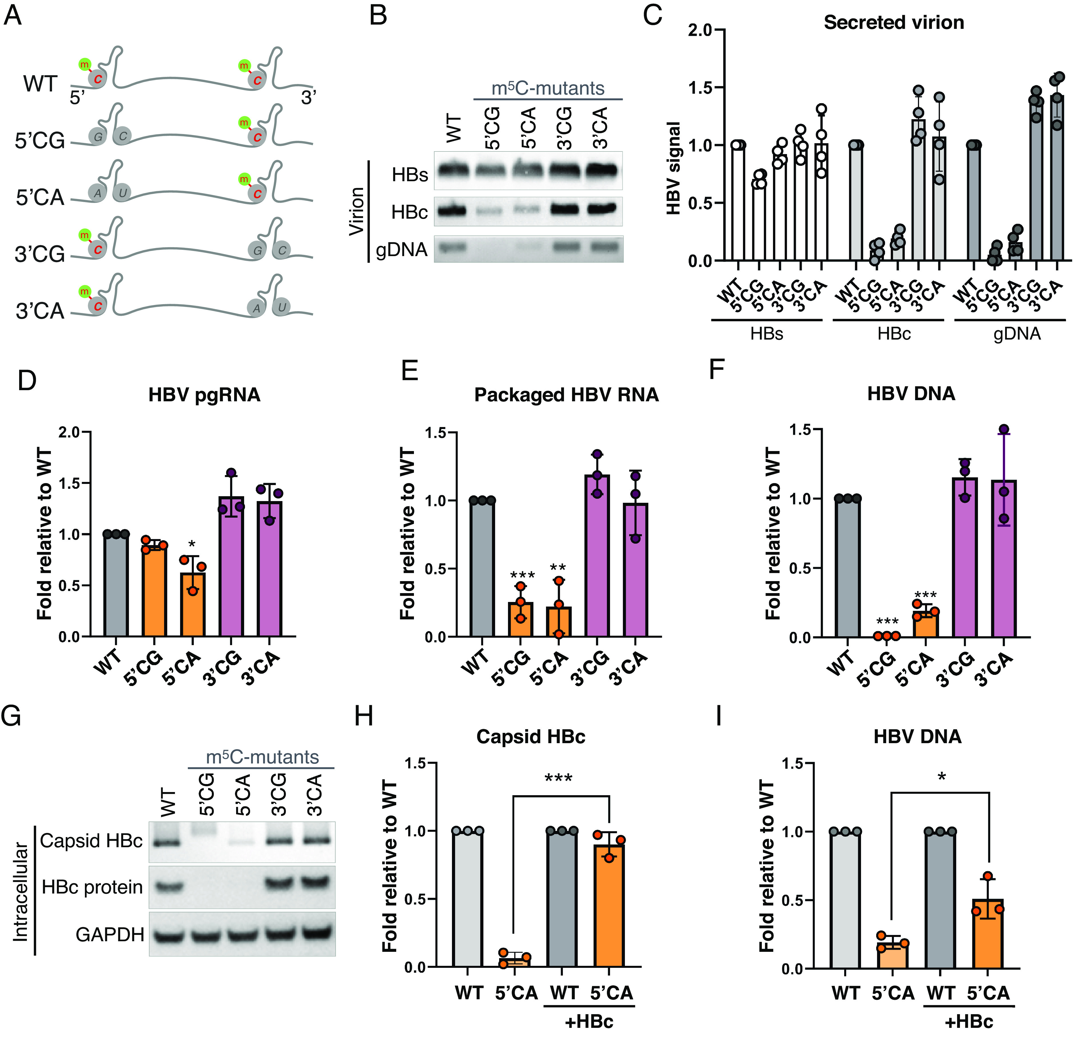
Epitranscriptomic RNA modifications have emerged as important regulators of the fate and function of viral RNAs. One prominent modification, the cytidine methylation 5-methylcytidine (m5C), is found on the RNA of HIV-1, where m5C enhances the translation of HIV-1 RNA. However, whether m5C functionally enhances the RNA of other pathogenic viruses remains elusive. Here, we surveyed a panel of commonly found RNA modifications on the RNA of hepatitis B virus (HBV) and found that HBV RNA is enriched with m5C as well as ten other modifications, at stoichiometries much higher than host messenger RNA (mRNA). Intriguingly, m5C is mostly found on the epsilon hairpin, an RNA element required for viral RNA encapsidation and reverse transcription, with these m5C mainly deposited by the cellular methyltransferase NSUN2. Loss of m5C from HBV RNA due to NSUN2 depletion resulted in a partial decrease in viral core protein (HBc) production, accompanied by a near-complete loss of the reverse transcribed viral DNA. Similarly, mutations introduced to remove the methylated cytidines resulted in a loss of HBc production and reverse transcription. Furthermore, pharmacological disruption of m5C deposition led to a significant decrease in HBV replication. Thus, our data indicate m5C methylations as a critical mediator of the epsilon elements' function in HBV virion production and reverse transcription, suggesting the therapeutic potential of targeting the m5C methyltransfer process on HBV epsilon as an antiviral strategy.

Keywords: NSUN2; RNA modifications; hepatitis B virus; m5C; reverse transcription.


Conflict of interest statement

Competing interests statement:The authors declare no competing interest.

Figures

Fig. 1.
Fig. 1.
HBV RNA is m5C methylated. (A and B) Quantification of RNA modifications on HBV virion RNA by UPLC–MS/MS, purified from HBV pol Y63D (RT mut.) virions, shown normalized to the levels of that on HuH-7 mRNA (A), with cytidine modifications also presented as percent total cytidine (B). (C and D) m6A and m5C sites on HBV RNA were mapped using PA-m6A/m5C-seq on HuH-7 cells transfected with or without (mock) the wildtype ayw strain HBV replicon. Accompanying total RNA-seqs shown in the top track as the pulldown input control. The whole HBV pgRNA depicted in (C), with the epsilon region enlarged in (D). Track heights scaled as the shown counts per million total reads (cpm), red-blue bars depict T>C conversions from 4SU-crosslinking.
Fig. 2.
Fig. 2.
HBV RNA m5C is deposited by the host methyltransferase NSUN2. (A and B) 5-AzaC-mediated RNA immunoprecipitation (Aza-IP) of NSUN proteins, the pulled down HBV RNA quantified by qRT-PCR (n = 3, error bars = SD) (A), with Western blot validation of pulldown (B). (C) Depiction of NSUN2-mediated cytidine (C) methylation to produce 5-methylcytidine (m5C). (D) NSUN2 was depleted from HuH-7 cells by CRISPR-Cas9 (ΔNSUN2), with a GFP-targeted Cas9+ cell line produced alongside as a control (Ctrl). NSUN2 depletion in two single-cell clones (#6 & #8) of ΔNSUN2 and Ctrl cells validated by Western blot. (E) PA-m5C-seq of HBV RNA from Ctrl or ΔNSUN2 HuH-7 cells (clone #8). (F) NSUN2 or GFP (RNA-nonbinding control) binding sites on HBV RNA in HuH-7 cells mapped via PAR-CLIP. RNA-seq tracks scaled as the cpm shown, red-blue bars denote T>C conversions from 4SU-crosslinking.
Fig. 3.
Fig. 3.
NSUN2 depletion diminishes virion secretion. (AC) CRISPR-mediated NSUN2-depleted clones (ΔNSUN2 #6 & #8) or Cas9 control (Ctrl) HuH-7 cells were transfected with a wild-type HBV replicon. The supernatants harvested at 5 days posttransfection (dpt) were assayed for genomic DNA (gDNA) by Southern blot (A) and secreted HBs proteins by Western blot (B) and ELISA (C). Dashed lines between ΔNSUN2 and Ctrl lanes in (A and B) represent lanes removed from between these lanes. (D and E) HuH-7 cells transduced with shRNAs against NSUN2 (shNSUN2) or scrambled control (shCtrl) were transfected with wild-type HBV replicon, and the supernatant virions assayed 5dpt for genomic DNA (gDNA) by Southern blot and secreted HBs and HBc proteins by Western blot, with NSUN2 knockdown verified by cell lysate Western, repeats quantified in (E). (FH) HepG2-NTCP-C4 cells with or without NSUN2-depletion by CRISPR (ΔNSUN2 or Cas9 control, Ctrl) were infected with wild-type HBV, the supernatant HBsAg measured by ELISA at 3 to 12 dpi (F). (G) Viral replication levels at 12 dpi were assayed for secreted virion gDNA, HBs, and HBc protein, with repeats quantified (H). (I) HBV-transfected HuH-7 cells were treated with the m5C methyltransfer disrupter 5-AzaC, secreted HBsAg assayed by ELISA at 3dpt. 5-AzaC toxicity at each dose assayed by lactate dehydrogenase (LDH) and CCK-8 assays to assess cell viability and proliferation. (J) 5-AzaC tested on HuH-7 cells transfected with HBV genotypes A & D, secreted virion HBc assayed by Western blot at 3 dpt with band intensities quantified. Error bars = SD with n = 3 to 4; ∗∗P < 0.01; ∗∗∗P < 0.001.
Fig. 4.
Fig. 4.
NSUN2 enhances HBV translation and is essential for reverse transcription. (A) Schematic of HBV replication cycle. ΔNSUN2 and control (Ctrl) HuH-7 cells were transfected with a wild-type HBV replicon (WT) or the pol Y63D mutant (RT mut.), and the following HBV products were measured. (B) Total intracellular HBV pgRNA levels measured by qRT-PCR at 2 dpt. (C) Intracellular core (HBc) protein at 3 dpt measured by Western blot, with band intensities quantified. (D) Intracellular HBc protein production from wild-type HBV in Ctrl and ΔNSUN2 cells, compared with NSUN2 add-back rescue in ΔNSUN2 cells (+N2). (EG) Intracellular assembled capsid particles purified and the packaged core-associated viral RNA quantified by qRT-PCR (E), assembled particle capsid proteins assayed via band-quantified Western blotting (F), and capsid-associated viral DNA assayed by qPCR at 5 dpt (G). Error bars = SD with n > 3; P < 0.05; ∗∗P < 0.01; ∗∗∗P < 0.001.
Fig. 5.
Fig. 5.
m5C at the 5′end epsilon is critical for HBV replication. (A) Schematic of m5C sites on HBV pgRNA epsilon hairpins. Lower four lanes depict replicons with the putative methylated cytidines (C1842, 1845, 1847, 1858, and 1859 in GenBank J02203 coordinates, SI Appendix, Fig. S6A) on the 5′ epsilons mutated to G or A (5′CG or 5′CA), or the 3′ epsilons (3′CG or 3′CA). G residues on the complimentary strand of the hairpin (G1899 & G1900, SI Appendix, Fig. S6A) were mutated to C or U to preserve the hairpin structure. (BG) WT and mutant replicons were transfected into HuH-7 cells, with the secreted virion HBs and HBc proteins measured via Western blot, and gDNA assayed via Southern blot at 5 dpt (B), repeats of panel B quantified in (C). (D) Intracellular pgRNA quantified by qRT-PCR at 2 dpt. (E) Capsid-packaged viral RNA quantified via qRT-PCR at 3 dpt. (F) Capsid-associated viral DNA assayed via qPCR at 5 dpt. (G) Intracellular capsid HBc and total HBc proteins assayed 3 dpt via native agarose gel electrophoresis and SDS-PAGE followed by Western blotting. (H and I) WT and mutant replicons were transfected into HuH-7 cells with or without HBc expression vector supplementation, with the capsid HBc protein assayed via band-quantified Western blots (H) and viral DNA quantified by qPCR (I). Error bars = SD, n > 3; P < 0.05; ∗∗P < 0.01; ∗∗∗P < 0.001.

Similar articles

Cited by

References

    1. Tsai K., Cullen B. R., Epigenetic and epitranscriptomic regulation of viral replication. Nat. Rev. Microbiol. 18, 559–570 (2020). - PMC - PubMed
    1. McIntyre W., et al. , Positive-sense RNA viruses reveal the complexity and dynamics of the cellular and viral epitranscriptomes during infection. Nucleic Acids Res. 46, 5776–5791 (2018). - PMC - PubMed
    1. Courtney D. G., et al. , Epitranscriptomic addition of m(5)C to HIV-1 transcripts regulates viral gene expression. Cell Host Microbe 26, 217–227.e6 (2019). - PMC - PubMed
    1. Kennedy E. M., et al. , Posttranscriptional m(6)A editing of HIV-1 mRNAs enhances viral gene expression. Cell Host Microbe 19, 675–685 (2016). - PMC - PubMed
    1. Tsai K., et al. , Epitranscriptomic addition of m(6)A regulates HIV-1 RNA stability and alternative splicing. Genes Dev. 35, 992–1004 (2021). - PMC - PubMed