. 2024 Jun 11;121(24):e2400163121.
doi: 10.1073/pnas.2400163121. Epub 2024 Jun 3.

Bispecific antibodies targeting two glycoproteins on SFTSV exhibit synergistic neutralization and protection in a mouse model

Affiliations

Bispecific antibodies targeting two glycoproteins on SFTSV exhibit synergistic neutralization and protection in a mouse model

Zhen Chang et al. Proc Natl Acad Sci U S A. .

Abstract

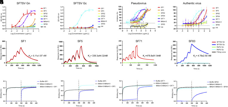
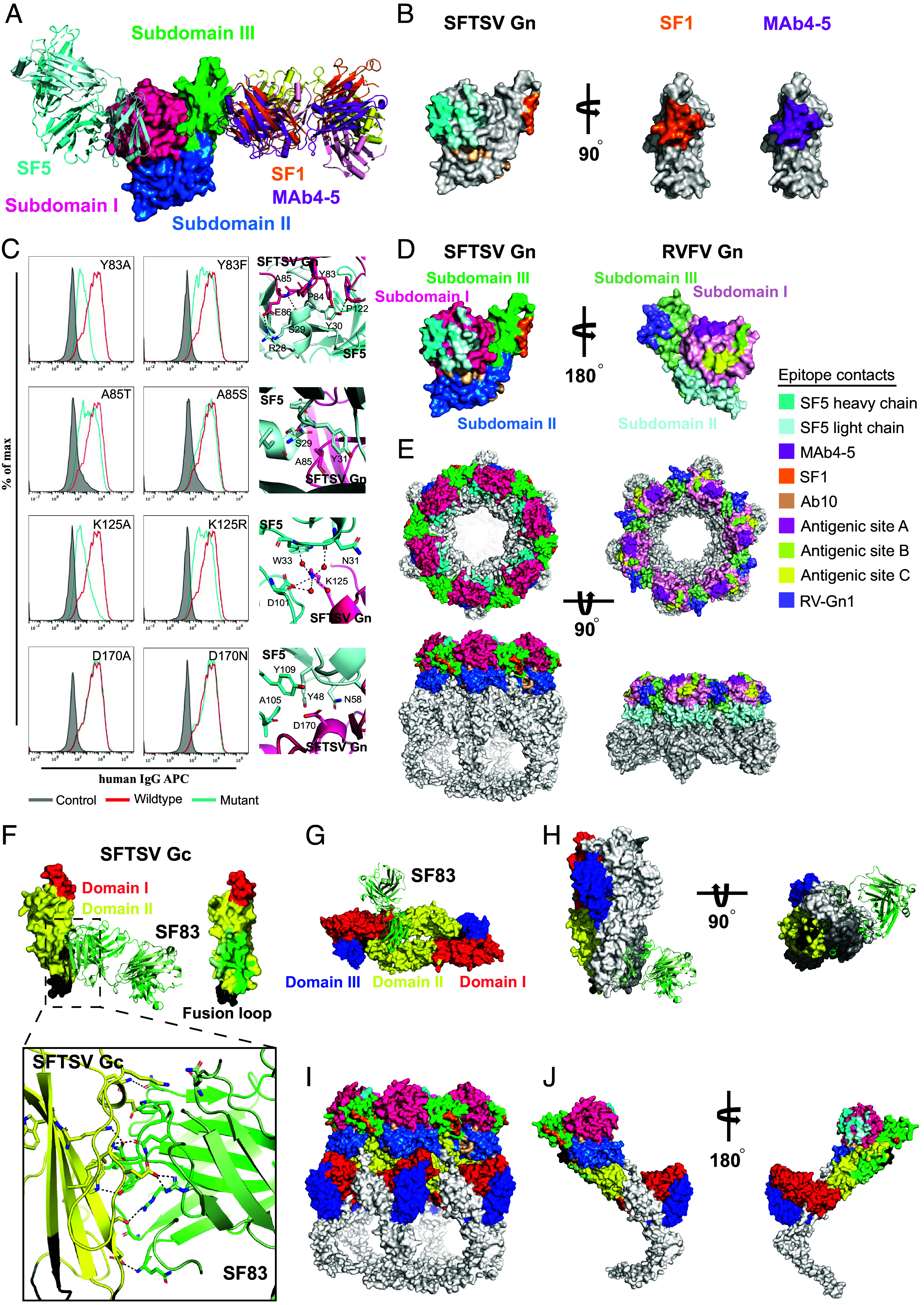
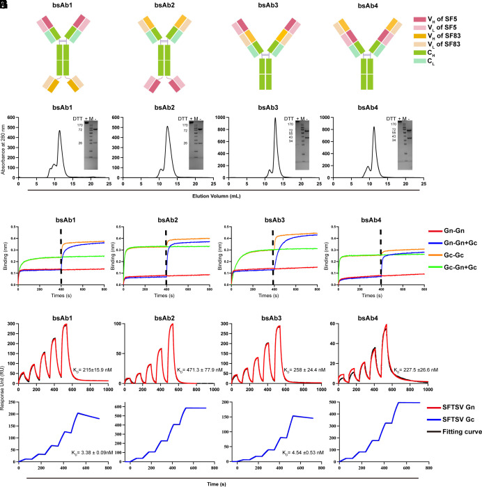
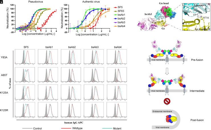
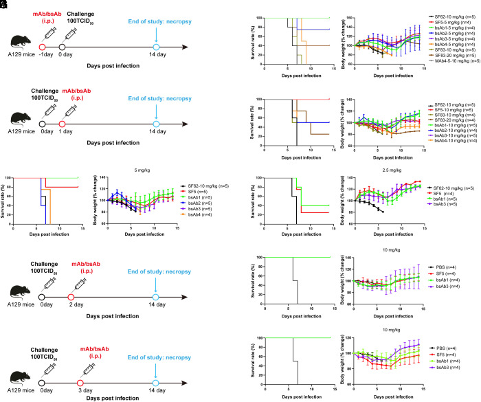
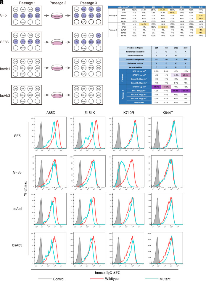
Severe fever with thrombocytopenia syndrome (SFTS) is an emerging infectious disease with a high fatality rate of up to 30% caused by SFTS virus (SFTSV). However, no specific vaccine or antiviral therapy has been approved for clinical use. To develop an effective treatment, we isolated a panel of human monoclonal antibodies (mAbs). SF5 and SF83 are two neutralizing mAbs that recognize two viral glycoproteins (Gn and Gc), respectively. We found that their epitopes are closely located, and we then engineered them as several bispecific antibodies (bsAbs). Neutralization and animal experiments indicated that bsAbs display more potent protective effects than the parental mAbs, and the cryoelectron microscopy structure of a bsAb3 Fab-Gn-Gc complex elucidated the mechanism of protection. In vivo virus passage in the presence of antibodies indicated that two bsAbs resulted in less selective pressure and could efficiently bind to all single parental mAb-escape mutants. Furthermore, epitope analysis of the protective mAbs against SFTSV and RVFV indicated that they are all located on the Gn subdomain I, where may be the hot spots in the phleboviruses. Collectively, these data provide potential therapeutic agents and molecular basis for the rational design of vaccines against SFTSV infection.

Keywords: SFTSV; bispecific antibody; bunyavirus; glycoproteins; structural basis.


Conflict of interest statement

Competing interests statement:Z. Chang, D.G., F.G., and Y.W. are listed as inventors on two pending patent applications. The other authors declare no competing interests.

Figures

Fig. 1.
Fig. 1.
Screening and characterization of mAbs against SFTSV. (A and B) Concentration-dependent binding of the mAbs to SFTSV Gn (A) or Gc (B). (C) Concentration-dependent neutralization of pseudovirus by mAbs. (D) Concentration-dependent neutralization of authentic SFTSV by mAbs. (EH) Characterization of specific binding between the indicated mAbs and specific antigens, including SFTSV Gn (red), SFTSV Gc (blue), RVFV Gn (green), and RVFV Gc (cyan). The fitting curves are indicated in black lines. The calculated KD for each mAb binding to the specific antigen is listed accordingly. (IL) Competitive binding to SFTSV Gn between MAb4-5 and another mAb by BLI. Biotinylated SFTSV Gn was immobilized on SA sensors and then saturated with MAb4-5, followed by SF1 (I), SF5 (J) SF71 (K), or SF64 (L) in the presence of an equimolar concentration of MAb4-5 (cyan) or without MAb4-5 (black). As a control, the immobilized biotinylated SFTSV Gn was exposed to PBST and then introduced to an equimolar concentration of the indicated mAbs (blue). All ELISAs, neutralization assays, and SPR assays were conducted in at least three independent experiments. The affinity data are represented as the mean ± SD.
Fig. 2.
Fig. 2.
Structural analysis of the epitopes of SF5 and SF83. (A) Superimposition of the Gn–SF1, Gn–SF5, and Gn–MAb4-5 structures. The three subdomains of the Gn head are shown in hot pink, blue, and green, respectively. (B) Footprints of SF1 (orange), SF5 (heavy chain in cyan and light chain in light cyan), and Mab4-5 (deep purple) on the SFTSV Gn head. (C) Evaluation of the key residues involved in Gn–SF5 binding using a flow cytometric assay. HEK293T cells were transfected with plasmids containing the coding regions of EGFP-fused SFTSV M (red) or the indicated mutants (cyan) and then tested with SF5 and goat anti-human IgG-APC. Cells transiently expressing M-EGFP stained with goat anti-human IgG-APC were used as a negative control (gray). (D) Footprints of mAbs on the SFTSV Gn head and RVFV Gn head. SF1 (orange), SF5 (heavy chain in cyan and light chain in light cyan), MAb4-5 (deep purple), and Ab10 (wheat) are mAbs against SFTSV. RV-Gn1 (purple) and mAbs targeting antigenic site A (magenta), B (limon), and C (yellow) recognize the RVFV Gn head. The colors of SFTSV Gn subdomains are consistent with (A). The colors of three RVFV Gn subdomains are light pink, pale cyan, and pale green, respectively. (E) The indicated RVFV mAbs are superimposed on the Gn–Gc glycoprotein shell of RVFV (PDB code: 6F9E). Four anti-SFTSV Gn mAbs are superimposed on the SFTSV Gn–Gc glycoprotein shell (PDB code: 8ILQ). The Top view (Up row) and side view (Bottom row) are shown. (F) Footprint of SF83 and the detailed interactions between SFTSV Gc and SF83. Domains I and domain II of Gc are colored red and yellow, respectively. The heavy chain and light chain of SF83 are green and pale green, respectively. (G) Superimposition of Gc–SF83 and the Gc dimer model (constructed based on the RVFV Gc dimer, PDB code: 4HJ1). (H) Superimposition of Gc–SF83 and the Gc trimer (PDB code: 6EGU). (I) Footprints of mAbs on a SFTSV glycoprotein shell. (J) The inner and outer side diagrams of the mAbs’ footprints on a Gn–Gc protomer.
Fig. 3.
Fig. 3.
Design and characterization of bsAbs. (AD) Schematic diagram of four engineering bsAbs. BsAb1 and bsAb2 are in IgG-(ScFv)2 format, whereas bsAb3 and bsAb4 are in DVD-Ig format. (E) Size-exclusion analysis of bsAbs. The SDS-PAGE profiles of the pooled samples under reducing and nonreducing conditions. The heavy and light chains of bsAb1 and bsAb2 are ~80 kDa and 25 kDa, respectively. The heavy and light chains of bsAb3 and bsAb4 are ~65 kDa and 40 kDa, respectively. (FI) Competitive binding of parental mAbs and bsAbs. The indicated bsAb was immobilized on the sensor and saturated with Gn, and then flowed through with Gn (red) or an equimolar mixture of Gn and Gc (blue). The indicated bsAb was immobilized on the sensor and saturated with Gc, and then flowed through with Gn (orange) or an equimolar mixture of Gn and Gc (green). The dashed line shows the starting time point of the mixture addition. (J) Binding kinetics of bsAbs to SFTSV Gn and Gc. BsAbs were immobilized on Protein A chips and tested for real-time association and dissociation of indicated antigens. The equilibrium dissociation constant (KD) is labeled accordingly. The solid red line (Gn) and blue line (Gc) are the actual operation curves, and the black line is the fitted curve. Each experiment was repeated three times, and the results shown are the means ± SD.
Fig. 4.
Fig. 4.
BsAbs showed potent neutralization against SFTSV and superior binding ability to mutants compared to mAbs. (A) Two parental mAbs and four bsAbs were analyzed using the pseudovirus system. (B) Two parental mAbs and four bsAbs were analyzed using the authentic virus system. All neutralization assays were conducted in at least three independent experiments. (C) Evaluation of the binding abilities between SF5 and bsAbs to different substitutions on Gn using a flow cytometric assay. HEK293T cells were transfected with plasmids containing coding regions of EGFP-fused SFTSV M (red) or indicated substitutions (cyan) and then tested with SF5 or bsAbs and goat anti-human IgG-APC. Cells transiently expressing M-EGFP stained with goat anti-human IgG-APC were used as a negative control (gray). This flow cytometric assay was performed in at least two independent experiments. (D) The complex structure of bsAb3 Fab–Gn–Gc and the detailed interaction between SF5 variable regions in bsAb3 and Gc. The Gn head and the variable regions of SF are shown in surface and cartoon representations, with the same color scheme as Fig. 2A. Gc is displayed in yellow cartoon format. The heavy chain and light chain of SF83 in bsAb3 are shown in purple and light pink, respectively. The detailed interaction between SF5 and Gc domain II is highlighted in the box. The hydrogen bond between T5 in the bsAb3 light chain and H716 in Gc domain II is labeled with a dashed line. (E) Model for bsAb3 neutralization against SFTSV. In the prefusion state, the SF5 on the bsAb3 can bind to Gn subdomain I, while the SF83 epitope is unexposed. At the intermediate state, the Gn and Gc move upward to expose the epitope of SF83, and bsAb3 can bind to both Gn and Gc, thereby preventing Gc trimerization and membrane fusion. The Gn and Gc domains are colored as in Fig. 2I, bsAb3 is colored as in (D), and the membrane is gray.
Fig. 5.
Fig. 5.
Prophylactic and therapeutic efficacies of the mAbs and bsAbs in a mouse model of SFTSV infection. (A) The prophylaxis strategy of mAbs and bsAbs in the SFTSV-infected A129 mouse model. (B) Mouse survival and body weight in the prophylaxis study were recorded daily. Mice in the SF62 group were set as an isotype control. (C) The therapeutic strategy of mAbs and bsAbs in the SFTSV-infected A129 mouse model. (DF) Mouse survival and body weight in the therapeutic study were recorded daily. Three doses were used for the therapeutic study, with 10 mg/kg (D), 5 mg/kg (E), and 2.5 mg/kg (F). Mice in the SF62 group were set as an isotype control. (G and I) The delayed treatment strategies of mAbs and bsAbs in the SFTSV-infected A129 mouse model. Mice were challenged with SFTSV and then were given the indicated antibodies in 2 d (G) or 3 d (I). (H and J) Mouse survival and body weight in the delayed treatment study were recorded daily. Mice in the PBS group were administered PBS instead of the indicated antibodies.
Fig. 6.
Fig. 6.
Escape mutant screening in the presence of antibodies. (A) Schematic of the escape mutant screening process. The VSV-GFP-based pseudovirus was passaged in the presence of antibodies with serial dilutions on Vero E6 cells. The light purple indicates the fluorescence value ratio in the indicated well and no-antibody well was >30%. The corresponding ratio values are indicated in (B). (B) Quantitative percentage of infected cells (monitored by transducing units) under different concentrations of antibodies during passage 1 and passage 3. Yellow boxes indicate the dilutions that were passaged and sequenced in passage 1 or passage 3. (C) NGS of virus genomes from passage 3. Variant nucleotides and corresponding residues are listed, and the qualitative percentage of infected cells (monitored by transducing units) was calculated (dark purple, >60%; pink, 30 to 60%; and light purple, 10 to 30%). (D) Evaluation of the binding between SF5, SF83, and bsAbs to screened substitutions using a flow cytometric assay. HEK293T cells were transfected with plasmids containing coding regions of EGFP-fused SFTSV M (red) or the indicated substitutions (cyan) and then tested with SF5, SF83, or bsAbs and goat anti-human IgG-APC. Cells transiently expressing M-EGFP stained with goat anti-human IgG-APC were used as a negative control (gray).

Similar articles

Cited by

References

    1. Yu X. J., et al. , Fever with thrombocytopenia associated with a novel bunyavirus in China. N. Engl. J. Med. 364, 1523–1532 (2011). - PMC - PubMed
    1. Liu Q., He B., Huang S. Y., Wei F., Zhu X. Q., Severe fever with thrombocytopenia syndrome, an emerging tick-borne zoonosis. Lancet Infect Dis. 14, 763–772 (2014). - PubMed
    1. Seo J. W., Kim D., Yun N., Kim D. M., Clinical update of severe fever with thrombocytopenia syndrome. Viruses 13, 1213 (2021). - PMC - PubMed
    1. Liu W., et al. , Case-fatality ratio and effectiveness of ribavirin therapy among hospitalized patients in china who had severe fever with thrombocytopenia syndrome. Clin. Infect. Dis. 57, 1292–1299 (2013). - PubMed
    1. Liu K., et al. , Epidemiologic features and environmental risk factors of severe fever with thrombocytopenia syndrome, Xinyang, China. PLoS Negl. Trop. Dis. 8, e2820 (2014). - PMC - PubMed

MeSH terms

LinkOut - more resources