

ZW10: an emerging orchestrator of organelle dynamics during the cell division cycle
- PMID: 38830800
- PMCID: PMC11757092
- DOI: 10.1093/jmcb/mjae026
ZW10: an emerging orchestrator of organelle dynamics during the cell division cycle
Abstract
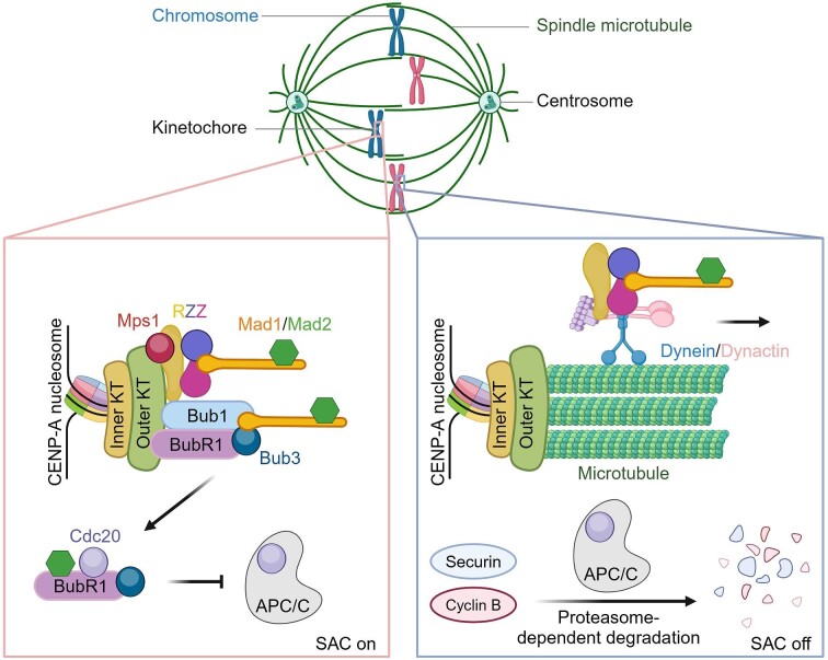
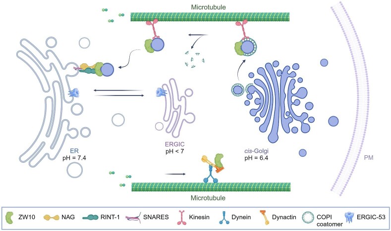
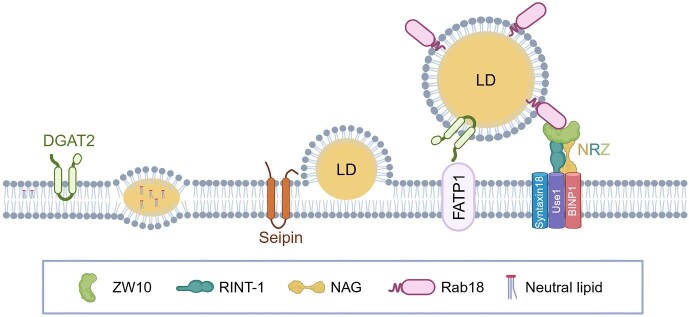
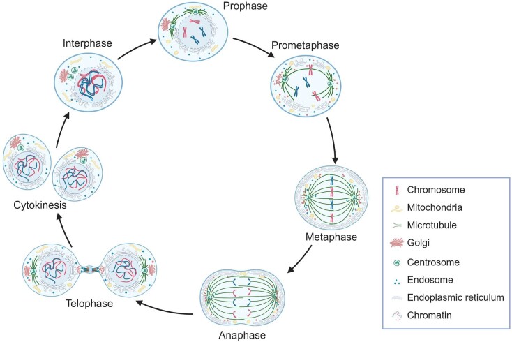
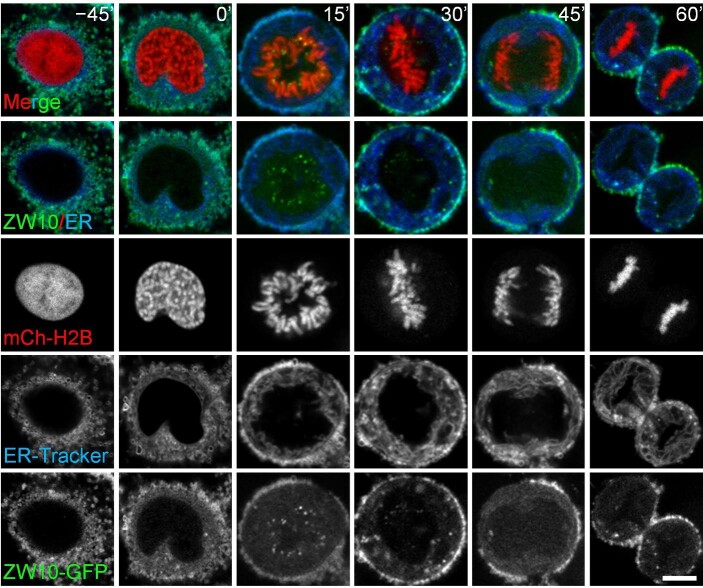
Zeste white 10 (ZW10) was first identified as a centromere/kinetochore protein encoded by the ZW10 gene in Drosophila. ZW10 guides the spindle assembly checkpoint signaling during mitotic chromosome segregation in metazoans. Recent studies have shown that ZW10 is also involved in membrane-bound organelle interactions during interphase and plays a vital role in membrane transport between the endoplasmic reticulum and Golgi apparatus. Despite these findings, the precise molecular mechanisms by which ZW10 regulates interactions between membrane-bound organelles in interphase and the assembly of membraneless organelle kinetochore in mitosis remain elusive. Here, we highlight how ZW10 forms context-dependent protein complexes during the cell cycle. These complexes are essential for mediating membrane trafficking in interphase and ensuring the accurate segregation of chromosomes in mitosis.
Keywords: ZW10; kinetochore; membrane-bound organelle; membraneless organelle; mitosis.
© The Author(s) (2024). Published by Oxford University Press on behalf of Journal of Molecular Cell Biology, CEMCS, CAS.
Figures
Similar articles
-
PLK1 phosphorylation of ZW10 guides accurate chromosome segregation in mitosis.J Mol Cell Biol. 2024 Jul 29;16(2):mjae008. doi: 10.1093/jmcb/mjae008. J Mol Cell Biol. 2024. PMID: 38402459 Free PMC article.
-
ZW10 function in mitotic checkpoint control, dynein targeting and membrane trafficking: is dynein the unifying theme?Cell Cycle. 2006 Nov 1;5(21):2447-51. doi: 10.4161/cc.5.21.3395. Epub 2006 Sep 12. Cell Cycle. 2006. PMID: 17102640 Free PMC article. Review.
-
Hec1 sequentially recruits Zwint-1 and ZW10 to kinetochores for faithful chromosome segregation and spindle checkpoint control.Oncogene. 2006 Nov 2;25(52):6901-14. doi: 10.1038/sj.onc.1209687. Epub 2006 May 29. Oncogene. 2006. PMID: 16732327
-
Bipolar spindle attachments affect redistributions of ZW10, a Drosophila centromere/kinetochore component required for accurate chromosome segregation.J Cell Biol. 1996 Sep;134(5):1127-40. doi: 10.1083/jcb.134.5.1127. J Cell Biol. 1996. PMID: 8794856 Free PMC article.
-
The RZZ complex and the spindle assembly checkpoint.Cell Struct Funct. 2009;34(1):31-45. doi: 10.1247/csf.08040. Cell Struct Funct. 2009. Retraction in: Cell Struct Funct. 2010;35(1):1. doi: 10.1247/csf.08040r. PMID: 19420794 Retracted. Review.
Cited by
-
Comprehensive Investigation of a Tyrosine Kinase Inhibitor-Resistant Gene Zeste White 10 in Hepatocellular Carcinoma.World J Oncol. 2025 Apr;16(2):210-226. doi: 10.14740/wjon2514. Epub 2025 Mar 9. World J Oncol. 2025. PMID: 40162104 Free PMC article.
References
-
- Allan B.B., Moyer B.D., Balch W.E. (2000). Rab1 recruitment of p115 into a cis-SNARE complex: programming budding COPII vesicles for fusion. Science 289, 444–448. - PubMed
-
- Andag U., Neumann T., Schmitt H.D. (2001). The coatomer-interacting protein Dsl1p is required for Golgi-to-endoplasmic reticulum retrieval in yeast. J. Biol. Chem. 276, 39150–39160. - PubMed
-
- Appenzeller C., Andersson H., Kappeler F. et al. (1999). The lectin ERGIC-53 is a cargo transport receptor for glycoproteins. Nat. Cell Biol. 1, 330–334. - PubMed
Publication types
MeSH terms
Substances
Grants and funding
LinkOut - more resources
Full Text Sources
Molecular Biology Databases
