. 2024 Jun 3;15(1):4728.
doi: 10.1038/s41467-024-48735-x.

Generation of nanobodies from transgenic 'LamaMice' lacking an endogenous immunoglobulin repertoire

Affiliations

Generation of nanobodies from transgenic 'LamaMice' lacking an endogenous immunoglobulin repertoire

Thomas Eden et al. Nat Commun. .

Abstract

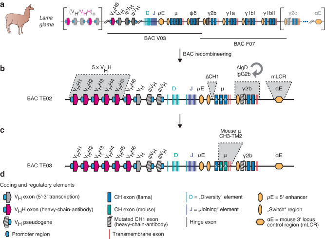
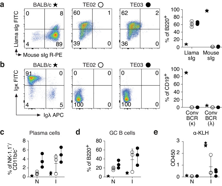
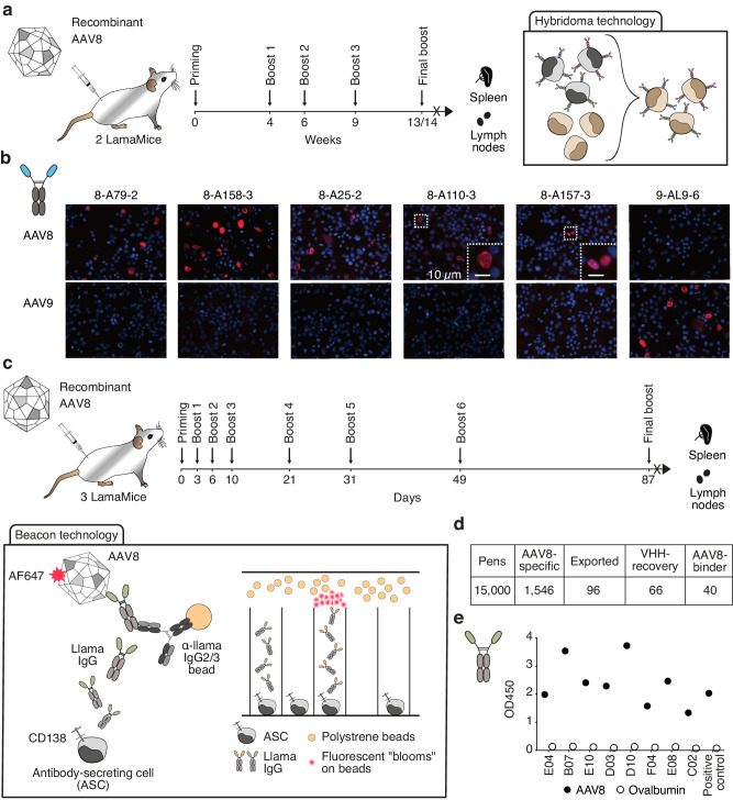
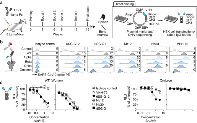
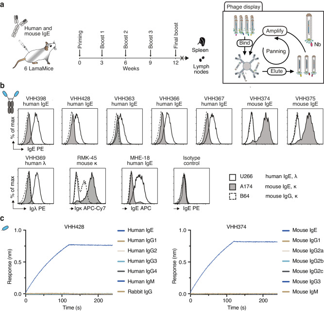
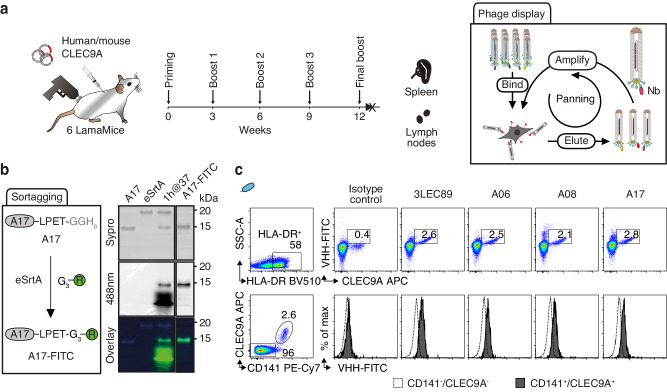
Due to their exceptional solubility and stability, nanobodies have emerged as powerful building blocks for research tools and therapeutics. However, their generation in llamas is cumbersome and costly. Here, by inserting an engineered llama immunoglobulin heavy chain (IgH) locus into IgH-deficient mice, we generate a transgenic mouse line, which we refer to as 'LamaMouse'. We demonstrate that LamaMice solely express llama IgH molecules without association to Igκ or λ light chains. Immunization of LamaMice with AAV8, the receptor-binding domain of the SARS-CoV-2 spike protein, IgE, IgG2c, and CLEC9A enabled us to readily select respective target-specific nanobodies using classical hybridoma and phage display technologies, single B cell screening, and direct cloning of the nanobody-repertoire into a mammalian expression vector. Our work shows that the LamaMouse represents a flexible and broadly applicable platform for a facilitated selection of target-specific nanobodies.


Conflict of interest statement

T.E., J.We., S.M., and F.K.-N. are co-inventors on a patent application WO2018/104528 on LamaMice. S.W., C.L.-W., M.S., I.B., A.W., Y.H., Le.S., La.S., D.Z., E.S., K.F.H., H.S., and J.F. work for companies that commercialize products in the field of nanobodies. The remaining authors declare no competing interests.

Figures

Fig. 1
Fig. 1. Recombineering of llama IgH BAC transgenes.
a Two bacterial artificial chromosomes (BAC V03, BAC F07) covering the core of the llama IgH locus were cloned from a liver genomic DNA library and fully sequenced. b BAC recombineering was used to insert five additional VHH-genes, to delete the CH1 exon of IgM, to replace the IgD pseudogene with the IgG2b gene, and to insert elements of the mouse 3’ locus control region (mLCR, αE), resulting in BAC TE02. c BAC recombineering was used to exchange the exons encoding the CH3, CH4, transmembrane, and cytosolic domains of IgM with corresponding murine sequences, resulting in BAC TE03. d Explanation of symbols used in the schematics.
Fig. 2
Fig. 2. LamaMice support the development of B cells that display llama heavy chain immunoglobulins.
a, b Spleen cells of 16-27-week-old mice (n = 3 per group) were stained with fluorochrome-conjugated antibodies and analyzed by flow cytometry. Gating was performed on B220+ (a) or CD19+ (b) cells. c–e 12-13-week-old mice (n = 3 per group) were immunized (I) with keyhole limpet hemocyanin (KLH). Naive (N) mice served as controls. c, d Four days after the second boost, spleen cells were stained with fluorochrome-conjugated antibodies and analyzed by flow cytometry. c Gating was performed on NK-1.1/CD11b/CD11c triple-negative cells. Plasma cells were identified as CD138+/PC-1+ cells. d Gating was performed on B220+ cells. Germinal center (GC) B cells were identified as CD95+/surface immunoglobulin (sIg)int cells. e Serum was analyzed for the presence of KLH-specific antibodies (α-KLH) by ELISA using peroxidase-conjugated mouse Ig-specific (BALB/c) or llama Ig-specific (LamaMice) secondary antibodies. a–e Asterisks indicate samples from BALB/c mice, open circles from TE02 LamaMice, closed circles from TE03 LamaMice. a, b Dot plots are from single representative animals. Numbers indicate the % of cells in the respective quadrant. a–e Bar diagrams show the corresponding results for all mice in a group. Data represent mean ± SD for n = 3 individuals.
Fig. 3
Fig. 3. Discovery of AAV-specific nanobodies from immunized LamaMice using classic hybridoma or single B cell screening technologies.
a LamaMice were immunized with AAV8 as indicated. Three days after the final boost, spleen and lymph node cells were fused with Sp2/0 mouse myeloma cells and cultured on 96-well plates in HAT-selection medium. Supernatants of hybridoma clones were screened by ELISA for reactivity with AAV8. Positive hybridomas were subcloned by limiting dilution. b The VHH-encoding region of positive clones was PCR-amplified, sequenced, and cloned into a mammalian expression vector upstream of the hinge, CH2 and CH3 of rabbit IgG. VHH-rabbit IgG hcAbs were produced in transiently transfected HEK-6E cells and cell supernatants were analyzed for reactivity with HEKAAV cells producing AAV by immunofluorescence microscopy. Bound hcAbs were detected with PE-conjugated anti-rabbit IgG. c, d LamaMice were immunized with AAV8 as indicated. Four days after the final boost, antibody-secreting cells (ASC) were sorted from spleen and lymph node cells using anti-CD138-coated beads, and cells were loaded into individual pens on a Berkley Lights Beacon® chip. Llama IgG heavy chain antibodies (hcAbs) captured on beads were detected with AF647-conjugated AAV8. Cells yielding an AAV8-specific signal were exported onto a 96-well plate and subjected to cDNA synthesis. e PCR-amplicons obtained with a VHH-specific primer pair were sequenced and cloned into a mammalian expression vector. VHHs were re-expressed as recombinant llama hcAbs and tested for specific binding to AAV8 by ELISA. Bound hcAbs were detected with PO-conjugated anti-llama IgG.
Fig. 4
Fig. 4. Discovery of RBD-specific nanobodies from immunized LamaMice using direct cloning technology.
a LamaMice were immunized with a fusion protein comprising the recombinant receptor-binding domain (RBD) of the SARS-CoV-2 spike protein fused to the hinge, CH2 and CH3 domains of llama IgG2b. Three days after the final boost, the VHH-repertoire was PCR-amplified from cDNA prepared from spleen and bone marrow cells. PCR-amplicons were cloned into the pCSE2.5 expression vector upstream of the hinge, CH2 and CH3 domains of rabbit IgG. Plasmid DNA was prepared from individual colonies grown in 96-well plates, sequenced, and transiently transfected into HEK-6E cells cultivated in serum-free medium in 96-well plates. Supernatants were harvested 5 days after transfection. A liquots of the supernatants were analyzed for the production of heavy chain antibodies (hcAbs) by SDS-PAGE (see Supplementary Fig. 6a). b VHH-rabbit IgG hcAbs in HEK-6E supernatants were screened for binding to HEK293T cells transiently co-transfected with expression vectors for GFP and the spike protein of various SARS-CoV-2-strains. Bound antibodies were detected with PE-conjugated anti-rabbit IgG. Numbers indicate the mean PE fluorescence intensities (MFI x 10-3) of the GFP+ cell population. Parallel stainings were performed with VHH-rabbit IgG hcAbs containing nanobodies discovered by other groups from nanomice immunized with recombinant RBD (Nb12, Nb30) and a llama immunized with the spike protein of SARS-CoV-1 (VHH-72). c HEK293T cells stably overexpressing human ACE2 were incubated with luciferase-encoding lentiviral gene ontology vectors pseudotyped with SARS-CoV-2 spike protein of the wild type (WT) (Wuhan) or Omicron BA.2 variant in the presence of titrated amounts of the indicated VHH-rabbit IgG hcAbs. Two days after transduction, luciferase activity was quantified on a luminometer, 20 min after addition of luciferin. Data represent mean ± SD for triplicates.
Fig. 5
Fig. 5. Discovery of IgE-specific nanobodies from immunized LamaMice using phage display technology.
a LamaMice were immunized with a cocktail of human IgE and mouse IgE. Three days after the final boost, the VHH-repertoire was PCR-amplified from cDNA prepared from spleen and lymph nodes. PCR-amplicons were cloned into the pHEN2 phagemid vector upstream of the coding sequences for a His6x-c-Myc tag, an amber stop codon followed by the region encoding the gp3 surface protein of the M13 phage. Phage display libraries prepared from transformed E. coli were panned in solution on biotinylated IgE and complexes were captured on streptavidin beads. b The VHH-encoding region of enriched phages was sequenced and subcloned into the pCSE2.5 expression vector upstream of the hinge, CH2 and CH3 domains of rabbit IgG as in Fig. 4a. Specific binding of VHH-rabbit IgG hcAbs to cell lines expressing B cell receptors composed of human IgE (myeloma U-266), mouse IgE (hybridoma M1-3A174), or mouse IgG2a (M261B64) was analyzed by flow cytometry. Bound antibodies were detected with PE-conjugated anti-rabbit IgG. Control stainings were performed with anti-human Igλ (VHH369-rabbit IgG hcAb) or a VHH-rabbit IgG hcAb isotype control followed by PE-conjugated anti-rabbit IgG, and with directly conjugated anti-mouse Igκ (RMK-45) or anti-human IgE (MHE-18). c Nanobodies VHH428 and VHH374 were biotinylated and captured on streptavidin-coated biosensors. Specific binding to IgE and the other indicated Ig isotypes was analyzed by biolayer interferometry.
Fig. 6
Fig. 6. Discovery of CLEC9A-specific nanobodies from cDNA-immunized LamaMice using phage display technology.
a LamaMice were immunized with a cDNA expression vector encoding human CLEC9A using a gene gun. Three days after the final boost, the VHH-repertoire was PCR-amplified from cDNA prepared from spleen and lymph nodes and cloned into the pHEN2 phagemid vector as in Fig. 5. Phage display libraries were panned on cells expressing cell surface CLEC9A. b The VHH-encoding region of enriched phages was subcloned upstream of the coding sequence for a sortase tag and VHHs were fused to a FITC-conjugated peptide using sortase (eSrtA). Unreacted product and sortase were removed with Ni-NTA resin and excess nucleophile was removed by spin filtration. In-gel Sypro (protein) and FITC fluorescence of reaction products were analyzed via SDS-PAGE. c Binding of FITC-conjugated VHHs to CLEC9A-expressing human peripheral blood cells was analyzed by flow cytometry. Gating was performed on MHCII HLA-DR+ cells (upper panels). Cells were counterstained with commercial CLEC9A-specific and CD141-specific mAbs. Gating was performed on HLA-DR+ cells that were either CD141+ or CD141- (lower panels).

References

    1. Voss JE. Engineered single-domain antibodies tackle COVID variants. Nature. 2021;595:176–178. doi: 10.1038/d41586-021-01721-5. - DOI - PubMed
    1. Saelens X, Schepens B. Single-domain antibodies make a difference. Science. 2021;371:681–682. doi: 10.1126/science.abg2294. - DOI - PubMed
    1. Koenig PA, et al. Structure-guided multivalent nanobodies block SARS-CoV-2 infection and suppress mutational escape. Science. 2021;371:eabe6230. doi: 10.1126/science.abe6230. - DOI - PMC - PubMed
    1. Sheridan C. Ablynx’s nanobody fragments go places antibodies cannot. Nat. Biotechnol. 2017;35:1115–1117. doi: 10.1038/nbt1217-1115. - DOI - PubMed
    1. Sheridan C. Llama-inspired antibody fragment approved for rare blood disorder. Nat. Biotechnol. 2019;37:333–334. doi: 10.1038/s41587-019-0101-7. - DOI - PubMed

MeSH terms

LinkOut - more resources