. 2024 Jun 3;14(1):12721.
doi: 10.1038/s41598-024-62484-3.

Effects of bone surface topography and chemistry on macrophage polarization

Affiliations

Effects of bone surface topography and chemistry on macrophage polarization

Birgün Özcolak et al. Sci Rep. .

Abstract

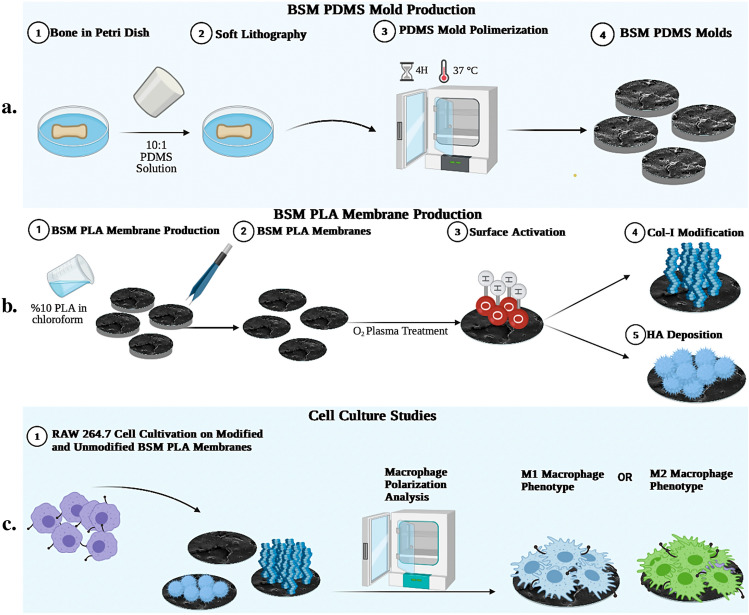
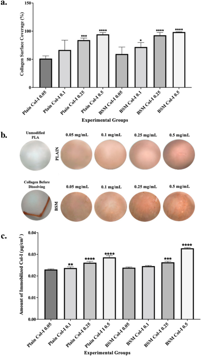
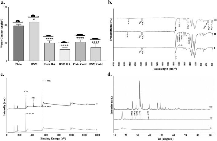
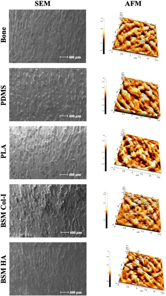
Surface structure plays a crucial role in determining cell behavior on biomaterials, influencing cell adhesion, proliferation, differentiation, as well as immune cells and macrophage polarization. While grooves and ridges stimulate M2 polarization and pits and bumps promote M1 polarization, these structures do not accurately mimic the real bone surface. Consequently, the impact of mimicking bone surface topography on macrophage polarization remains unknown. Understanding the synergistic sequential roles of M1 and M2 macrophages in osteoimmunomodulation is crucial for effective bone tissue engineering. Thus, exploring the impact of bone surface microstructure mimicking biomaterials on macrophage polarization is critical. In this study, we aimed to sequentially activate M1 and M2 macrophages using Poly-L-Lactic acid (PLA) membranes with bone surface topographical features mimicked through the soft lithography technique. To mimic the bone surface topography, a bovine femur was used as a model surface, and the membranes were further modified with collagen type-I and hydroxyapatite to mimic the bone surface microenvironment. To determine the effect of these biomaterials on macrophage polarization, we conducted experimental analysis that contained estimating cytokine release profiles and characterizing cell morphology. Our results demonstrated the potential of the hydroxyapatite-deposited bone surface-mimicked PLA membranes to trigger sequential and synergistic M1 and M2 macrophage polarizations, suggesting their ability to achieve osteoimmunomodulatory macrophage polarization for bone tissue engineering applications. Although further experimental studies are required to completely investigate the osteoimmunomodulatory effects of these biomaterials, our results provide valuable insights into the potential advantages of biomaterials that mimic the complex microenvironment of bone surfaces.

Keywords: Biomimetic; Bone surface topography; Macrophage polarization; Macrophages; Soft lithography; Surface modification osteoimmunomodulation.


Conflict of interest statement

The authors declare no competing interests.

Figures

Figure 1
Figure 1
Schematic of BSM membrane production. (a) BSM-PDMS Molds preparation process. Negative molds were initially created using bone samples, and these molds were subsequently utilized to fabricate BSM-PLA membranes. (b) Unmodified and Modified BSM-PLA membrane production process. (c) RAW 264.7 cell culture studies. RAW 264.7 cells were cultured on unmodified, and Col-I and HA modified plain and BSM membranes to investigate macrophage phenotypes using SEM and ELISA.
Figure 2
Figure 2
(a) Immunohistostaining analysis of unmodified and Col-I modified plain and BSM PLA membranes by using Image J, (b) Immunohistostaining of unmodified and Col-I modified plain and BSM PLA membranes, (c) Quantification of Col-I content by Sirius Red assay (*p < 0.05, **p < 0.01, ***p < 0.001, ****p < 0.0001).
Figure 3
Figure 3
Characterization of surface wettability, chemical composition, and crystallinity of membranes. (a) WCA analysis, (b) FTIR analysis; I. HA modified PLA, II. Col-I modified PLA, III. Unmodified PLA (c) XPS analysis; I. Col-I modified PLA, II. Unmodified PLA, (d) XRD Analysis I. Unmodified PLA, II. HA modified PLA, III. Pure HA.
Figure 4
Figure 4
SEM and AFM images of the surface 3-D structure of the bone, BSM PDMS mold, unmodified, Col-I and HA modified plain and BSM PLA membranes.
Figure 5
Figure 5
Viability of RAW264.7 cells adherent on unmodified, Col-I modified, and HA modified plain PLA and BSM PLA surfaces were measured at day 1, day 3, and day 6 (*p < 0.05, **p < 0.01, ***p < 0.001, ****p < 0.0001).
Figure 6
Figure 6
SEM images of unmodified, and Col-I and HA modified, plain and BSM membranes on day 1, day 3, and day 6 (Scale bars: 2 µm).
Figure 7
Figure 7
Pro-Inflammatory and Anti-Inflammatory factor content and amount analysis on unmodified and Col-I and HA modified, plain PLA and BSM PLA membranes by days. (a) IL-6 Concentration, (b) IL-10 Concentration, (c) IL-1β Concentration, d. IL-1ra Concentration (*p < 0.05, **p < 0.01, ***p < 0.001, ****p < 0.0001).

Similar articles

Cited by

References

    1. Dowling DP, Miller IS, Ardhaoui M, Gallagher WM. Effect of surface wettability and topography on the adhesion of osteosarcoma cells on plasma-modified polystyrene. J. Biomater. Appl. 2011;26(3):327–347. doi: 10.1177/0885328210372148. - DOI - PubMed
    1. Saino E, Focarete ML, Gualandi C, et al. Effect of electrospun fiber diameter and alignment on macrophage activation and secretion of proinflammatory cytokines and chemokines. Biomacromolecules. 2011 doi: 10.1021/bm200248h. - DOI - PubMed
    1. Brown BN, Ratner BD, Goodman SB, Amar S, Badylak SF. Macrophage polarization: an opportunity for improved outcomes in biomaterials and regenerative medicine. Biomaterials. 2012;33(15):3792–3802. doi: 10.1016/j.biomaterials.2012.02.034. - DOI - PMC - PubMed
    1. Bota PCS, Collie AMB, Puolakkainen P, et al. Biomaterial topography alters healing in vivo and monocyte/macrophage activation in vitro. J. Biomed. Mater. Res. Part A. 2010;95(2):649–657. doi: 10.1002/jbm.a.32893. - DOI - PMC - PubMed
    1. Graney PL, Lurier EB, Spiller KL. Biomaterials and bioactive factor delivery systems for the control of macrophage activation in regenerative medicine. ACS Biomater. Sci. Eng. 2018 doi: 10.1021/acsbiomaterials.6b00747. - DOI - PubMed

MeSH terms