. 2024 Jun 3;14(1):12738.
doi: 10.1038/s41598-024-62974-4.

Proton-driven sodium secretion in a saline water animal

Affiliations

Proton-driven sodium secretion in a saline water animal

Marjorie L Patrick et al. Sci Rep. .

Erratum in

Abstract

Aquatic animals residing in saline habitats either allow extracellular sodium concentration to conform to environmental values or regulate sodium to lower levels. The latter strategy requires an energy-driven process to move sodium against a large concentration gradient to eliminate excess sodium that diffuses into the animal. Previous studies of invertebrate and vertebrate species indicate a sodium pump, Na+/K+ ATPase, powers sodium secretion. We provide the first functional evidence of a saline-water animal, Aedes taeniorhynchus mosquito larva, utilizing a proton pump to power this process. Vacuolar-type H+ ATPase (VHA) protein is highly expressed on the apical membrane of the posterior rectal cells, and in situ sodium flux across this epithelium increases significantly in larvae held in higher salinity and is sensitive to Bafilomycin A1, an inhibitor of VHA. We also report the first evidence of splice variants of the sodium/proton exchanger, NHE3, with both high and low molecular weight variants highly expressed on the apical membrane of the posterior rectal cells. Evidence of NHE3 function was indicated with in situ sodium transport significantly inhibited by a NHE3 antagonist, S3226. We propose that the outward proton pumping by VHA establishes a favourable electromotive gradient to drive sodium secretion via NHE3 thus producing a hyperosmotic, sodium-rich urine. This H+- driven Na+ secretion process is the primary mechanism of ion regulation in salt-tolerant culicine mosquito species and was first investigated over 80 years ago.


Conflict of interest statement

The authors declare no competing interests.

Figures
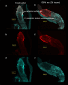
Figure 1
Figure 1
Expression of V-type H+ ATPase and P-type Na+/K+ ATPase protein in rectum of A. taeniorhynchus larvae. Whole mount of the two-part rectum from larval A. taeniorhynchus acclimated to 1% (AC) and 24 h post-transfer from 1 to 100% seawater (DF) showing immunolocalization of V-type H+ ATPase protein (Cy5 blue A, C, D, F) to both the anterior and posterior rectum. P-type Na+/K+ ATPase protein (Cy3 red A, B, D, E) was immunolocalized to the anterior rectum. Figures A and D contain overlays of Cy3 and Cy5 signals. (B and E) contain Cy3 signal only and Figures C and F contain Cy 5 only. Scale bars, 100·μm.
Figure 2
Figure 2
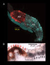
Expression of V-type H+ ATPase protein on the apical membrane of posterior rectal cells and P-type Na+/K+ ATPase protein expressed on basolateral membrane of anterior rectal cells of A. taeniorhynchus larvae. (A) Whole mount of the two-part rectum from larval A. taeniorhynchus acclimated to 100% seawater showing immunolocalization of V-type H+ ATPase protein (blue) to the posterior rectum (PR) and P-type Na+/K+ ATPase protein (red) to the anterior rectum (AR). (B) Sectional preparation of segmented rectum from a larva acclimated to 100% seawater indicating expression of the V-type H+ ATPase protein (dark grey staining, indicated by black arrows), on the apical membrane of PR facing the rectal lumen. P-type Na+/K+ ATPase protein (red staining, indicated by red arrows) was localized to the basolateral membrane of the AR that would face the hemocoele. Scale bars, 100 μm (A) and 25 μm (B).
Figure 3
Figure 3
Expression of V-type H+ ATPase protein in A. taeniorhynchus larvae. Western blot analysis of (VHA) protein with molecular mass standards (left lane) and homogenate of Malpighian tubules (second lane) and posterior recta (third lane) from 1% seawater held larvae and posterior recta (fourth lane) from 150% seawater held larvae revealing a band of ∼ 50 kDa.
Figure 4
Figure 4
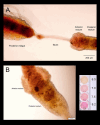
Hindgut lumen pH visualized with phenol red indicator dye of A. taeniorhynchus larvae reared in 100% seawater. Intact alimentary canal was dissected from larvae following one hour incubation ion 0.1% phenol red in 100% seawater medium. Scale of pH colour with Phenol Red diluted in 100% seawater titrated to pH 6.5, 7.0, 7.5 and untitrated seawater with a pH of 8.2.
Figure 5
Figure 5
Expression of NHE3 protein on the apical membrane of posterior rectal cells and P-type Na+/K+ ATPase protein expressed on basolateral membrane of anterior rectal cells of A. taeniorhynchus larvae. Sectional preparation of segmented rectum of larval A. taeniorhynchus acclimated to 100% seawater showing immunolocalization of NHE3 protein (red) using NHE3 polyclonal antibody (A) and NHE3 peptide (C), on the apical membrane of PR facing the rectal lumen (indicated by white arrows). P-type Na+/K+ ATPase protein (green) was immunolocalized to the basolateral membrane of the AR (indicated by white arrows) that would face the hemocoele. (B, D). Scale bars, 50 μm (A, C, D) and 25 μm (B).
Figure 6
Figure 6
Expression of Na+/H+ exchanger isoform 3 (NHE3) protein in A. taeniorhynchus larvae. Western blot analysis of (NHE3) protein with molecular mass standards (right lane) comparing homogenates of posterior recta (first and second lane), anterior recta (third and fourth lane) and Malpighian tubules (fifth and sixth lane) from A. taeniorhynchus larvae acclimated to 1% and 150% seawater. Two bands were detected at 75 and 130 kDa in each tissue. The polyclonal NHE3 antibody was used.
Figure 7
Figure 7
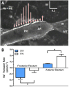
Na+ transport by the rectum of A. taeniorhynchus larvae. (A) A representative scan of Na+ flux at locations along the rectum of larval A. taeniorhynchus. The length and direction of the arrows represent the magnitude and direction of net Na+ transepithelial flux respectively (IN = intestine; MT = Malpighian tubule; Na+ ME = Na+ selective microelectrode). Scale bar units are in pmol cm−2 s−1 (vertical = flux) and mm (horizontal). (B) The rate of Na+ transport at the posterior rectum and the anterior rectum of A. taeniorhynchus larvae reared in freshwater (FW) or seawater (SW). A positive rate of transport indicates efflux (from lumen to bath) and a negative rate of transport represents influx (from bath to lumen). Values are expressed as mean ± SEM with n = 6 for FW and n = 12 for SW. A comparison of Na+ transport rates in FW and SW larvae was performed with unpaired t-test for both posterior (p = 0.02) and anterior rectum (p < 0.001).
Figure 8
Figure 8
Inhibition of Na+ transport at the posterior rectum by V-type H+-ATPase, NHE3 and NKCC inhibitors. Na+ transport rate was measured at the posterior rectum of A. taeniorhynchus larvae that were reared in seawater before and after application of bafilomycin A1 (V-type H+-ATPase inhibitor, 8 μmol l−1), S3226 (NHE3 inhibitor, 1 μmol l−1), bumetanide (NKCC inhibitor, 10 µM) or dimethylsulfoxide (DMSO, inhibitor solvent, 0.8%). Values are expressed as mean ± SEM with n = 9 for bafilomycin, n = 11 for S3226, n = 9 for bumetanide and n = 7 for DMSO. Effects of each treatment were assessed by comparing Na+ transport rates before (Control) and after inhibitor or solvent with a paired t-test (p = 0.024 for bafilomycin; p = 0.025 for S3226; p = 0.017 for bumetanide; p = 0.754 for DMSO). Na+ transport rates of before inhibitor/solvent did not statistically differ from one another (ANOVA, p = 0.502).
Figure 9
Figure 9
Model of Na+ secretion mechanism in the posterior rectum of larval A. taeniorhynchus held in high salinity water. A cation-chloride co-transporter, CCC, transporter is the site of Na+ entry into the cell across the basolateral membrane. The apical V-type H+-ATPase (VHA) pumps protons from the posterior rectal cell into the rectal lumen to establish an electromotive gradient (inside negative). Lumenal protons diffuse down this inward gradient through the apical Na+/H+ exchanger, NHE3, to drive the secretion of Na+ via this exchanger against the large Na+ gradient. This H+- driven Na+ secretion generates the hyperosmotic, Na+-rich urine.

Similar articles

References

    1. Hill, R. W., Wyse, G. A. & Anderson, M. Animal Physiology 4th edn. (Sinauer Associates, Inc. Publishers, 2016).
    1. Nation, J. L. Insect Physiology and Biochemistry 3rd edn. (CRC Press, Taylor & Francis Group, 2016).
    1. Sutcliffe, D. W. Osmotic regulation in the larvae of some euryhaline diptera. Nature187, 331–332. 10.1038/187331a0 (1960). - PubMed
    1. Stobbart, R. H. & Shaw, J. in The Physiology of Insecta, Vol. 3, 190–255 (ed Rockstein, M.) Ch. 4 (Academic Press, 1964).
    1. Beadle, L. C. Regulation of the haemolymph in the saline water mosquito larvae Aedes detritus Edw. J. Exp. Biol.16, 346–362 (1939).