

Potent activity of polymyxin B is associated with long-lived super-stoichiometric accumulation mediated by weak-affinity binding to lipid A
- PMID: 38830951
- PMCID: PMC11148078
- DOI: 10.1038/s41467-024-49200-5
Potent activity of polymyxin B is associated with long-lived super-stoichiometric accumulation mediated by weak-affinity binding to lipid A
Abstract
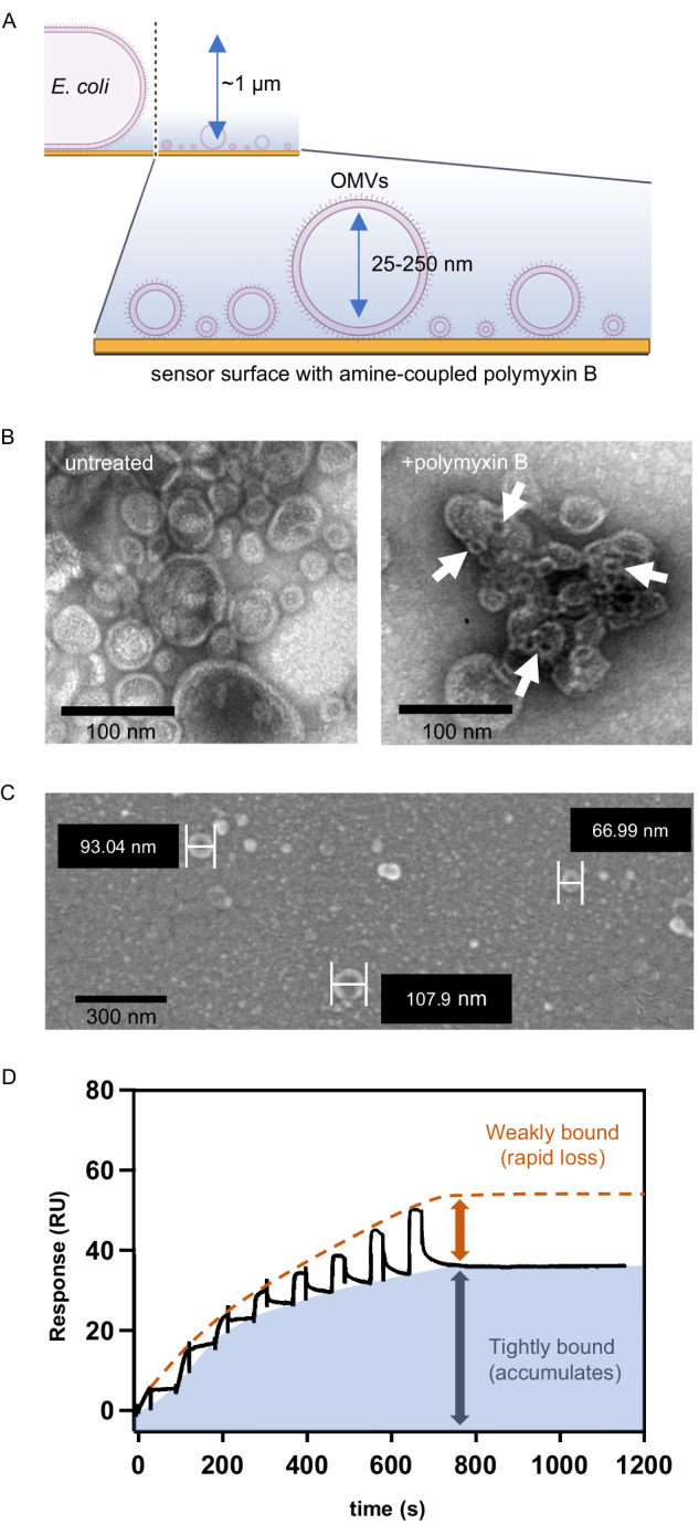
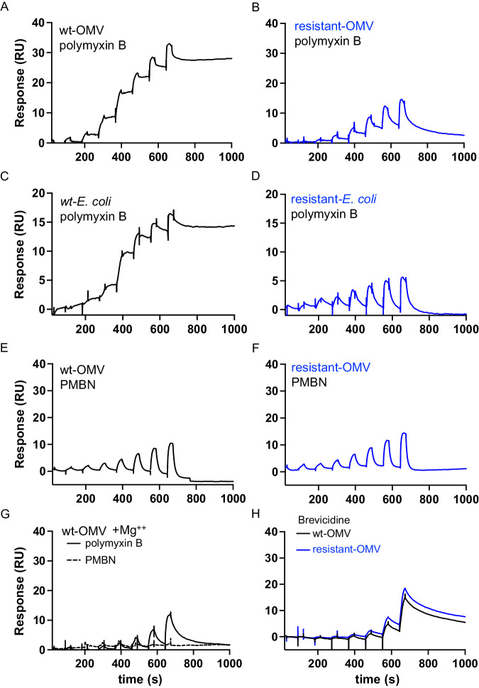
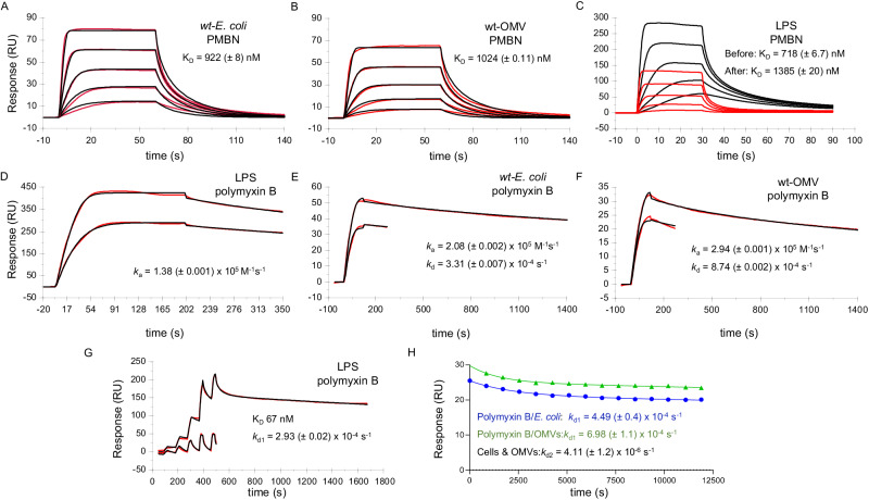
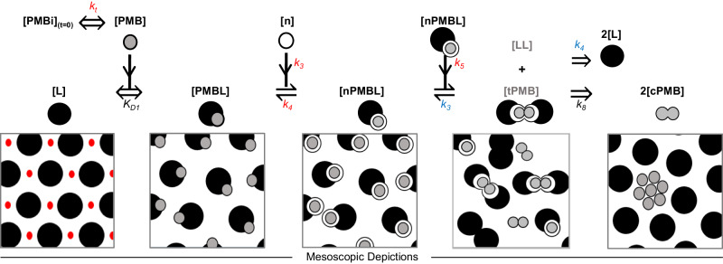
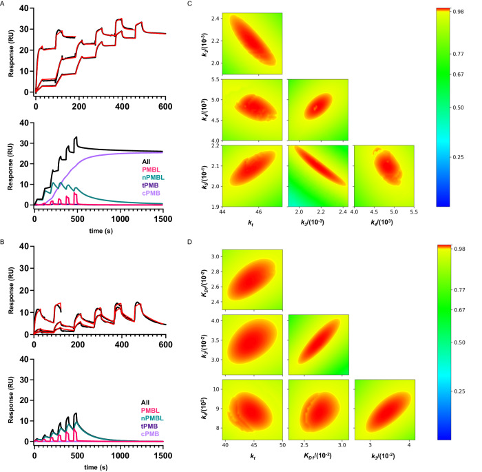
Polymyxins are gram-negative antibiotics that target lipid A, the conserved membrane anchor of lipopolysaccharide in the outer membrane. Despite their clinical importance, the molecular mechanisms underpinning polymyxin activity remain unresolved. Here, we use surface plasmon resonance to kinetically interrogate interactions between polymyxins and lipid A and derive a phenomenological model. Our analyses suggest a lipid A-catalyzed, three-state mechanism for polymyxins: transient binding, membrane insertion, and super-stoichiometric cluster accumulation with a long residence time. Accumulation also occurs for brevicidine, another lipid A-targeting antibacterial molecule. Lipid A modifications that impart polymyxin resistance and a non-bactericidal polymyxin derivative exhibit binding that does not evolve into long-lived species. We propose that transient binding to lipid A permeabilizes the outer membrane and cluster accumulation enables the bactericidal activity of polymyxins. These findings could establish a blueprint for discovery of lipid A-targeting antibiotics and provide a generalizable approach to study interactions with the gram-negative outer membrane.
© 2024. The Author(s).
Conflict of interest statement
All authors, except P.A.S., are employees of Genentech, Inc., a member of the Roche Group. P.A.S. is a former employee of Genentech, Inc., and is currently an employee of Revagenix, Inc.
Figures
Similar articles
-
Molecular basis for the increased polymyxin susceptibility of Klebsiella pneumoniae strains with under-acylated lipid A.Innate Immun. 2013 Jun;19(3):265-77. doi: 10.1177/1753425912459092. Epub 2012 Sep 24. Innate Immun. 2013. PMID: 23008349 Free PMC article.
-
Impact of the cAMP-cAMP Receptor Protein Regulatory Complex on Lipopolysaccharide Modifications and Polymyxin B Resistance in Escherichia coli.J Bacteriol. 2023 May 25;205(5):e0006723. doi: 10.1128/jb.00067-23. Epub 2023 Apr 18. J Bacteriol. 2023. PMID: 37070977 Free PMC article.
-
Pseudomonas aeruginosa MipA-MipB envelope proteins act as new sensors of polymyxins.mBio. 2024 Mar 13;15(3):e0221123. doi: 10.1128/mbio.02211-23. Epub 2024 Feb 12. mBio. 2024. PMID: 38345374 Free PMC article.
-
Prevalence of polymyxin resistance through the food chain, the global crisis.J Antibiot (Tokyo). 2022 Apr;75(4):185-198. doi: 10.1038/s41429-022-00502-0. Epub 2022 Jan 26. J Antibiot (Tokyo). 2022. PMID: 35079146 Review.
-
Mechanisms of bactericidal action and resistance of polymyxins for Gram-positive bacteria.Appl Microbiol Biotechnol. 2020 May;104(9):3771-3780. doi: 10.1007/s00253-020-10525-y. Epub 2020 Mar 10. Appl Microbiol Biotechnol. 2020. PMID: 32157424 Review.
Cited by
-
Depth-Resolved Temperature-Dependent Penetration of Polymyxin B in Phospholipids/Lipopolysaccharide Asymmetric Bilayers.ACS Omega. 2025 Jan 14;10(3):2616-2627. doi: 10.1021/acsomega.4c07648. eCollection 2025 Jan 28. ACS Omega. 2025. PMID: 39895715 Free PMC article.
-
Synergistic Antibacterial Activity of Amorolfine Combined with Colistin Against Acinetobacter baumannii.Int J Mol Sci. 2025 Apr 2;26(7):3312. doi: 10.3390/ijms26073312. Int J Mol Sci. 2025. PMID: 40244182 Free PMC article.
-
Antibiotic-mediated dysbiosis leads to activation of inflammatory pathways.Front Immunol. 2025 Jan 9;15:1493991. doi: 10.3389/fimmu.2024.1493991. eCollection 2024. Front Immunol. 2025. PMID: 39850904 Free PMC article.
-
A critical role for Vibrio parahaemolyticus LPS to mediate evasion of host immune response during infection.Proc Natl Acad Sci U S A. 2025 Aug 19;122(33):e2426547122. doi: 10.1073/pnas.2426547122. Epub 2025 Aug 13. Proc Natl Acad Sci U S A. 2025. PMID: 40802686 Free PMC article.
-
Unlocking the power of antimicrobial peptides: advances in production, optimization, and therapeutics.Front Cell Infect Microbiol. 2025 Apr 28;15:1528583. doi: 10.3389/fcimb.2025.1528583. eCollection 2025. Front Cell Infect Microbiol. 2025. PMID: 40365533 Free PMC article. Review.
References
MeSH terms
Substances
LinkOut - more resources
Full Text Sources
Medical
