

doi: 10.1186/s13089-024-00381-6.
Ultrasound-guided percutaneous tracheostomy: a risk-based protocol
Affiliations
- PMID: 38831088
- PMCID: PMC11147987
- DOI: 10.1186/s13089-024-00381-6
Item in Clipboard
Ultrasound-guided percutaneous tracheostomy: a risk-based protocol
Ultrasound J.
.
No abstract available
Conflict of interest statement
The authors declare that they have no competing interests.
Figures
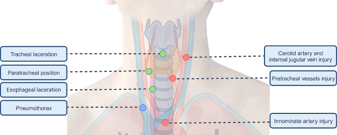
Anatomical relationships of complications associated with percutaneous dilatational tracheostomy. Establishing a safety margin for puncture and dilation is difficult based only on anatomical references and bronchoscopy due to the number of adjacent structures and their anatomical variations (Created with BioRender.com)

Tracheostomy kit

A Step 1: ultrasound exploration with linear transducer, absence of high-riding innominate artery. B Step 1 ultrasound exploration with sector transducer, innominate artery is identified with color Doppler. C Step 2: pretracheal structures identified with color Doppler along the transversal axis of the airway. D Step 3: Lateral margins are defined measuring the distance from the midpoint of the trachea to the lateral margin of the vessel. E The safety area is delimited (Created with BioRender.com)
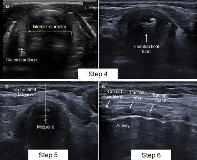
A Step 4: Cricoid cartilage and endotracheal tube visualization in transversal axis. B Step 5. Pretracheal and midpoint distance measurement. C Step 6 Airway longitudinal axis

A Step 7: Endotracheal tube is visualized as two parallel hyperechoic line beyond the trachea. B Step 7: Real-time visualization of tube displacement. C Step 7: Distal end of the tube above cricoid cartilage. D Step 8: Air flush test

A Step 9: Transducer 90° rotation in longitudinal axis and real-time visualization of the needle. B Step 10: Guidewire path between second and third tracheal rings. C Step 11: Tube cuff. D Step 12: Assessment of lung sliding on B-mode and M-Mode, and lung point sign in M-Mode
References
LinkOut - more resources
Full Text Sources