. 2024 Jun 4;23(1):120.
doi: 10.1186/s12943-024-02034-7.

Diversifying the anthracycline class of anti-cancer drugs identifies aclarubicin for superior survival of acute myeloid leukemia patients

Affiliations

Diversifying the anthracycline class of anti-cancer drugs identifies aclarubicin for superior survival of acute myeloid leukemia patients

Xiaohang Qiao et al. Mol Cancer. .

Abstract

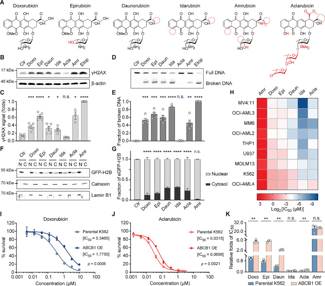
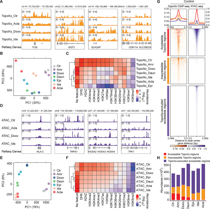
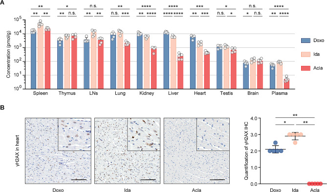
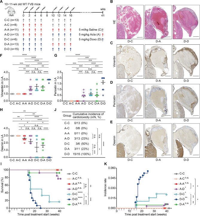
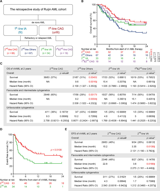
The efficacy of anthracycline-based chemotherapeutics, which include doxorubicin and its structural relatives daunorubicin and idarubicin, remains almost unmatched in oncology, despite a side effect profile including cumulative dose-dependent cardiotoxicity, therapy-related malignancies and infertility. Detoxifying anthracyclines while preserving their anti-neoplastic effects is arguably a major unmet need in modern oncology, as cardiovascular complications that limit anti-cancer treatment are a leading cause of morbidity and mortality among the 17 million cancer survivors in the U.S. In this study, we examined different clinically relevant anthracycline drugs for a series of features including mode of action (chromatin and DNA damage), bio-distribution, anti-tumor efficacy and cardiotoxicity in pre-clinical models and patients. The different anthracycline drugs have surprisingly individual efficacy and toxicity profiles. In particular, aclarubicin stands out in pre-clinical models and clinical studies, as it potently kills cancer cells, lacks cardiotoxicity, and can be safely administered even after the maximum cumulative dose of either doxorubicin or idarubicin has been reached. Retrospective analysis of aclarubicin used as second-line treatment for relapsed/refractory AML patients showed survival effects similar to its use in first line, leading to a notable 23% increase in 5-year overall survival compared to other intensive chemotherapies. Considering individual anthracyclines as distinct entities unveils new treatment options, such as the identification of aclarubicin, which significantly improves the survival outcomes of AML patients while mitigating the treatment-limiting side-effects. Building upon these findings, an international multicenter Phase III prospective study is prepared, to integrate aclarubicin into the treatment of relapsed/refractory AML patients.

Keywords: Aclarubicin; Anthracycline; Bio-distribution; Cardiotoxicity; Chromatin damage; Cross-resistance; DNA damage; Doxorubicin; Histone eviction; Refractory/relapsed acute myeloid leukemia.


Conflict of interest statement

J.N. is a shareholder in NIHM that aims to produce Acla for clinical use. The authors do not declare any other competing interests.

Figures

Fig. 1
Fig. 1
Acla differs from other anthracyclines in mechanisms of action and cross-resistance. (A) Structures of anthracyclines used in this study. Chemical features divergent from Doxo are depicted in red. (B) DNA damage examined by γH2AX Western blot in THP-1 cells. (C) Quantification of the γH2AX signal normalized to β-actin. Data are mean ± SEM; n = 4 biological replicates; Student’s t-test. (D) DSBs analyzed by CFGE in THP-1 cells. (E) Quantification of relative broken DNA in (D). Data are mean ± SEM; n = 4 biological replicates; Student’s t-test. (F) Histone eviction revealed by cell fractionation assay in K562-eGFP-H2B cells. N, nuclear fraction; C, cytosolic fraction. Calnexin and Lamin B1 are the loading control of each fraction. (G) The distribution of eGFP-H2B was quantified for both compartments. Data are mean ± SD; n = 4 biological replicates; two-way ANOVA. (H) IC50 of each anthracycline in different leukemia cell lines. (I, J) Cell viability of parental K562 cells and ABCB1-overexpressing K562 cells upon Doxo (I) and Acla (J) treatment. Data are mean ± SD; n = 3 biological replicates; two-way ANOVA. (K) Relative IC50 folds of each condition compared to that of Doxo in parental K562 cells. Data are mean ± SEM; n = 3 biological replicates; Student’s t-test. *P < 0.05, **P < 0.01, ***P < 0.001, and ****P < 0.0001; n.s., not significant
Fig. 2
Fig. 2
Epigenetic selectivity of TopoIIα redistribution and histone eviction of anthracyclines. (A) Illustration of drug-specific TopoIIα redistribution revealed by ChIP-seq. (B) Principal component analysis (PCA) of TopoIIα ChIP-seq data. Two independent biological replicates were included for each condition. (C) Heatmap of TopoIIα peak abundance associated with specific histone features derived from Roadmap Epigenomics Project. (D) Illustration of drug-specific accessible chromatin regions revealed by ATAC-seq. (E) PCA analysis of ATAC-seq data. Two independent biological replicates were included for each condition. (F) Heatmap of ATAC peak abundance associated with specific histone. (G) Density plots showing the accessible and inaccessible TopoIIα regions, and TopoIIα-excluded accessible regions surrounding ± 2 kb from the center of the detected peaks in the untreated K562 cells. (H) The abundance of each category in different conditions
Fig. 3
Fig. 3
Bio-distribution of clinically used anthracyclines in mice. (A) Drug bio-distribution was determined 4 h after i.v. injection of indicated drug at 5 mg/kg. Data are represented as mean ± SD from 5 mice per group. Student’s t-test. (B) Representative microscopic images of γH2AX IHC staining of the hearts. Scale bars, 100 μm. Quantification is represented as mean ± SD, n = 5, Mann-Whitney test. *P < 0.05, **P < 0.01, ***P < 0.001, and ****P < 0.0001; n.s., not significant
Fig. 4
Fig. 4
Acla is safe and well tolerated following Doxo treatment. (A) Wild-type FVB mice were i.v. injected with Doxo (D), Acla (A), or saline (C) as indicated. (B-E) Representative microscopic images of the left atrium (LA) of heart. Lesions caused by Doxo treatment represent as impairment of the wall, thrombosis, inflammation, fibrosis/calcification, and disappearing of the lumen of atrium (filled by a large thrombus). Scale bars, 500 μm. (F-H) Quantification of the indicated IHC staining in LA. Data are mean ± SEM, Mann-Whitney test. (I) Animal survival is plotted in Kaplan-Meier curves. Log-rank test. (J) Cumulative incidence of cardiotoxicity. Fisher’s exact test. (K) Incidence rate of cardiotoxicity. Two-way ANOVA with repeated measures, two-sided. *P < 0.05, **P < 0.01, ***P < 0.001, and ****P < 0.0001; n.s., not significant
Fig. 5
Fig. 5
CAG is effective in treating r/rAML patients. (A) The therapy overview of Ruijin AML cohort. CAG: cytarabin, aclarubicin, G-CSF; IA: idarubicin, cytarabine; VA: venetoclax, azacitidine; Others: chemotherapy regimens other than CAG. (B) Overall survival of the r/rAML patients from the start of r/rAML induction treatment to the date of death. Log-rank test. (C) The statistical analysis of OS of r/rAML patients at 2 years. Data are n/N (%), * compared with 2nd-line CAG group, † Fisher’s exact test, ‡ Log-rank test, § Mantel-Haenszel test, # compared with 2nd-line CAG patients with favorable and intermediate cytogenetics. (D) Event-free survival of the r/rAML patients from the start of r/rAML induction treatment to the date of relapse/refractory/death. (E) The statistical analysis of EFS of r/rAML patients at 2 years. Data are n/N (%), * compared with 2nd-line CAG group, † Fisher’s exact test, ‡ Log-rank test, § Mantel-Haenszel test, # compared with 2nd-line CAG patients with favorable and intermediate cytogenetics

Similar articles

Cited by

References

    1. Tewey KM, Rowe TC, Yang L, Halligan BD, Liu LF. Adriamycin-induced DNA damage mediated by mammalian DNA topoisomerase II. Science. 1984;226(4673):466–8. doi: 10.1126/science.6093249. - DOI - PubMed
    1. Pang B, Qiao X, Janssen L, Velds A, Groothuis T, Kerkhoven R, et al. Drug-induced histone eviction from open chromatin contributes to the chemotherapeutic effects of doxorubicin. Nat Commun. 2013;4:1908. doi: 10.1038/ncomms2921. - DOI - PMC - PubMed
    1. Pang B, de Jong J, Qiao X, Wessels LF, Neefjes J. Chemical profiling of the genome with anti-cancer drugs defines target specificities. Nat Chem Biol. 2015;11(7):472–80. doi: 10.1038/nchembio.1811. - DOI - PubMed
    1. Yang F, Kemp CJ, Henikoff S. Doxorubicin enhances nucleosome turnover around promoters. Curr Biol. 2013;23(9):782–7. doi: 10.1016/j.cub.2013.03.043. - DOI - PMC - PubMed
    1. Qiao X, van der Zanden SY, Wander DPA, Borras DM, Song JY, Li X, et al. Uncoupling DNA damage from chromatin damage to detoxify doxorubicin. Proc Natl Acad Sci USA. 2020;117(26):15182–92. doi: 10.1073/pnas.1922072117. - DOI - PMC - PubMed