Case Reports
. 2024 Jun 3;19(1):223.
doi: 10.1186/s13023-024-03169-y.

Whole genome sequencing of HER2-positive metastatic extramammary Paget's disease: a case report

Affiliations
Case Reports

Whole genome sequencing of HER2-positive metastatic extramammary Paget's disease: a case report

Boon Yee Lim et al. Orphanet J Rare Dis. .

Abstract

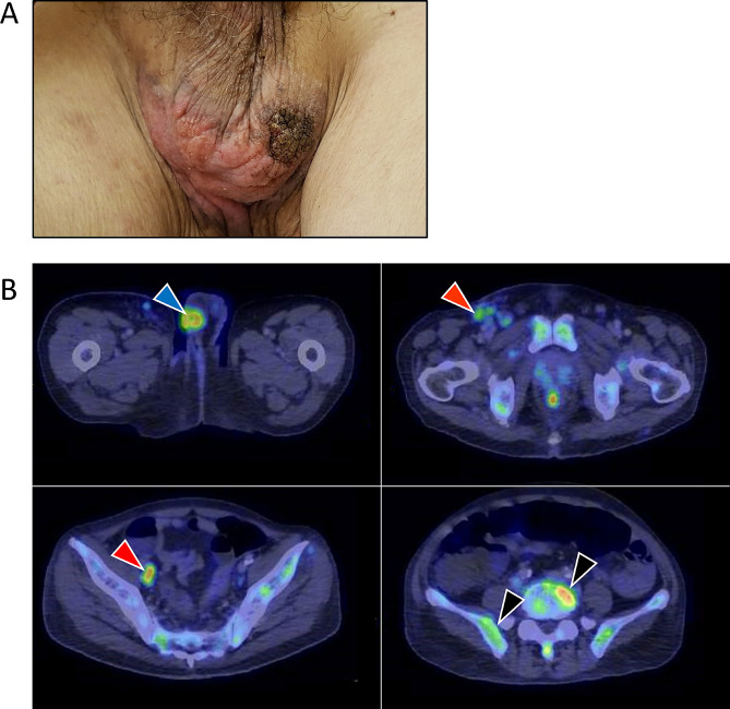
Background: Extramammary Paget's disease (EMPD) is a rare cancer that occurs within the epithelium of the skin, arising predominantly in areas with high apocrine gland concentration such as the vulva, scrotum, penis and perianal regions. Here, we aim to integrate clinicopathological data with genomic analysis of aggressive, rapidly-progressing de novo metastatic EMPD responding to HER2-directed treatment in combination with other agents, to attain a more comprehensive understanding of the disease landscape.

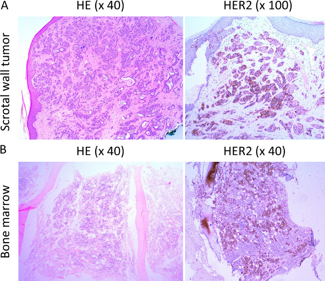
Methods: Immunohistochemical staining on the scrotal wall tumor and bone marrow metastasis demonstrated HER2 overexpression. Whole genome sequencing of the tumor and matched blood was performed.

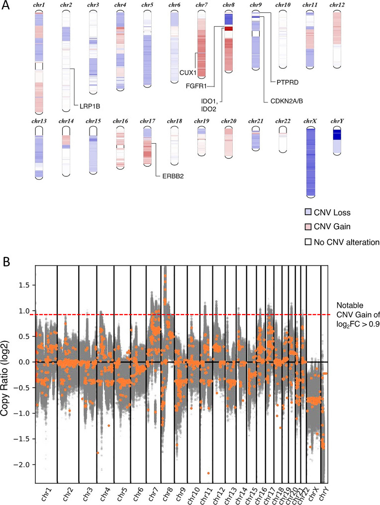
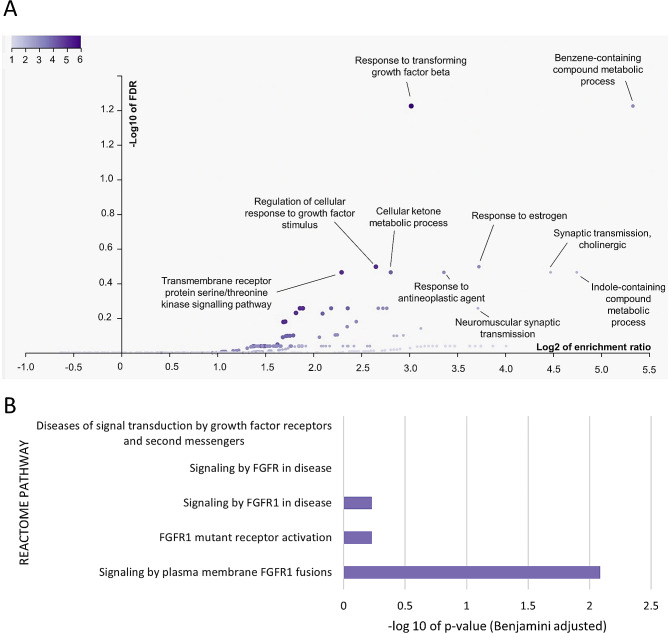
Results: Notable copy number gains (log2FC > 0.9) on chromosomes 7 and 8 were detected (n = 81), with 92.6% of these unique genes specifically located on chromosome 8. Prominent cancer-associated genes include ZNF703, HOOK3, DDHD2, LSM1, NSD3, ADAM9, BRF2, KAT6A and FGFR1. Interestingly, ERBB2 gene did not exhibit high copy number gain (log2FC = 0.4) although 90% of tumor cells stained HER2-positive. Enrichment in pathways associated with transforming growth factor-beta (TGFβ) (FDR = 0.0376, Enrichment Ratio = 8.12) and fibroblast growth factor receptor (FGFR1) signaling (FDR = 0.0082, Enrichment Ratio = 2.3) was detected. Amplicon structure analysis revealed that this was a simple-linear amplification event.

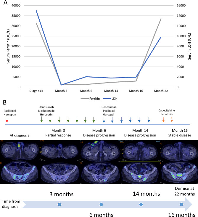
Conclusion: Whole genome sequencing revealed the underlying copy number variation landscape in HER2-positive metastatic EMPD. The presence of alternative signalling pathways and genetic variants suggests potential interactions with HER2 signalling, which possibly contributed to the HER2 overexpression and observed response to HER2-directed therapy combined with other agents in a comprehensive treatment regimen.

Keywords: ERBB2; Next generation sequencing; Precision oncology; Targeted therapy; Trastuzumab.


Conflict of interest statement

The authors declare no conflict of interest.

Figures

Fig. 1
Fig. 1
Clinical and diagnostic features of the patient with metastatic EMPD. (A) Erythematous hyperkeratotic rash with central ulceration over the scrotum. (B) PET/CT imaging. Blue arrow indicates an FDG-avid scrotal wall mass measuring 2.8 cm x 2.5 cm (SUVmax 6.9). Red arrows indicate right iliac and inguinal lymph nodes involvement (SUVmax 6.7). Black arrows indicate extensive FDG-avid mixed lytic-sclerotic bony lesions involving the ribs (not shown), spine and pelvis (SUVmax 9.3)
Fig. 2
Fig. 2
Immunohistochemical (IHC) staining of the scrotal wall tumor and bone marrow presenting HER2 positivity. (A) Representative image of HER2-positive scrotal wall tumor with strong membranous staining on 90% of tumor cells at 100X magnification. (B) Representative image of HER2-positive bone marrow showing HER2 staining on approximately 40% of cells, indicating a case of metastatic EMPD
Fig. 3
Fig. 3
Overall timeline of patient’s disease history. (A) Serial monitoring of serum ferritin and LDH levels in the patient upon diagnosis and at months 3, 6, 14, 16 and 22, presenting changes in a potential serum biomarker levels over time. (B) Chronological representation of the patient’s PET/CT imaging results and drug therapy administered accordingly upon diagnosis and at months 3, 6, 14, and 16. Patient met with eventual demise at month 22 following disseminated metastases
Fig. 4
Fig. 4
WGS was performed on the EMPD tumor and copy number alterations were analysed by CNVkit. (A) Schematic diagram of chromosomal regions exhibiting copy number variation (CNV) in the patient’s genome. Gene loci with copy number losses (Log2FC < 1) are denoted by blue bands located on the chromosome while those with copy number gains (Log2FC > 1) are denoted by red bands. Darker hues of blue indicate the presence of multiple gene loci with copy number losses within that chromosomal region; similarly, darker hues of red indicate the presence of multiple gene loci with copy number gains. (B) Copy numbers of the variants were presented using segmented log2 ratios (y-axis) on the respective chromosomal locations (x-axis). Gray spots indicate each variant and variants with indicative genes denote the orange spots. The orange spots above the dotted red line denote genes exhibiting notable CNV gains (log2FC value greater than 0.9), present on chromosome 7 and 8
Fig. 5
Fig. 5
Functional enrichment analysis of genes in chromosome 8 with notable copy number gain. (A) Gene Ontology Over Representation Analysis (WebGestalt) of genes on chromosome 8 with log2FC > 0.9 reported an enrichment in the Response to TGFβ Pathway (GO:0071559, FDR = 0.0376, Enrichment Ratio = 8.12; genes involved: ADAM9, FNTA, HTRA4, SFRP1, STAR, ZNF703) and the Benzene-containing Compound Metabolic process (GO:0042537, FDR = 0.0376, Enrichment Ratio = 40.2; genes involved: IDO1, IDO2, STAR) (B) DAVID functional annotation clustering analysis identified a FGFR1 fusion pathway (REACTOME Pathway ID: R-HSA-8,853,336, FDR = 0.0082, Enrichment Ratio = 2.3; genes involved: BAG4, ERLIN2, FGFR1).
Fig. 6
Fig. 6
COSMIC mutational signatures identified in the case of EMPD. Proportion of SBS (Single Base Substitutions), ID (Insertion-Deletions) and DBS (Double Base Substitutions) mutational signatures from the WGS data of the EMPD tumor, as determined by mSigAct.

References

    1. Ohara K, Fujisawa Y, Yoshino K, Kiyohara Y, Kadono T, Murata Y, et al. A proposal for a TNM staging system for extramammary Paget disease: retrospective analysis of 301 patients with invasive primary tumors. J Dermatol Sci. 2016;83:234–9. doi: 10.1016/j.jdermsci.2016.06.004. - DOI - PubMed
    1. Zattarin E, Nichetti F, Ligorio F, Mazzeo L, Lobefaro R, Fucà G et al. Case Report: prolonged clinical benefit with sequential trastuzumab-containing treatments in a patient with advanced extramammary paget disease of the groin. Front Oncol 2022;12. - PMC - PubMed
    1. Caruso G, Barcellini A, Mazzeo R, Gallo R, Vitale MG, Passarelli A, et al. Vulvar Paget’s Disease: a systematic review of the MITO Rare Cancer Group. Cancers. 2023;15:1803. doi: 10.3390/cancers15061803. - DOI - PMC - PubMed
    1. St. Claire K, Hoover A, Ashack K, Khachemoune A. Extramammary Paget disease. Dermatol Online J 2019;25. - PubMed
    1. Chow A, Arteaga CL, Wang SE. When Tumor suppressor TGFβ meets the HER2 (ERBB2) Oncogene. J Mammary Gland Biol Neoplasia. 2011;16:81–8. doi: 10.1007/s10911-011-9206-4. - DOI - PMC - PubMed

Publication types