. 2024 Jun 4;29(1):309.
doi: 10.1186/s40001-024-01872-x.

Long non-coding RNA Small Nucleolar RNA Host Gene 4 ameliorates cigarette smoke-induced proliferation, apoptosis, inflammation, and airway remodeling in alveolar epithelial cells through the modulation of the mitogen-activated protein kinase signaling pathway via the microRNA-409-3p/Four and a Half LIM Domains 1 axis

Affiliations

Long non-coding RNA Small Nucleolar RNA Host Gene 4 ameliorates cigarette smoke-induced proliferation, apoptosis, inflammation, and airway remodeling in alveolar epithelial cells through the modulation of the mitogen-activated protein kinase signaling pathway via the microRNA-409-3p/Four and a Half LIM Domains 1 axis

Meng Liu et al. Eur J Med Res. .

Abstract

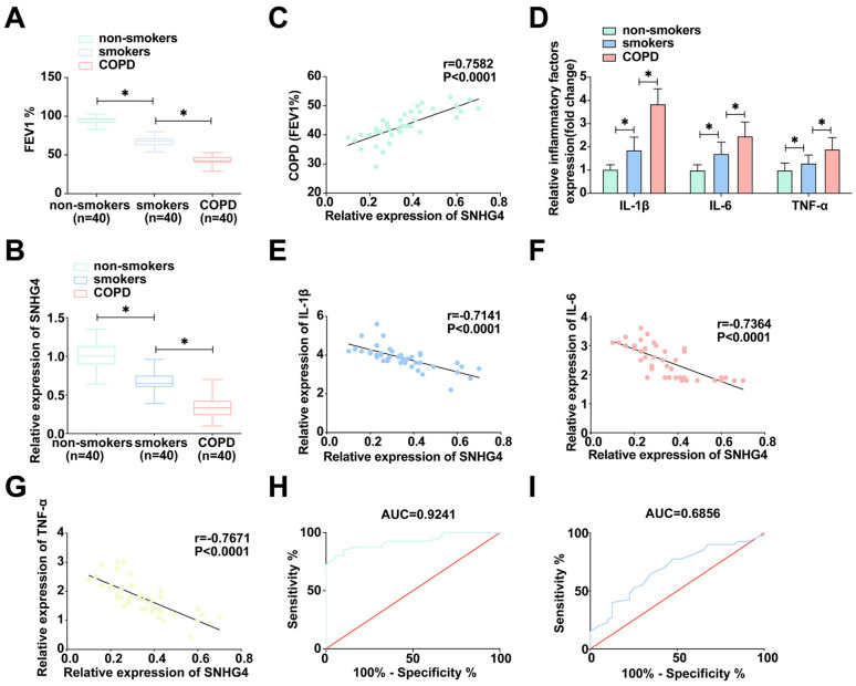
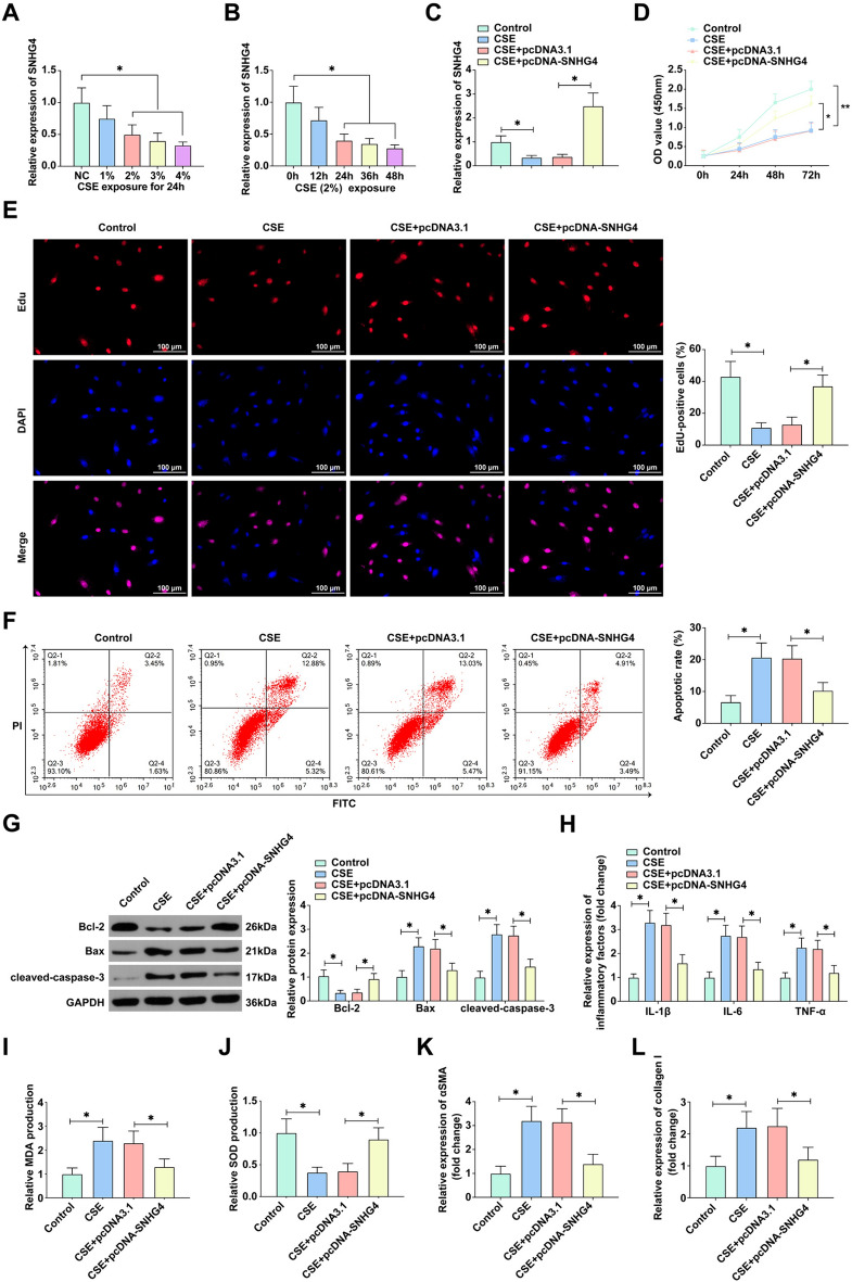
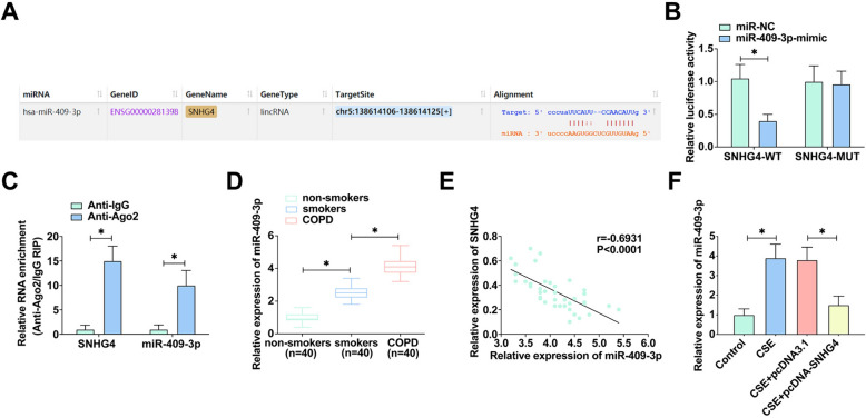
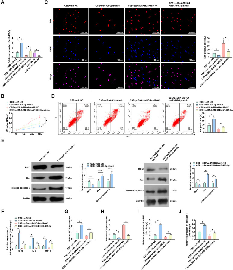
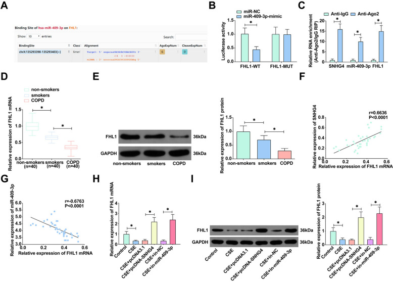
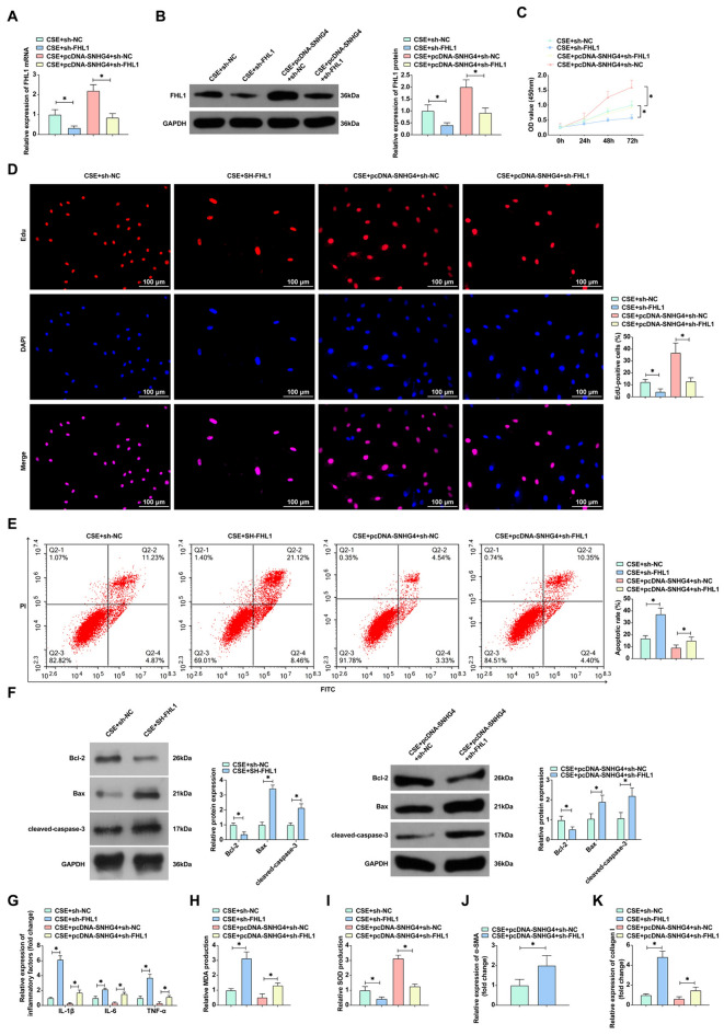
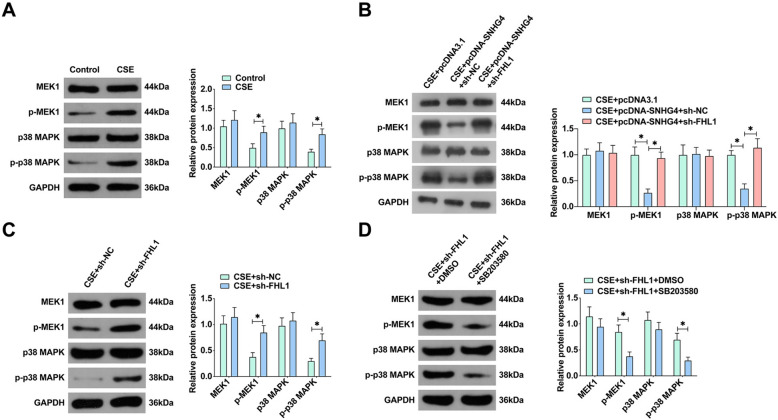
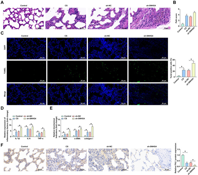
The long non-coding RNA (lncRNA) Small Nucleolar RNA Host Gene 4 (SNHG4) has been demonstrated to be significantly downregulated in various inflammatory conditions, yet its role in chronic obstructive pulmonary disease (COPD) remains elusive. This study aims to elucidate the biological function of SNHG4 in COPD and to unveil its potential molecular targets. Our findings reveal that both SNHG4 and Four and a Half LIM Domains 1 (FHL1) were markedly downregulated in COPD, whereas microRNA-409-3p (miR-409-3p) was upregulated. Importantly, SNHG4 exhibited a negative correlation with inflammatory markers in patients with COPD, but a positive correlation with forced expiratory volume in 1s percentage (FEV1%). SNHG4 distinguished COPD patients from non-smokers with high sensitivity, specificity, and accuracy. Overexpression of SNHG4 ameliorated cigarette smoke extract (CSE)-mediated inflammation, apoptosis, oxidative stress, and airway remodeling in 16HBE bronchial epithelial cells. These beneficial effects of SNHG4 overexpression were reversed by the overexpression of miR-409-3p or the silencing of FHL1. Mechanistically, SNHG4 competitively bound to miR-409-3p, mediating the expression of FHL1, and consequently improving inflammation, apoptosis, oxidative stress, and airway remodeling in 16HBE cells. Additionally, SNHG4 regulated the miR-409-3p/FHL1 axis to inhibit the activation of the mitogen-activated protein kinase (MAPK) pathway induced by CSE. In a murine model of COPD, knockdown of SNHG4 exacerbated CSE-induced pulmonary inflammation, apoptosis, and oxidative stress. In summary, our data affirm that SNHG4 mitigates pulmonary inflammation, apoptosis, and oxidative damage mediated by COPD through the regulation of the miR-409-3p/FHL1 axis.

Keywords: COPD; FHL1; Lnc SNHG4; MAPK; miR-409-3p.


Conflict of interest statement

Authors declared no conflict of interest.

Figures

Fig. 1
Fig. 1
Regulation of SNHG4 in COPD and its correlation with inflammation. A Pulmonary function tests measure FEV1% levels in clinical lung specimens. B The expression of SNHG4 in clinical lung samples is quantified. C Pearson correlation analysis assesses the relationship between SNHG4 and FEV1%. D ELISA quantifies the expression of IL-1β, IL-6, and TNF-α in clinical lung samples. EG Pearson correlation analysis evaluates the correlations between SNHG4 and IL-1β, IL-6, TNF-α, respectively. H ROC analysis of SNHG4 in differentiating COPD patients from non-smokers. I ROC analysis of SNHG4 in distinguishing COPD patients from smokers. Data are presented as mean ± SD (n = 40). *P < 0.05
Fig. 2
Fig. 2
SNHG4 mitigates CSE-induced effects on 16HBE Cell proliferation, apoptosis, inflammation, and airway remodeling. A RT-qPCR assesses SNHG4 expression in 16HBE cells exposed to various concentrations of CSE (1%, 2%, 3%, and 4%) for 24 h. B RT-qPCR measures SNHG4 expression at different time points under 2.5% CSE exposure. C RT-qPCR evaluates the impact of transfection with pcDNA 3.1-SNHG4 on SNHG4 expression in 16HBE cells. D, E Cell proliferation assessed by CCK-8 and EdU assays. F Flow cytometry detects cell apoptosis. G Western blot analyzes the levels of apoptosis-related proteins (Bcl-2, Bax, and cleaved-caspase-3). H ELISA measures the levels of inflammatory cytokines (IL-1β, IL-6, and TNF-α). I, J Oxidative stress response evaluated by MDA assay and SOD activity. K, L ELISA quantifies airway remodeling markers (α-SMA and collagen I). Data are presented as mean ± SD (n = 3). *P < 0.05
Fig. 3
Fig. 3
SNHG4 acts as a sponge for miR-409-3p. A The binding sites between SNHG4 and miR-409-3p predicted by the bioinformatics website https://starbase.sysu.edu.cn. B, C The interaction between SNHG4 and miR-409-3p assessed by dual-luciferase reporter assay and RIP experiment. D RT-qPCR analyzes miR-409-3p expression in various tissues. E Pearson correlation analysis evaluates the correlation between miR-409-3p and SNHG4. F RT-qPCR measures miR-409-3p expression in 16HBE cells under different treatments. Data are presented as mean ± SD (n = 3). *P < 0.05
Fig. 4
Fig. 4
Overexpression of SNHG4 alleviates CSE effects on 16HBE cells by suppressing miR-409-3p expression. A RT-qPCR measures miR-409-3p expression in 16HBE cells under various treatments. B, C Cell proliferation assessed by CCK-8 and EdU assays. D Flow cytometry detects cell apoptosis. E Western blot analyzes expression of apoptosis-related proteins (Bcl-2, Bax, and cleaved-caspase-3). F ELISA measures levels of inflammatory cytokines (IL-1β, IL-6, and TNF-α). G, H Oxidative stress response evaluated by ROS assay and SOD activity. I, J ELISA quantifies airway remodeling markers (α-SMA and collagen I). Data are presented as mean ± SD (n = 3). *P < 0.05
Fig. 5
Fig. 5
miR-409-3p directly targets FHL1. A The binding sites between FHL1 and miR-409-3p predicted by the bioinformatics website https://starbase.sysu.edu.cn. B, C The interaction between FHL1 and miR-409-3p assessed by dual-luciferase reporter assay and RIP experiment. D, E RT-qPCR and Western blot analyze FHL1 expression in clinical lung samples. F, G Pearson correlation analysis evaluates the correlation between FHL1, miR-409-3p, and SNHG4. H, I RT-qPCR and Western blot measure FHL1 expression in 16HBE cells under various treatments. Data are presented as mean ± SD (n = 3). *P < 0.05
Fig. 6
Fig. 6
SNHG4 regulates COPD progression via the miR-409-3p/FHL1 axis. A, B RT-qPCR and Western blot assess the impact of transfection with pcDNA 3.1-SNHG4 and sh-FHL1 on FHL1 mRNA and protein expression. C, D Cell proliferation evaluated by CCK-8 and EdU assays ( ×60). E Flow cytometry measures cell apoptosis. F Western blot analyzes the levels of apoptosis-related proteins (Bcl-2, Bax, and cleaved-caspase-3). G ELISA quantifies the levels of inflammatory cytokines (IL-1β, IL-6, and TNF-α). H, I Oxidative stress response assessed by MDA assay and SOD activity. J, K ELISA measures airway remodeling markers (α-SMA and collagen I). Data are presented as mean ± SD (n = 3). *P < 0.05
Fig. 7
Fig. 7
SNHG4 regulates the MAPK signaling pathway via the miR-409-3p/FHL1 axis. A Western blot analyzes the expression of MAPK pathway proteins in 16HBE cells treated with CSE. B Western blot evaluates the expression of MAPK pathway proteins following co-transfection with pcDNA-SNHG4 and sh-FHL1. C Western blot examines the expression of MAPK pathway proteins after FHL1 knockdown. D Western blot analysis of MAPK pathway protein expression in 16HBE cells treated with CSE, FHL1 knockdown, and MAPK pathway inhibitor SB203580 co-treatment. Data are presented as mean ± SD (n = 3). *P < 0.05
Fig. 8
Fig. 8
Knockdown of SNHG4 exacerbates pathological damage in COPD mice. A Representative images of mouse lung tissues stained with HE (×40); B functional assessment of mouse lung tissues using Penh score; C representative images of TUNEL staining for apoptosis in mouse lung tissues (×40); D ELISA measures the levels of TNF-α, IL-1β, IL-6 in mouse lung tissues BALF; E ELISA and commercial kits assess the levels of MDA, α-SMA, collagen I, SOD in mouse lung tissues; F IHC staining detects FHL1 expression in lung tissues (×40). Data are presented as mean ± SD (n = 6). *P < 0.05

Similar articles

Cited by

References

    1. Tu YH, Guo Y, Ji S, Shen JL, Fei GH. The influenza A virus H3N2 triggers the hypersusceptibility of airway inflammatory response via activating the lncRNA TUG1/miR-145-5p/NF-kappaB pathway in COPD. Front Pharmacol. 2021;12:604590. doi: 10.3389/fphar.2021.604590. - DOI - PMC - PubMed
    1. Zhu B, Wang Y, Ming J, Chen W, Zhang L. Disease burden of COPD in China: a systematic review. Int J Chron Obstruct Pulmon Dis. 2018;13:1353–1364. doi: 10.2147/COPD.S161555. - DOI - PMC - PubMed
    1. Zhang H, Guan R, Zhang Z, Li D, Xu J, Gong Y, et al. LncRNA Nqo1-AS1 attenuates cigarette smoke-induced oxidative stress by upregulating its natural antisense transcript Nqo1. Front Pharmacol. 2021;12:729062. doi: 10.3389/fphar.2021.729062. - DOI - PMC - PubMed
    1. Chen S, Yao Y, Lu S, Chen J, Yang G, Tu L, et al. CircRNA0001859, a new diagnostic and prognostic biomarkers for COPD and AECOPD. BMC Pulm Med. 2020;20(1):311. doi: 10.1186/s12890-020-01333-1. - DOI - PMC - PubMed
    1. Lei Z, Guo H, Zou S, Jiang J, Kui Y, Song J. Long non-coding RNA maternally expressed gene regulates cigarette smoke extract induced lung inflammation and human bronchial epithelial apoptosis via miR-149-3p. Exp Ther Med. 2021;21(1):60. doi: 10.3892/etm.2020.9492. - DOI - PMC - PubMed

MeSH terms