

Evaluation of sacral hiatus changes in children using ultrasound
- PMID: 38831844
- PMCID: PMC11145470
- DOI: 10.1016/j.heliyon.2024.e31526
Evaluation of sacral hiatus changes in children using ultrasound
Abstract
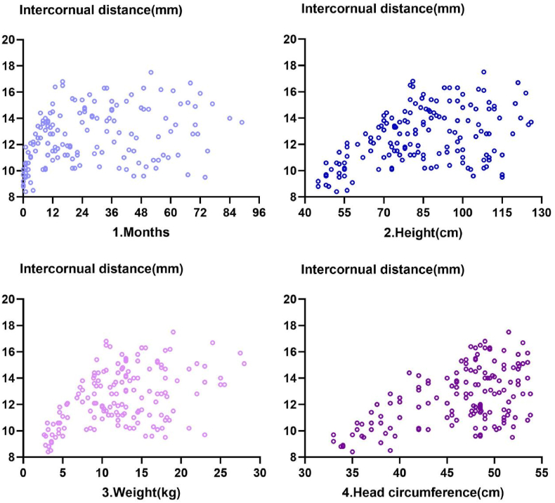
Background and objectives: The intercornual distance in the sacral hiatus has yet to be studied precisely in children. This age-stratified, observational study aimed to clarify the changes in sacral hiatus dimensions and to quantify the correlations between the intercornual distance of the sacral hiatus and age, height, weight, and head circumference by using real-time ultrasonography.
Methods: The patients were stratified into three groups: neonates and infants, toddlers, and schoolchildren. In the operating room, the ultrasonic probe was placed at the sacral cornua to obtain a transverse view of the sacral hiatus, and the intercornual distance was measured three times in millimetres.
Results: The study included a total of 156 patients. The mean ± SD (95%CI) of intercornual distance in neonates and infants (<12 months) was 11.58 ± 1.79 (11.11-12.04) mm, 13.29 ± 1.97 (12.71-13.86) mm in toddlers (13-36 months), and 13.36 ± 2.49 (12.64-14.08) mm in schoolchildren (>36 months).The mean values of neonates and infants were different from those of toddlers and schoolchildren (p < 0.001), but it was similar between toddlers and schoolchildren (p > 0.05, 95 % CI mean difference -1.10 to 0.95).Intercornual distance was correlated with age, height, weight, and head circumference before one year of age (Spearman's R values > 0.7), but there was no correlation thereafter (Spearman's p value > 0.05).
Conclusion: In the first year after birth, the intercornual distance increases rapidly with body growth; after one year of age, the sacral hiatus dimension changes significantly. Ultrasound is superior for assessing the gradually ossified cartilage components in older children.
Keywords: Intercornual distance; Paediatric caudal block; Sacral hiatus dimension; Ultrasound.
© 2024 The Authors. Published by Elsevier Ltd.
Conflict of interest statement
The authors declare that they have no known competing financial interests or personal relationships that could have appeared to influence the work reported in this paper.
Figures


Similar articles
-
Anatomic Differences in the Sacral Hiatus During Caudal Epidural Injection Using Ultrasound Guidance.J Ultrasound Med. 2015 Dec;34(12):2143-8. doi: 10.7863/ultra.14.12032. Epub 2015 Oct 21. J Ultrasound Med. 2015. PMID: 26491092
-
Ultrasonographic Evaluation of Anatomic Variations in the Sacral Hiatus: Implications for Caudal Epidural Injections.Spine (Phila Pa 1976). 2016 Jul 1;41(13):E759-E763. doi: 10.1097/BRS.0000000000001448. Spine (Phila Pa 1976). 2016. PMID: 27340767
-
The myth of the equiangular triangle for identification of sacral hiatus in children disproved by ultrasonography.Reg Anesth Pain Med. 2013 May-Jun;38(3):243-7. doi: 10.1097/AAP.0b013e31828e8a1a. Reg Anesth Pain Med. 2013. PMID: 23558373
-
Surgical anatomy of the sacral hiatus for caudal access to the spinal canal.Acta Neurochir Suppl. 2011;108:1-3. doi: 10.1007/978-3-211-99370-5_1. Acta Neurochir Suppl. 2011. PMID: 21107930 Review.
-
Caudal epidural anesthesia: an anesthetic technique exclusive for pediatric use? Is it possible to use it in adults? What is the role of the ultrasound in this context?Rev Bras Anestesiol. 2011 Jan-Feb;61(1):95-109. doi: 10.1016/S0034-7094(11)70011-3. Rev Bras Anestesiol. 2011. PMID: 21334512 Review.
References
-
- Klocke R., Jenkinson T., Glew D. Sonographically guided caudal epidural steroid injections. J. Ultrasound Med. 2003;22:1229–1232. - PubMed
-
- Giaufré E., et al. Epidemiology and morbidity of regional anaesthesia in children: a one-year prospective Survey of the French language society of pediatric anesthesiologists. Paediatr. Anaesth. 2010;20:1061–1069. - PubMed
-
- Polaner D.M., et al. A multi-institutional study of the use and incidence of complications of pediatric regional anaesthesia. Anesth. Analg. 2012;115:1353–1364. - PubMed
-
- Crighton I.M., Barry B.P., Hobbs G.J. A study of the anatomy of the caudal space using magnetic resonance imaging. Br. J. Anaesth. 1997;78:391–395. - PubMed
-
- Dae H.K., Jung H.P., Sang C.L. Ultrasonographic evaluation of anatomic variations in the sacral hiatus: implications for caudal epidural injections. Spine. 2016;41:759–763. - PubMed
LinkOut - more resources
Full Text Sources
