

Mating-induced Ecdysone in the testis disrupts soma-germline contacts and stem cell cytokinesis
- PMID: 38832826
- PMCID: PMC11190578
- DOI: 10.1242/dev.202542
Mating-induced Ecdysone in the testis disrupts soma-germline contacts and stem cell cytokinesis
Abstract
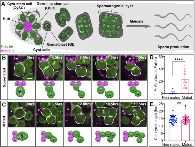
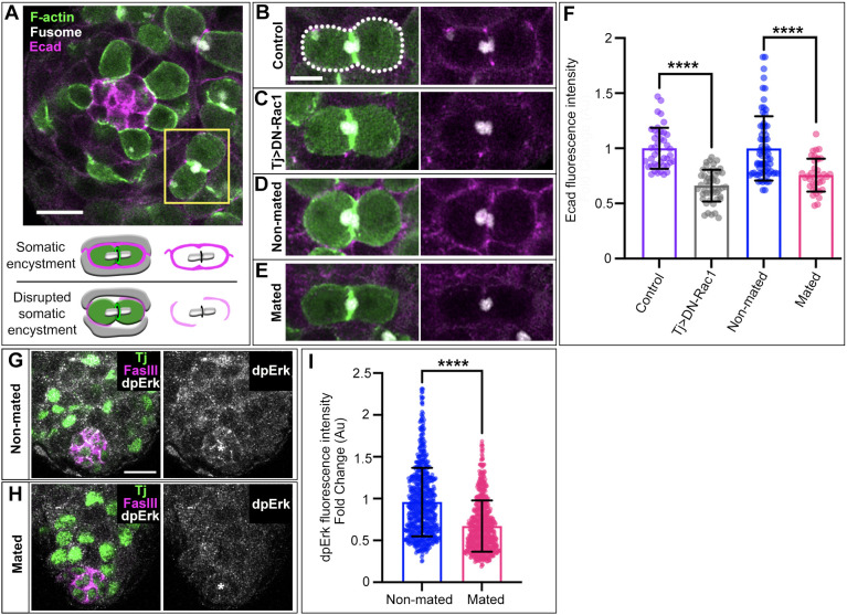
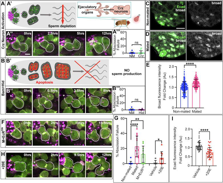
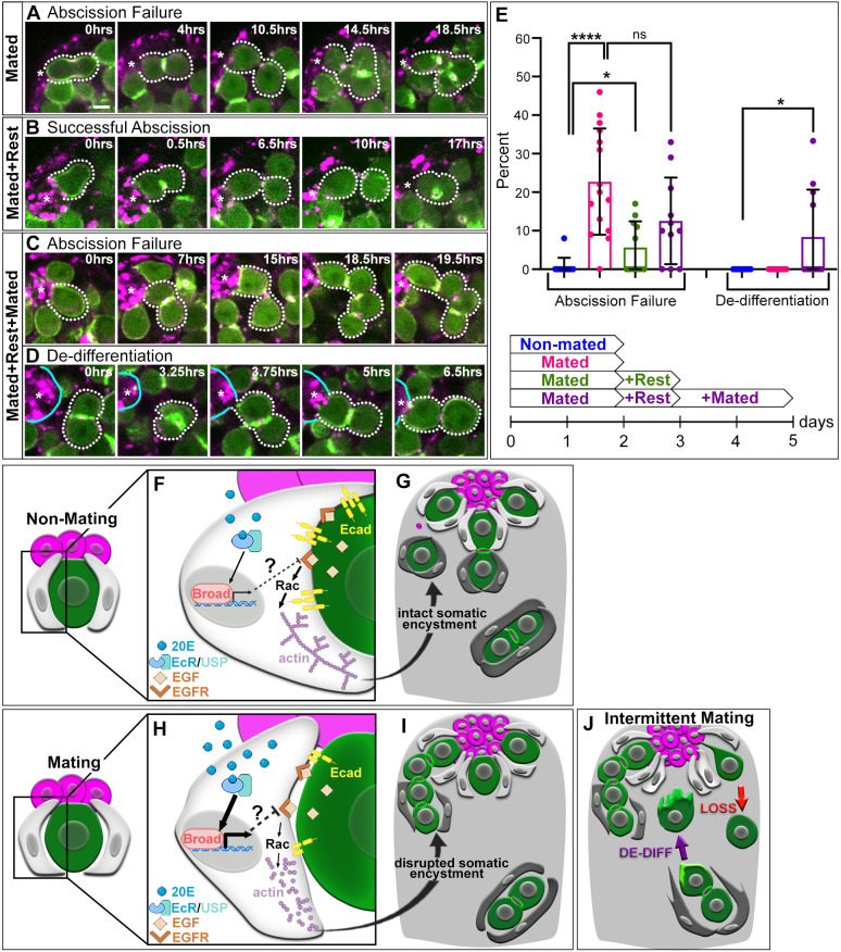
Germline maintenance relies on adult stem cells to continually replenish lost gametes over a lifetime and respond to external cues altering the demands on the tissue. Mating worsens germline homeostasis over time, yet a negative impact on stem cell behavior has not been explored. Using extended live imaging of the Drosophila testis stem cell niche, we find that short periods of mating in young males disrupts cytokinesis in germline stem cells (GSCs). This defect leads to failure of abscission, preventing release of differentiating cells from the niche. We find that GSC abscission failure is caused by increased Ecdysone hormone signaling induced upon mating, which leads to disrupted somatic encystment of the germline. Abscission failure is rescued by isolating males from females, but recurs with resumption of mating. Importantly, reiterative mating also leads to increased GSC loss, requiring increased restoration of stem cells via symmetric renewal and de-differentiation. Together, these results suggest a model whereby acute mating results in hormonal changes that negatively impact GSC cytokinesis but preserves the stem cell population.
Keywords: Drosophila; Cytokinesis; Ecdysone; Encystment; Mating; Stem cells.
© 2024. Published by The Company of Biologists Ltd.
Conflict of interest statement
Competing interests The authors declare no competing or financial interests.
Figures
Update of
-
Mating-induced ecdysone in the testis disrupts soma-germline contacts and stem cell cytokinesis.bioRxiv [Preprint]. 2023 Nov 10:2023.10.16.562562. doi: 10.1101/2023.10.16.562562. bioRxiv. 2023. Update in: Development. 2024 Jun 1;151(11):dev202542. doi: 10.1242/dev.202542. PMID: 37905121 Free PMC article. Updated. Preprint.
Similar articles
-
Mating-induced ecdysone in the testis disrupts soma-germline contacts and stem cell cytokinesis.bioRxiv [Preprint]. 2023 Nov 10:2023.10.16.562562. doi: 10.1101/2023.10.16.562562. bioRxiv. 2023. Update in: Development. 2024 Jun 1;151(11):dev202542. doi: 10.1242/dev.202542. PMID: 37905121 Free PMC article. Updated. Preprint.
-
Somatic cell encystment promotes abscission in germline stem cells following a regulated block in cytokinesis.Dev Cell. 2015 Jul 27;34(2):192-205. doi: 10.1016/j.devcel.2015.05.003. Epub 2015 Jul 2. Dev Cell. 2015. PMID: 26143993 Free PMC article.
-
Coordinated regulation of niche and stem cell precursors by hormonal signaling.PLoS Biol. 2011 Nov;9(11):e1001202. doi: 10.1371/journal.pbio.1001202. Epub 2011 Nov 22. PLoS Biol. 2011. PMID: 22131903 Free PMC article.
-
Molecular control of the female germline stem cell niche size in Drosophila.Cell Mol Life Sci. 2019 Nov;76(21):4309-4317. doi: 10.1007/s00018-019-03223-0. Epub 2019 Jul 12. Cell Mol Life Sci. 2019. PMID: 31300869 Free PMC article. Review.
-
Restricting self-renewal signals within the stem cell niche: multiple levels of control.Curr Opin Genet Dev. 2011 Dec;21(6):684-9. doi: 10.1016/j.gde.2011.07.008. Epub 2011 Aug 19. Curr Opin Genet Dev. 2011. PMID: 21862315 Review.
Cited by
-
Tbx1 ortholog org-1 is required to establish testis stem cell niche identity in Drosophila.bioRxiv [Preprint]. 2025 May 15:2025.05.09.653127. doi: 10.1101/2025.05.09.653127. bioRxiv. 2025. PMID: 40463141 Free PMC article. Preprint.
References
MeSH terms
Substances
Grants and funding
LinkOut - more resources
Full Text Sources
Molecular Biology Databases
Research Materials
