

Analysis of PD-L1 promoter methylation combined with immunogenic context in pancreatic ductal adenocarcinoma
- PMID: 38833018
- PMCID: PMC11150339
- DOI: 10.1007/s00262-024-03745-y
Analysis of PD-L1 promoter methylation combined with immunogenic context in pancreatic ductal adenocarcinoma
Abstract
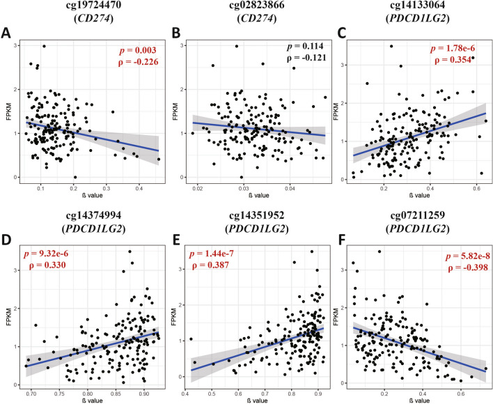
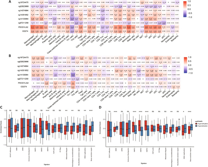
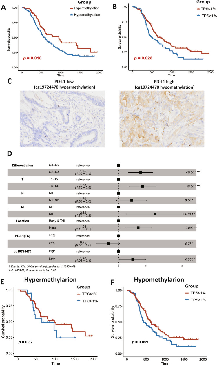
Despite the successful application of programmed cell death ligand 1 (PD-L1)-blocking strategies in some types of cancers and well-established prognostic indicators in pancreatic ductal adenocarcinoma (PDAC), the biological and clinical implications of the methylation status of PD-L1/PD-L2 in PDAC remain largely unknown. Therefore, this study aimed to explore the biological role of PD-L1/PD-L2 methylation and its association with clinicopathological features, clinical outcomes, and the immune microenvironment by analyzing the data on PD-L1/PD-L2 methylation and mRNA expression in PDAC cohorts obtained from the Cancer Genome Atlas and International Cancer Genome Consortium. The correlation between PD-L1 promoter methylation and PD-L1 expression and survival was further validated in an independent validation cohort (Peking Union Medical College Hospital [PUMCH] cohort) using pyrosequencing and immunohistochemistry. These results demonstrated that hypomethylation of the PD-L1 promoter was strongly associated with upregulated PD-L1 expression and shorter overall survival in PDAC. Multivariate Cox regression analyses revealed that the PD-L1 promoter methylation was an independent prognostic factor. PD-L1 promoter hypomethylation and high expression were related to aggressive clinical phenotypes. Moreover, both PD-L1 and PD-L2 methylation correlated with immune cell infiltration and the expression of immune checkpoint genes. PD-L1 promoter methylation status was further validated as an independent prognostic biomarker in patients with PDAC using the PUMCH cohort. The prognostic significance of PD-L1 promoter methylation was more discriminative in tumors with perineural/lymphovascular invasion and distant metastasis than in those without perineural/lymphovascular invasion and distant metastasis. In summary, the methylation status of the PD-L1 promoter is a promising biomarker for survival outcomes, immune infiltration, and the potential immune benefits of immunotherapy in PDAC.
Keywords: Epigenetic biomarker; Methylation; PD-L1; PD-L2; Pancreatic ductal adenocarcinoma; Survival.
© 2024. The Author(s).
Conflict of interest statement
The authors declare that they have no competing interests.
Figures


Similar articles
-
PD-L1 and PD-L2 expression in pancreatic ductal adenocarcinoma and their correlation with immune infiltrates and DNA damage response molecules.J Pathol Clin Res. 2022 May;8(3):257-267. doi: 10.1002/cjp2.259. Epub 2022 Jan 17. J Pathol Clin Res. 2022. PMID: 35037417 Free PMC article.
-
Expression of classical human leukocyte antigen class I antigens, HLA-E and HLA-G, is adversely prognostic in pancreatic cancer patients.Cancer Sci. 2020 Aug;111(8):3057-3070. doi: 10.1111/cas.14514. Epub 2020 Jul 9. Cancer Sci. 2020. PMID: 32495519 Free PMC article.
-
Dipeptidyl peptidase like 6 promoter methylation is a potential prognostic biomarker for pancreatic ductal adenocarcinoma.Biosci Rep. 2020 Jul 31;40(7):BSR20200214. doi: 10.1042/BSR20200214. Biosci Rep. 2020. PMID: 32701143 Free PMC article.
-
The clinicopathological and prognostic significance of PD-L1 expression in pancreatic cancer: A meta-analysis.Hepatobiliary Pancreat Dis Int. 2018 Apr;17(2):95-100. doi: 10.1016/j.hbpd.2018.03.007. Epub 2018 Mar 13. Hepatobiliary Pancreat Dis Int. 2018. PMID: 29576277 Review.
-
Immune cell infiltrates as prognostic biomarkers in pancreatic ductal adenocarcinoma: a systematic review and meta-analysis.J Pathol Clin Res. 2021 Mar;7(2):99-112. doi: 10.1002/cjp2.192. Epub 2021 Jan 22. J Pathol Clin Res. 2021. PMID: 33481339 Free PMC article.
Cited by
-
Programmed Cell Death Ligand as a Biomarker for Response to Immunotherapy: Contribution of Mass Spectrometry-Based Analysis.Cancers (Basel). 2025 Mar 17;17(6):1001. doi: 10.3390/cancers17061001. Cancers (Basel). 2025. PMID: 40149335 Free PMC article. Review.
-
Epigenetic Landscape of DNA Methylation in Pancreatic Ductal Adenocarcinoma.Epigenomes. 2024 Nov 3;8(4):41. doi: 10.3390/epigenomes8040041. Epigenomes. 2024. PMID: 39584964 Free PMC article. Review.
-
Advances in pancreatic cancer epigenetics: From the mechanism to the clinic.World J Gastrointest Oncol. 2025 Jul 15;17(7):106238. doi: 10.4251/wjgo.v17.i7.106238. World J Gastrointest Oncol. 2025. PMID: 40697230 Free PMC article. Review.
References
MeSH terms
Substances
Grants and funding
LinkOut - more resources
Full Text Sources
Medical
Research Materials
