. 2024 Jun 4;11(1):16.
doi: 10.1186/s40708-024-00230-1.

Brain age gap estimation using attention-based ResNet method for Alzheimer's disease detection

Affiliations

Brain age gap estimation using attention-based ResNet method for Alzheimer's disease detection

Atefe Aghaei et al. Brain Inform. .

Abstract

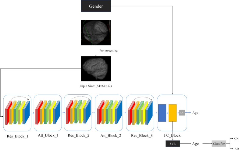
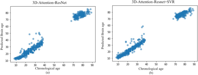
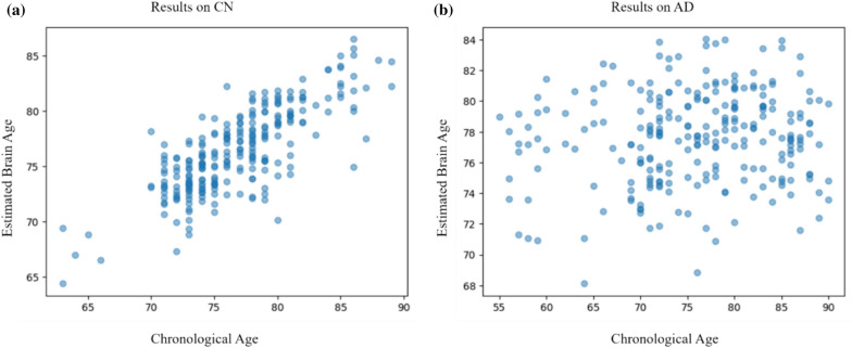
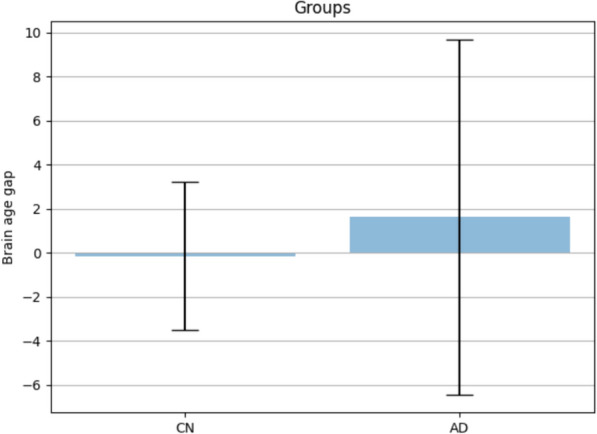
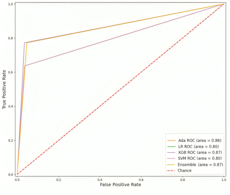
This study investigates the correlation between brain age and chronological age in healthy individuals using brain MRI images, aiming to identify potential biomarkers for neurodegenerative diseases like Alzheimer's. To achieve this, a novel attention-based ResNet method, 3D-Attention-Resent-SVR, is proposed to accurately estimate brain age and distinguish between Cognitively Normal (CN) and Alzheimer's disease (AD) individuals by computing the brain age gap (BAG). Unlike conventional methods, which often rely on single datasets, our approach addresses potential biases by employing four datasets for training and testing. The results, based on a combined dataset from four public sources comprising 3844 data points, demonstrate the model's efficacy with a mean absolute error (MAE) of 2.05 for brain age gap estimation. Moreover, the model's generalizability is showcased by training on three datasets and testing on a separate one, yielding a remarkable MAE of 2.4. Furthermore, leveraging BAG as the sole biomarker, our method achieves an accuracy of 92% and an AUC of 0.87 in Alzheimer's disease detection on the ADNI dataset. These findings underscore the potential of our approach in assisting with early detection and disease monitoring, emphasizing the strong correlation between BAG and AD.

Keywords: 3D-Resnet; Alzheimer’s disease; Attention; Brain age gap; Structural MRI.


Conflict of interest statement

The authors declare no competing interests.

Figures

Fig. 1
Fig. 1
An overview of the proposed method
Fig. 2
Fig. 2
preprocessing of the images. The results on the sagittal side of one image from ABIDE dataset. (To show the results better, we show some slices of the 3D volume). a; the original image, b: after brain extraction using the brain mask, c: After N4 bias field correction
Algorithm 1
Algorithm 1
Brain age prediction using attention based 3DResNet and SVR
Algorithm 2
Algorithm 2
Alzheimer’s disease detection using brain age gap
Fig. 3
Fig. 3
the distribution of the chronological age of the combination of the introduced datasets. The horizontal axis is the age group and the vertical axis is the count of each age group a: the distribution of the chronological age of 2883 training data and b the distribution of the chronological age of 961 test data
Fig. 4
Fig. 4
The Heatmap (The most important parts of the brain which are involved in brain age estimation in our proposed method) of the brain of three different subjects, which the age of subject a and b are more than 60 and subject c is between 30 and 40 years old. The most important parts for brain age estimation are marked with a circle which are ventricles, hippocampus, and some parts of parietal lobe
Fig. 5
Fig. 5
the scatter plot of the chronological age and the brain age estimated using the proposed method on 961 test data. a the results without the SVR and b the results using the SVR model
Fig. 6
Fig. 6
Comparison of FLU and ReLU activation function. a: ELU, b: ReLU
Fig. 7
Fig. 7
the scatter plot of the chronological age and the brain age estimated using the proposed method on 500 test data of ADNI dataset. a the results on Cognitively 250 Normal subjects and b the results on 250 Alzheimer’s disease patients
Fig. 8
Fig. 8
Brain Age Gap of the 250 Cognitively Normal (CN) and 250 Alzheimer’s disease (AD) subjects
Fig. 9
Fig. 9
The ROC curve of Alzheimer’s disease classification using Logistic Regression, Adaboost, XGboost, SVM and Ensemble learning

References

    1. Fox NC, Schott JM. Imaging cerebral atrophy: normal ageing to Alzheimer’s disease. Lancet. 2004;363:392–394. doi: 10.1016/S0140-6736(04)15441-X. - DOI - PubMed
    1. Cole JH. Multimodality neuroimaging brain-age in UK biobank: relationship to biomedical, lifestyle, and cognitive factors. Neurobiol Aging. 2020;92:34–42. doi: 10.1016/j.neurobiolaging.2020.03.014. - DOI - PMC - PubMed
    1. Peng H, Gong W, Beckmann CF, Vedaldi A, Smith SM. Accurate brain age prediction with lightweight deep neural networks. Med Image Anal. 2021 doi: 10.1016/j.media.2020.101871. - DOI - PMC - PubMed
    1. Jonsson BA, et al. Brain age prediction using deep learning uncovers associated sequence variants. Nat Commun. 2019 doi: 10.1038/s41467-019-13163-9. - DOI - PMC - PubMed
    1. Franke K, Ziegler G, Klöppel S, Gaser C. Estimating the age of healthy subjects from T1-weighted MRI scans using kernel methods: exploring the influence of various parameters. Neuroimage. 2010 doi: 10.1016/j.neuroimage.2010.01.005. - DOI - PubMed

LinkOut - more resources